, Hans Guski2 and Glen Kristiansen3
(1)
Carl-Thiem-Klinikum, Institut für Pathologie, Cottbus, Germany
(2)
Vivantes Klinikum Neukölln, Institut für Pathologie, Berlin, Germany
(3)
Universität Bonn, UKB, Institut für Pathologie, Bonn, Germany
15.1 Diagnostic Antibody Panel for Mesothelial Tumors
15.2 Diagnostic Antibody Panel for Epithelial Tumors of Müllerian Type
Cytokeratin profile, CEA, CA125, PAX-8, WT-1, p53, and p16 (see ovarian tumors).
15.3 Diagnostic Antibody Panel for Smooth Muscle Tumors
Actin, h-caldesmon, calponin, and cytokeratin profile.
15.4 Diagnostic Antibody Panel for Tumors of Uncertain Origin and Miscellaneous Peritoneal Primary Tumors
CD34, CD99, DOG-1, actin, h-caldesmon, desmin, ALK, and cytokeratin profile.
Cytokeratin Profile
All mesothelial tumors are positive for pan-cytokeratin and the cytokeratins 5/6/7/8/10/14/18 but typically lack the expression of cytokeratin 20. Consequently, the cytokeratin profile alone cannot discriminate between mesotheliomas and metastatic carcinomas. It is important to consider that submesothelial fibroblasts are usually positive for pan-cytokeratin and other keratins that maybe a source of misinterpretation.
Calretinin | ||
|---|---|---|
Expression pattern: cytoplasmic | ||
Main diagnostic use | Expression in other tumors | Expression in normal cells |
Mesothelioma, adrenocortical tumors, ovarian sex cord-stromal tumors | Squamous cell carcinoma, ameloblastoma, thymic tumors, transitional cell carcinoma, colonic carcinoma, granular cell tumor, fibrosarcoma, PEComa, myxoid chondrosarcoma, synovial sarcoma, desmoplastic small round cell tumor, atrial myxoma, lipogenic tumors, mast cell lesions | Central and peripheral neural cells, ganglion cells, neuroendocrine cells, mesothelial cells, mast cells, steroid-producing cells (Leydig and Sertoli cells, adrenal cortex cells, ovarian theca interna, and surface cells), endometrium, eccrine glands, thymus, adipose tissue |
Positive control: appendix | ||
Diagnostic Approach
Calretinin is an intracellular neuron-specific calcium-binding vitamin D-dependent protein expressed in various epithelial, mesenchymal, and central and peripheral neurogenic tissue types. Calretinin is strongly expressed in normal and neoplastic mesothelial cells and considered as an important mesothelioma marker (Fig. 15.1). Calretinin is also a marker for mast cells and steroid-producing cells and tumors derived from these cells, namely, sex cord-stromal tumors including granulosa cell tumor, Sertoli and Leydig cell tumors, gonadoblastoma, and gynandroblastoma in addition to adrenocortical tumors. About one third of squamous cell carcinomas shows also different calretinin expression intensity. Calretinin is also widely expressed in different soft tissue tumors such as synovial sarcoma, chondrosarcoma, desmoplastic small round cell tumor, lipoma, and liposarcoma [4, 5]. Moreover, calretinin is an optimal marker to highlight ganglion cells in colonic biopsies for the diagnosis of Hirschsprung disease.
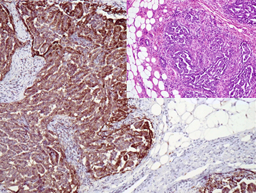

Fig. 15.1
Calretinin highlighting mesothelioma cells infiltrating the chest wall
Diagnostic Pitfalls
Calretinin has a wide expression spectrum, and the calretinin positivity alone is not enough for the diagnosis of mesothelioma.
Thrombomodulin (CD141) | ||
|---|---|---|
Expression pattern: membranous | ||
Main diagnostic use | Expression in other tumors | Expression in normal cells |
Mesothelioma, transitional cell carcinoma | Squamous cell carcinoma, trophoblastic tumors, vascular tumors, synovial sarcoma | Endothelial cells, urothelium, mesothelial cells, keratinizing epithelial cells, monocytes, neutrophils, platelets/megakaryocytes, meningeal cells, smooth muscle cells, syncytiotrophoblasts, synovial lining cells, osteoblasts |
Positive control: appendix | ||
Diagnostic Approach
Thrombomodulin (also known as endothelial anticoagulant protein, clustered as CD141) is a transmembrane glycoprotein expressed on the surface of endothelial cells and taking part in the regulation of intravascular coagulation. The expression of thrombomodulin is characteristic for other cell and tissue types including mesothelial cells, squamous epithelial cells, and transitional epithelium of the urinary tract. Thrombomodulin is a useful screening antibody for mesothelioma, transitional cell carcinoma, and squamous cell carcinoma in addition to vascular tumors (Fig. 15.2). Thrombomodulin is usually negative in sarcomatoid mesothelioma. To discriminate between thrombomodulin-positive tumors, it is important to use other more specific markers. Thrombomodulin is constantly negative in renal cell carcinoma, prostatic carcinoma, gastrointestinal adenocarcinoma, and endometrioid carcinoma.
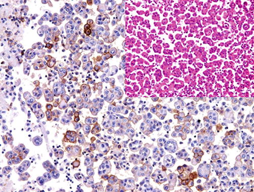

Fig. 15.2
Thrombomodulin labeling mesothelioma cells in malignant pleural effusion
Mesothelin | ||
|---|---|---|
Expression pattern: membranous | ||
Main diagnostic use | Expression in other tumors | Expression in normal cells |
Mesothelioma, non-mucinous ovarian surface carcinomas | Adenocarcinoma of different origin, acinar cell carcinoma and squamous cell carcinoma | Mesothelial cells, renal tubules, tracheal and tonsil epithelial cells, fallopian tube mucosa |
Positive control: appendix | ||
Diagnostic Approach
Mesothelin is a glycoprotein located on the cell surface of mesothelial cells in addition of some other types of epithelial cells.
Diagnostic Pitfalls
Mesothelin labels mesothelioma in addition to other carcinoma types including ovarian, pancreatic, and pulmonary carcinoma and adenocarcinomas. Generally, mesothelin is a screening antibody and cannot be considered as a specific mesothelioma marker. Sarcomatoid mesothelioma is negative for mesothelin.
WT-1:
WT-1 is one of the important mesothelioma markers discussed in a previous chapter. In mesothelioma cells, WT-1 has a nuclear expression pattern and can be used as the double stain in combination with other markers exhibiting membranous stain.
Podoplanin:
Podoplanin (also known as D2-40) is a mucoprotein expressed on the membrane of lymphatic endothelium discussed in the chapter of vascular tumors. Podoplanin is not specific for lymphatic endothelium but also expressed in other cell and tumor types such as meningeal cells, germ cells and germ cell tumors, mesothelial cells and mesothelioma in addition to many other mesenchymal tumors (Fig. 15.3) [6, 7].
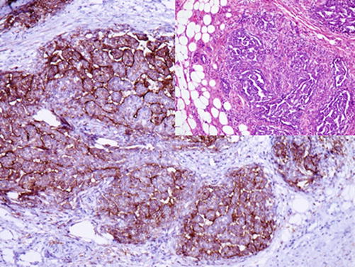

Fig. 15.3
Podoplanin (D2-40) labeling the cells of mesothelioma
GLUT1 | ||
|---|---|---|
Expression pattern: membranous | ||
Main diagnostic use | Expression in other tumors | Expression in normal cells |
Malignant mesothelioma vs. reactive mesothelial hyperplasia Benign endometrial hyperplasia vs. atypical hyperplasia | Perineurioma, hemangioma, chordoma, epithelioid sarcoma, wide range of carcinomas of different origin | Red blood cells, testicular germinal cells, renal tubules, placental trophoblasts, brain capillaries, perineural cells |
Positive control: mesothelioma | ||
Diagnostic Approach
Glucose transporter 1 (GLUT1) is a member of the GLUT transporter family and a membrane-associated erythrocyte glucose transport protein maintaining the basal glucose transport in most cell types. GLUT1 is not a tissue-specific marker but expressed in a wide range of epithelial and non-epithelial tumors. In diagnostic histopathology, GLUT1 is a potential marker for malignant transformation as it is overexpressed in many types of malignant epithelial and non-epithelial tumors. It is helpful marker to discriminate between malignant mesothelioma and reactive proliferation of mesothelial cells. GLUT1 is a helpful marker to distinguish between hemangioma usually positive for GLUT1 and vascular malformation, pyogenic granuloma, and granulation tissue lacking the expression of GLUT1.
Diagnostic Pitfalls
GLUT1 is a hypoxia-inducible factor (HIF) target gene which is also induced by the hypoxia-inducible factor 1α (HIF-1α) [8]. Consequently, hypoxic areas will also overexpress GLUT1.
Insulin-Like Growth Factor II mRNA-Binding Protein 3:
IMP3 is a cytoplasmic oncofetal protein mediating RNA trafficking and cell growth expressed in fetal tissue and different premalignant and malignant lesions. Benign adult tissue usually lacks the expression of IMP3 with the exception of the ovarian and testicular tissue, placenta, endocrine cells, and brain. In routine immunohistochemistry, IMP3 is used to discriminate between malignant and reactive proliferative lesions. Similar to GLUT1, IMP3 is a helpful marker to discriminate between mesothelioma and reactive mesothelial proliferation, as the majority of benign mesothelial cells are negative for IMP3 (Fig. 15.4) [9].
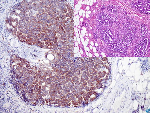

Fig. 15.4
IMP3 expression in malignant mesothelioma
IMP3 is also a selective marker for Hodgkin cells; however, it can be also found some extrafollicular blasts or cells of B-cell lymphoma. Furthermore, IMP3 is a helpful marker to discriminate between serous endometrial carcinoma positive for IMP3 and endometrioid carcinoma negative for IMP3 [10].
Stay updated, free articles. Join our Telegram channel

Full access? Get Clinical Tree


