, Hans Guski2 and Glen Kristiansen3
(1)
Carl-Thiem-Klinikum, Institut für Pathologie, Cottbus, Germany
(2)
Vivantes Klinikum Neukölln, Institut für Pathologie, Berlin, Germany
(3)
Universität Bonn, UKB, Institut für Pathologie, Bonn, Germany
Diagnostic Antibody Panel for Fibroblastic, Myofibroblastic, and Fibrohistiocytic Tumors
Vimentin, actin, desmin, CD34, and CD68
Vimentin | ||
|---|---|---|
Expression pattern: cytoplasmic | ||
Main diagnostic use | Expression in other tumors | Expression in normal cells |
Mesenchymal tumors | Metaplastic carcinoma, endometrioid carcinoma, carcinomas of salivary glands, follicular thyroid carcinoma, clear cell renal cell carcinoma, hepatocellular carcinoma, poorly differentiated carcinomas of different origin | Cells of mesenchymal origin: fibrocytes and fibroblasts, lipocytes, smooth muscle cells, endothelium, macrophages, myoepithelial cells, thyroid follicular cells, adrenal cortex, renal tubules, mesangial cells of renal glomerulus, pancreatic acinar cells, melanocytes, lymphocytes, astrocytes, Schwann cells |
Positive control: appendix | ||
Diagnostic Approach
Vimentin is a 57-kDa protein, a member of the type III family of intermediate filaments, expressed in all mesenchymal cells forming an important part of the cytoskeleton of these cells. The type III family of intermediate filaments includes vimentin, desmin, GFAP, and peripherin. Vimentin is generally expressed in all primitive cells in the early embryogenesis and be replaced by other intermediate filaments during maturation and differentiation.
Diagnostic Pitfalls
The use of vimentin as a single marker is of limited diagnostic value as the co-expression of vimentin with other different cytokeratins has been demonstrated in many types of epithelial cells and tumors such as carcinomas of the lung, salivary glands, liver and biliary tract, thyroid gland, adrenal cortex, kidney, endometrium, gonads and meningioma (Fig. 22.1). Generally, poorly differentiated carcinomas may acquire vimentin expression with loss of keratins and finally resulting a sarcomatoid phenotype. For diagnostic purposes, vimentin can be only used as a part of diagnostic antibody panel.
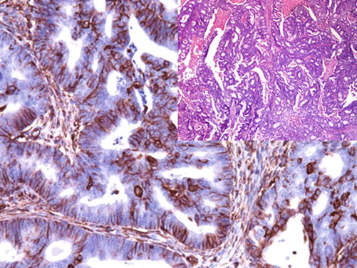

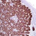
Fig. 22.1
Neoplastic glands of endometrioid carcinoma exhibiting strong vimentin expression
STAT-6:
STAT-6 is a member of the STAT family of cytoplasmic transcription factors involved in the modulation of signal transmission between DNA promoters and cell receptors. The inv. (12)(q13;q13) is a chromosomal aberration characteristic for solitary fibrous tumor generating the NAB2-STAT6 fusion transcript causing the overexpression of the STAT-6 protein, which is a characteristic immunohistochemical marker for solitary fibrous tumor (Fig. 22.2). This chromosomal abnormality affects also the promoter of the ERG-1 gene causing the overexpression of the ERG-1 transcription factor, which can be a further marker for this tumor identity [1, 2].
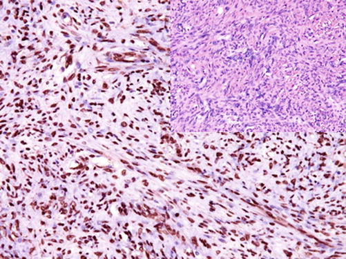

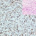
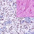
Fig. 22.2
Strong nuclear STAT-6 expression in the cells of solitary fibrous tumor
Diagnostic Pitfalls
Mucin-4:
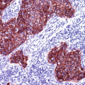
MUC-4 is a transmembrane mucoprotein mentioned in a previous chapter with other mucins. In addition to glandular epithelial tumors, the expression of MUC-4 is also a characteristic marker for low-grade fibromyxoid sarcoma, sclerosing epithelioid fibrosarcoma, and glandular components in biphasic synovial sarcoma [6, 7].
Immunoprofile of fibroblastic and myofibroblastic tumors | ||||
|---|---|---|---|---|
Tumor type | + in >90% (+) | + in 50–90% (+/−) | + in 10–50% (−/+) | + in < 10% (−) |
Nodular fasciitis : | Vimentin | Actin, CD68 | Desmin, S100, CD34, pan-CK, EMA | |
Proliferative fasciitis : | Vimentin, myoglobin
Stay updated, free articles. Join our Telegram channel
Full access? Get Clinical Tree
 Get Clinical Tree app for offline access

| |||
