, Hans Guski2 and Glen Kristiansen3
(1)
Carl-Thiem-Klinikum, Institut für Pathologie, Cottbus, Germany
(2)
Vivantes Klinikum Neukölln, Institut für Pathologie, Berlin, Germany
(3)
Universität Bonn, UKB, Institut für Pathologie, Bonn, Germany
27.1 Diagnostic Antibody Panel for Tumors of the Central Nervous System
GFAP, MAP2, NeuN, Olig-2, neurofilaments, synaptophysin, pan-cytokeratin, Ki-67.
Glial fibrillary acidic protein (GFAP) | ||
|---|---|---|
Expression pattern: cytoplasmic | ||
Main diagnostic use | Expression in other tumors | Expression in normal cells |
CNS tumors (astrocytoma, glioblastoma, oligodendroglioma, medulloblastoma, ependymoma), retinoblastoma, neurilemoma, neurothekeoma, MPNST | Salivary gland tumors (myoepithelial tumors, basal cell adenoma/carcinoma, pleomorphic adenoma), neuroblastoma, osteosarcoma, chondrosarcoma | Astrocytes, subset of CNS ependymal cells, cells of choroid plexus, Schwann cells, Kupffer cells, myoepithelial cells, chondrocytes |
Positive control: brain tissue | ||
Diagnostic Approach
Glial fibrillary acidic protein (GFAP) is a member of class III of intermediate filament proteins. GFAP is mainly expressed in neuroglia including astrocytes and ependymal cells. Lower expression levels are found in Schwann cells, paraganglial cells, enteric glial cells, Kupffer cells of the liver, osteocytes, chondrocytes, and myoepithelial cells. GFAP is a marker of neoplastic glial cells and glial differentiation. Lower GFAP expression level is also found in neurilemoma and neuroblastoma.
Diagnostic Pitfalls
GFAP is an important marker to discriminate between primary brain and metastatic tumors; however, it can be expressed in non-glial tumors such as myoepithelioma and myoepithelial component of different types of salivary gland tumors, osteosarcoma, chondrosarcoma, and angiosarcoma.
Microtubule-Associated Protein 2 (MAP2):
MAP2 is one of the five members of the microtubule-associated protein family. This protein is a neuron-specific cytoskeletal protein found in three isoforms a, b, and c expressed in neurons and reactive astrocytes. MAP2 labels the cytoplasm of the neuronal cell body and basal dendrites and is considered as an early marker for neuronal differentiation. In immunohistochemistry, MAP2 is used as a marker of neuronal differentiation. Positive stain is found in glial tumors, medulloblastoma, neuroblastoma, pulmonary neuroendocrine tumors, a subset of melanomas, and some carcinoma types (mainly thyroid and prostate).
Neuronal Nuclear Antigen (NeuN):
NeuN (also known as FOX-3 protein) is a low molecular weight protein localized in the nuclei and cytoplasm of most neuronal cells of the central and peripheral nervous system and tumors derived from these cells. NeuN is a marker for central neurocytoma and gangliogliomas. The majority of PNETs of the CNS and medulloblastoma are also NeuN positive. Less than 5% of astrocytic and oligodendroglial tumors show NeuN expression.
Oligodendrocyte Lineage Transcription Factor 2 (Olig-2):
Olig-2 is a transcription factor involved in the regulation of neuroectodermal progenitor cells and development of oligodendrocytes and motoneurons. Normally, Olig-2 is strongly expressed in oligodendroglial cells and oligodendroglioma. Weak to moderate Olig-2 expression is also found in all other gliomas including glioblastoma. Olig-2 expression is also reported in neuroendocrine carcinomas and in a small subset of central neurocytoma and supratentorial ependymoma.
27.2 Diagnostic Antibody Panel for Meningeal Tumors
S100, podoplanin, nestin, claudin-1, pan-cytokeratin, EMA, CEA, vimentin, Ki-67.
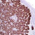
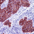
Characteristic for meningeal tumors is the co-expression of EMA, pan-cytokeratin, and S100. Other markers such as podoplanin (D2 40) are useful to confirm the diagnosis mainly in aggressive tumor types such as atypical and anaplastic meningioma (Fig. 27.1). For the assessment of tumor grade, the estimation of Ki-67 proliferation index is essential. The CEA expression is characteristic for the pseudopsammoma bodies found in secretory meningioma (Fig. 27.2).
Immunoprofile of central nervous system tumors | ||||
|---|---|---|---|---|
Tumor type | + in >90% (+) | + in 50–90% (±) | + in 10–50% (∓) | + in <10% (−) |
A. Astrocytic tumors | ||||
− Pilocytic astrocytoma (grade I) − Diffuse astrocytoma − (grade II) − Anaplastic astrocytoma (grade III) − Glioblastoma (grade IV) | GFAP, S100, NSE, Olig-2, bcl-2 (only in gemistocytic astrocytoma) Proliferation index (Ki-67): Diffuse astrocytoma: <5% Anaplastic astrocytoma: 5–10% Glioblastoma: >15% (5–40%) | CD56, CD99, HER-2 | Synaptophysin, pan-CKa | Chromogranin, CK7, CK20, neurofilaments |
Diffuse midline glioma (grade IV) | CD56, Olig-2, S100
Stay updated, free articles. Join our Telegram channel
Full access? Get Clinical Tree
 Get Clinical Tree app for offline access

| |||
