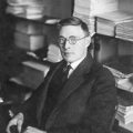
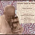
Felix Mandl. (Reprinted from [15], Copyright 2000, with permission from Elsevier)
Early Life and Training
Felix Mandl M.D. son of industrialist Emil Mandl and Linda Basch Mandl, was born on November 8, 1892, in the city of Brno (Brünn in German) located in the modern-day Czech Republic [2]. After graduating from his local German high school, Mandl entered the medical school of the University of Vienna in 1910 [2]. His medical training was disrupted during World War I when he served in the Austro-Hungarian army from 1914 to 1919 [2]. Shortly thereafter, he received his M.D. degree in 1919 and entered surgical residency under Julius Von Hochenegg in the Department of Surgery II at Vienna General Hospital [1]. He worked as a surgical trainee assuming the position of chief resident in 1923 and was promoted to lecturer in surgery at the University of Vienna in 1928. In 1932, he became the director of the surgery department at the S Canning-Childs Hospital and Research Institute in Vienna [3].
Albert Jahne and the First Parathyroidectomy
It was during his early surgical training that Felix Mandl made surgical history by performing the first successful parathyroidectomy for a patient suffering from osteitis fibrosa cystica (von Recklinghausen’s disease of the bone). In 1924, Albert Jahne, a 34-year-old streetcar conductor in Vienna , was admitted to the Department of Surgery II, University of Vienna. Jahne had begun developing progressive lower-extremity weakness and bone pain in 1919. By October 1924, the 38-year-old Jahne required crutches to ambulate and roentgen (X-ray) analysis demonstrated cystic lesions in his pelvis and femurs [3]. The prevailing hypothesis for Jahne’s bone lesions at that time was based on Viennese pathologist Jacob Erdheim’s (1874–1937) theory that osteitis fibrosa cystica was due to a deficiency of parathyroid hormone and that parathyroid hyperplasia was secondary to increased calcium metabolism in bone [4]. Erdheim first recognized an association between the parathyroid glands and calcium levels in 1906. Experimentally, he determined that cauterization of the parathyroid glands of laboratory rats resulted in both tetany and defective mineralization of teeth. Additionally, he observed devascularization and destruction of the parathyroid glands at postmortem examination in three patients who died of postoperative tetany following thyroidectomy for goiter and later identified parathyroid hyperplasia in patients with osteomalacia [3].
Given Erdheim’s prevailing hypothesis of compensatory parathyroid hyperplasia in response to osteitis fibrosa cystica , Albert Jahne was initially treated with “parathyreoidin tablets,” a concentrate of parathyroid extract [3]. However, his symptoms continued to worsen, and he developed a pathologic fracture of his left femur on December 12, 1924. His fracture healed by February 1925 but over the next 4 months he became cachectic, was no longer able to ambulate due to lower-extremity pain, and he developed a white precipitate in his urine. On June 22, 1925, he was readmitted to the hospital. On July 2, 1925, still operating under Erdheim’s hypothesis of compensatory parathyroid hyperplasia , Mandl transplanted four parathyroid glands from a patient who died from trauma into Jahne’s preperitoneal space [3]. Although Jahne’s symptoms did not improve, Mandl had directly tested Erdheim’s hypothesis in a patient, thereby providing evidence against the theory of compensatory parathyroid hyperplasia in the pathophysiology of osteitis fibrosa cystica .
Consequently, Mandl hypothesized that Jahne’s bone lesions were not compensatory due to a deficiency of parathyroid hormone but rather due to an excess of parathyroid hormone . On July 30, 1925, Mandl set out to test his hypothesis by taking Jahne to the operating room to perform a cervical exploration for a parathyroid tumor [3]. In the operating room, Mandl discovered that “in the rim between larynx and esophagus, a dark, partly grayish nodule separate from the thyroid was located in the inferior parathyroid position close to the inferior thyroid artery and between its branches, adhering to the left recurrent nerve.” The 2.5 × 1.5 × 1.2-cm grayish-white nodule was surgically removed, and further exploration of the neck revealed that the remaining three parathyroid glands were macroscopically normal [3]. Pathologic analysis by Erdheim and his colleagues concluded that
the cell structure is typical for parathyroid tissue…it is most likely a so-called ‘atypical (fetal) adenoma’ of the parathyroid gland with polymorphous cell structure which may be a ‘malignant degeneration.’ In one part of the tumor there is a hyaline round-shaped patch with numerous pigmented cells (remnants of a previous hemorrhage). There is no evidence of a rim of normal parathyroid tissue. [3]
Subsequently, Jahne’s urinary calcium excretion decreased from a preoperative level of 54–7.6 mg % by postoperative day 11 and he did not develop tetany [3]. He was discharged from the hospital on August 7, 1925, and made a rapid recovery: His strength improved, his body weight increased, his lower-extremity pain subsided, he was able to begin ambulating with a cane, and X-rays of his bones demonstrated healing of his cystic lesions and an increase in his bone density. In effect, Mandl had performed the first successful parathyroidectomy . He presented the case at the Vienna Congress of Physicians on December 4, 1925, and subsequently published his findings [4–8]. At clinical follow-up in 1929, Jahne remained asymptomatic and biochemical analysis demonstrated “blood calcium levels between 13 and 14 mg/100 mL (normal range, 8.4 to 10.4 mg/100 mL) and urinary calcium excretions between 264 and 300 mg/100 mL (normal range, < 200 mg %)” [3]. Ultimately, Mandl had demonstrated that Jahne’s osteitis fibrosa cystica was secondary to a hyperfunctioning parathyroid gland, thereby disproving Erdheim’s compensatory parathyroid hyperplasia hypothesis that had stood for nearly 20 years.
Unfortunately, Jahne’s symptoms began to return in 1931, and, by January 1933, he was bedridden. He was diagnosed clinically and biochemically with recurrent hyperparathyroidism and on October 18, 1933, Mandl took Jahne to the operating room for a repeat cervical exploration to examine for another parathyroid tumor. According to Mandl, “there was neither a local recurrence nor another adenoma in the entire neck and in the mediastinum explored through the neck” [3]. Jahne’s symptoms and biochemical parameters did not improve over the next 3 years and in February 1936, he passed away from uremia. Postmortem examination demonstrated generalized cystic fibrosis with brown tumors in multiple bones and bilateral hydronephrosis with nephrocalcinosis [3]. Ultimately, Jahne’s clinical course raised the question of parathyroid carcinoma but no metastatic parathyroid tissue was found at autopsy [4]. An excellent and detailed English translation of the operative, pathology, and clinical reports regarding the case of Albert Jahne was published by Niederle et al. in 2006 [3].
Stay updated, free articles. Join our Telegram channel

Full access? Get Clinical Tree


