Fig. 18.1
MRI of the abdomen with intravenous contrast showing hepatic metastases indicated by red arrows measuring approximately 1.2 × 1 cm (posterior lesion) and 0.8 × 0.9 cm (anterior lesion)
Staging of MTC
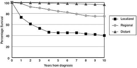
The staging system most commonly used for MTC is the Tumor-Node-Metastases (TNM) staging system adopted by the American Joint Committee on Cancer. This staging system is based on tumor size, invasion of the tumor, the presence of nodal metastases, and the presence of distant metastases. Overall survival in all cases of MTC is approximately 75 %, with the TNM staging system providing good discrimination as follows in one short-term study: stage I with 0 % mortality, stage II with 13 % mortality, stage III with 56 % mortality, and stage IV with 100 % mortality. An alternate staging system developed by the National Thyroid Cancer Treatment Cooperative Study Group provides similar separation of outcomes for a population affected by MTC. Survival is significantly affected by the whether disease is confined to the thyroid, affects local and regional lymph nodes only, or involves distant sites (see Fig. 18.2). A comparison of several of these staging systems found the European Organization for Research and Treatment of Cancer (EORTC) staging system to have the best predictive value [5]. In retrospect, our patient was stage IV by all staging systems at the time of her diagnosis. Approximately 50 % of similarly staged patients are alive at 5 years. However, such staging systems do not incorporate other important prognostic indicators such as age and calcitonin doubling time.


Fig. 18.2
Disease specific survival for patients with medullary thyroid cancer according to site of disease metastases based on Surveillance, Epidemiology, and End Results (SEER) data. From Roman et al., Cancer 107: 2134–2142, 2006, with permission
Calcitonin Doubling Time
The calcitonin doubling time is a powerful prognostic indicator of survival in MTC. In one study when calcitonin doubling time was less than 6 months, as it was in our patient after her initial therapy, 25 % and 8 % of such patients survived 5 years and 10 years, respectively. In the same study, although TNM stage, EORTC score, and calcitonin doubling time were significant predictors of survival by univariate analysis, only calcitonin doubling time remained an independent predictor of survival by multivariate analysis [6]. A tool that can be used to calculate calcitonin doubling time is available on the website of the American Thyroid Association (http://www.thyroid.org/thyroid-physicians-professionals/calculators/thyroid-cancer-carcinoma/).
Side Effects of Tyrosine Kinase Inhibitors
Molecularly targeted and orally active antineoplastic agents are increasingly used for the treatment of MTC [7]. Tyrosine kinases are involved in signaling, and thereby stimulate tumor proliferation, angiogenesis, invasion, and metastases. TKIs may have inhibitory effects at multiple levels, but inhibition of angiogenesis seems to be an important part of their action. Both vandetanib and cabozantinib are approved agents for the treatment of patients with progression of structural disease. As these agents are now available outside of clinical trials, a restricted distribution program has been put in place to monitor their use and provide oversight. Common side effects of these agents include anorexia, transaminitis, diarrhea, fatigue, weight loss, and QT interval prolongation. A comprehensive review of the experience of the University of Texas MD Anderson Cancer Center with the use of these agents for MTC and non-MTC has recently been published [8]. Our patient suffered the typical side effects of vandetanib, including anorexia, diarrhea, fatigue, and QT prolongation. The hand and foot syndrome typically affects approximately 30 % of patient taking sorafenib and sunitinib, but does not affect those taking vandetanib.
Common and Rare Complications of External Beam Radiotherapy
Radiotherapy to the neck and mediastinum are often considered after initial surgery in those with residual disease. For example the National Comprehensive cancer Network recommends that external beam radiotherapy (EBRT) is considered for “gross extrathyroidal extension (T4a or T4b) with positive margins after resection of all gross disease and following resection of moderate- to high-volume disease in the central or lateral neck lymph nodes with extra-nodal soft tissue extension.” Our patient certainly fell into that category. Slight improvements have been reported in local disease-free survival after EBRT for selected patients. In one study, high-risk patients with microscopic residual disease, extraglandular invasion, or lymph node involvement had a higher local/regional relapse free rate of 86 % at 10 years compared with a relapse free rate of 52 % in those who did not receive postoperative external beam radiation [9].
Unfortunately, there are significant toxicities associated with external beam radiotherapy. These include mucositis, radiation dermatitis, dysphagia, fatigue, taste changes, xerostomia, and hoarseness. Sometimes such side effects necessitate a break from treatment. Patients undergoing radiation therapy may require a tracheostomy or percutaneous endoscopic gastrostomy tube placement. Although this is typically due to progressive local disease, occasionally this may be due to pharyngeal or laryngeal edema associated with the therapy. Our patient suffered a catastrophic but rare complication associated with both the progression and management of head and neck cancers, namely rupture of the carotid artery [10]. This carotid “blowout” is associated with radiotherapy, nodal metastases, and neck dissection. Radiation therapy has been implicated in the pathophysiology of carotid blowout because it causes changes in the vasa vasorum and adventitia that lead to subsequent weakening of the arterial wall. In one retrospective study 14 of 17 patients with carotid artery rupture had received radiation therapy, with the rate of rupture being about eightfold higher in radiated compared with nonirradiated patients.
Stay updated, free articles. Join our Telegram channel

Full access? Get Clinical Tree


