Fig. 11.1
Atypical femur fracture treated with intramedullary nail showing sclerotic margins consistent with nonunion
Complete fractures are treated most commonly with intramedullary devices; more proximal fractures that are difficult to control with nails are considered for plating [20]. When selecting intramedullary fixation, surgeons must be very careful with the choice of device as fracture union is not as reliable as in the patient without bisphosphonate exposure. Problems with outcomes from atypical fracture nailing can be attributed in part to start point selection, nail design, and shaft/nail mismatch.
The insertion point of the nail can be the piriformis fossa or the greater trochanter, and the design of the nail may or may not allow for fixation into the neck. Extension of fixation into the neck (cephalomedullary nailing) theoretically extends the length of bone protected by the device and alleviates the concern of initiating a secondary femoral neck or basal fracture (as with a piriformis entry nail). The difficulty with cephalomedullary devices , however, is that the patients have a thickened proximal cortex and a narrowed canal. The large size of the proximal part of the nail encounters the thickened cortex on insertion and may force a proximal fracture into a varus deformity. If left malreduced, healing time and outcome results suffer significantly in this patient population [18]. In addition to varus deformity, iatrogenic fracture of the proximal femur on nail insertion is more common in patients with atypical fractures [20]. If selecting a greater trochanteric start point, surgeons should stay medial on the trochanter with the guidewire to decrease the chances of this complication, as well as help prevent a varus malreduction. High subtrochanteric fractures may require piriformis nails to avoid the varus driving force that is present in greater trochanteric entry cephalomedullary devices; plate and screw constructs are also a viable option in these difficult patterns.
Once start point and device have been carefully selected, obtaining and maintaining an anatomic reduction are paramount to the success of treatment in atypical femur fractures [18]. If any evidence of malreduction exists after closed reduction attempts, surgeons should be prepared to perform an open reduction, whether on a fracture table or a flat radiolucent table. Reductions should be maintained after passing the guidewire; this leads to symmetrical reaming of the proximal and distal segments of the fracture. Reaming with a malreduction in place reinforces the poor alignment and may prevent future attempts to restore anatomy. As reaming progresses, over-reaming the canal beyond the typical 1.5 mm may be necessary to have ample room for the proximal component of the nail. Over-reaming also helps if there is bowing of the femur. Matching the femur to the curve of the nail is critical to prevent anterior perforation of the shaft on nail insertion. Femoral bowing is related to age and ethnicity; Asian populations in particular have been found to have shorter radii of curvature [21, 22].
The postoperative course with intramedullary devices should allow the patient to bear weight as soon as comfortable. Calcium, vitamin D, and perhaps anabolic agent should be started. There is evidence in the literature that administration of a bone-forming agent such as teriparatide in the postoperative course leads to faster union times [14, 15, 23]. Bisphosphonate drugs should be stopped. The opposite femur should be continuously monitored secondary to the increasing weight requirements in the setting of a healing contralateral limb. Atypical fractures have occurred even when the original screening X-rays were negative at the time of the contralateral fracture.
If there is a delay in healing, further augmentation or revision surgery may be warranted. The Hernigou technique, which applies mesenchymal stem cells possibly coupled with an anabolic agent, or a formal bone grafting procedure can be tried in the setting of delayed union [24]. There is no evidence to suggest that BMP will augment delayed union in this group of patients. If the patient shows no progressive bone formation over a 3-month period, they are diagnosed with nonunion. Usually, the nonunion appears atrophic without appreciable callous formation, likely secondary to the metabolic derangements in the setting of bisphosphonates. Infection should always be considered, and cultures should always be taken at the time of revision. Options for treatment include exchange nailing of the femur or conversion to a plate and screw construct (Fig. 11.2 ). In either setting, the ability of the body to produce bone, not just the hardware, needs revision and augmentation. Locally, bone grafting should be used to enhance the regional environment; systemically, ensuring proper calcium and vitamin D levels along with consideration for anabolic pharmacologic therapy optimizes the systemic bone-forming capability of the patient.
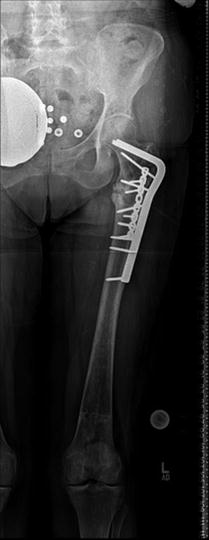

Fig. 11.2
The patient has undergone revision surgery for prior nonunion with blade plate and screws. Also treated with Forteo postoperatively with evidence of periosteal bridging and callous formation
From a surgical standpoint, both exchange nailing and conversion to a plate and screw construct have pitfalls. Exchange nailing should only be considered in the setting of anatomic alignment in the diaphysis. Varus deformity, even if subtle, lengthens the healing time and is a risk factor for nonunion [18]. Correcting a varus nonunion deformity of the proximal femur after a cephalomedullary nailing procedure with an exchange nailing is dangerous because the varus positioning often persists; the new nail falls into the same reamed track as the first device. In the setting of a varus deformity, blade plating with correction of deformity and compression across the fracture is a better option. Bone grafting and systemic anabolic medication should be employed as discussed above. Compression is usually gained through an articulated tensioning device, as the fracture often has a near transverse plane. The downside of plating is the lengthy period of weight-bearing restriction that follows; 3 months of non-weight bearing is advised with plates secondary to lengthy expected union times. Because of this delay, a second plate orthogonal to the first may be added to give additional fixation and torque control to the construct. There is no data as to whether further augmentation with allograft struts would be beneficial at this period of time.
Incomplete Fractures
Patients with an incomplete atypical femoral fracture commonly present with several months of prodromal thigh pain similar to an initial atypical fracture. Prompt diagnosis and management of an incomplete fracture are critical to prevent progression to a complete fracture. However, because approximately 2 % of asymptomatic patients on greater than 3 years of bisphosphonate treatment may have incomplete atypical femoral fractures, careful radiographic screening must be performed in patients with histories of long-term bisphosphonate use [25].
Clinical imaging provides diagnostic information that is essential in planning a treatment strategy. On plain radiographs, the lateral femoral cortex characteristically shows focal thickening as a consequence of the underlying stress reaction (Fig. 11.3 ). Additionally, a transverse radiolucent line may or may not be present [26]. The presence of this finding, known as the “dreaded black line,” portends a poor prognosis for spontaneous healing and suggests the impending progression to complete fracture. MRI and CT can also be used both as a confirmatory test and to detect fracture line, bone marrow, and periosteal edema indicative of an active stress reaction (Fig. 11.4 ) [26]. Collectively, these radiographic findings can guide whether to manage conservatively or to surgically intervene.
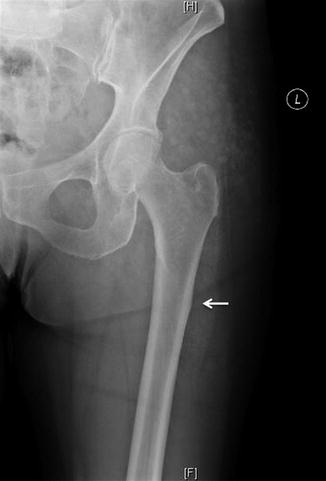
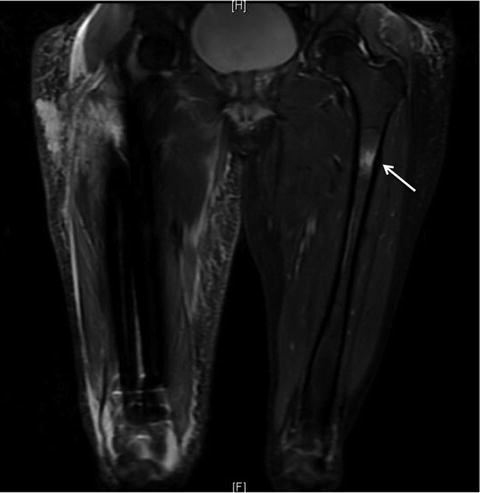

Fig. 11.3
X-ray of the left femur showing thickening of cortex indicative of bisphosphonate-related incomplete fracture

Fig. 11.4
Impending atypical femur fracture demonstrating bone marrow edema pattern on fat suppressed MRI
Symptomatic patients presenting with pain, a transverse radiolucent line on plain film, and bone marrow edema on MRI are unlikely to improve with conservative medical therapy. When managed conservatively, these patients are likely to have persistent pain, poor radiographic evidence of healing, and a high probability of fracture completion within one year. One level III study focused solely on patients with incomplete atypical femoral fractures and saw improved clinical outcomes with prophylactic surgery compared to medical management [27]. Surgical fixation via intramedullary nailing is the recommended treatment due to the high rate of union and rare progression to complete fracture [27–30].
Certain considerations must be addressed regarding surgical technique. Because the fracture is incomplete, it is critical that the nail entry port be on the medial aspect of the trochanter. Furthermore, due to the difficulty in aligning the nail with the anatomic bow of the femur, the medullary canal must be over-reamed to limit nail deformity upon insertion. Over-reaming also helps to decrease the risk of intraoperative comminuted fracture during nail placement, which is the most common complication of this procedure. Postoperatively, the medical management of these patients is identical to that of complete atypical femoral fractures.
There is no consensus treatment strategy for symptomatic patients without a fracture line who have bone marrow edema on MRI. Conservative medical management may be effective but is highly controversial due to insufficient evidence and the concern for fracture completion [27, 30, 31]. Nonsurgical treatment consists of immediate cessation of the bisphosphonate, calcium, and vitamin D supplementation to correct any abnormalities and non-weight-bearing restriction. There are conflicting reports regarding the use of an anabolic agent such as teriparatide, which may facilitate healing and improve bone quality [32, 33]. Studies supporting the use of teriparatide saw increased bone activity in fragility fractures of the distal radius and tibia compared to conservative or surgical management, which indicates a potential pharmacologic intervention to treat these fractures [33]. Patients managed conservatively must be closely monitored because of the risk for fracture progression. An MRI should be repeated after 2–3 months to assess for diminution of marrow edema. If no improvement is seen on imaging or there is persistent pain, prophylactic intramedullary nailing should be considered. However, conservative therapy should be continued if there is an interval decrease in bone marrow edema and pain reduction. Full weight bearing can be resumed upon complete resolution of marrow edema and pain improvement to less than two out of ten on the visual analog scale. When managed conservatively, incomplete fractures can take up to 1–2 years to fully heal. Patients who are unwilling to endure a prolonged period of limited activity, reduced weight bearing, and medical therapy should be considered for intramedullary fixation.
Stay updated, free articles. Join our Telegram channel

Full access? Get Clinical Tree


