IX.C.001 Precursor T-Cell Acute Lymphoblastic Leukemia/Lymphoma
IX.C.001
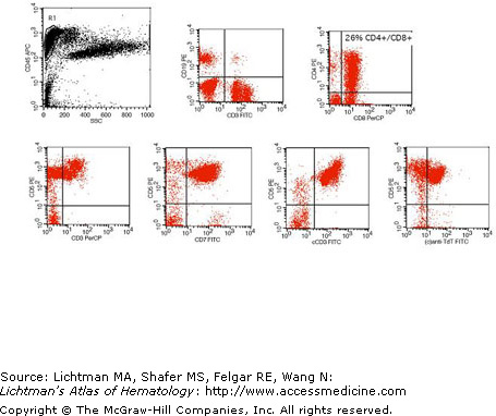
Precursor T-cell acute lymphoblastic leukemia/lymphoma. Flow cytometry. Staining pattern seen in a marrow cells from a patient with acute lymphoblastic leukemia/lymphoma, precursor T-cell type. The upper left panel shows the pattern of leukocyte common antigen (CD45) versus side-scatter analysis. Lymphoblasts have slightly dimmer CD45 staining than mature lymphocytes but merges with the normal lymphocyte population. The upper middle and upper right panels show that the bright CD45+ population (indicated by R1 and shown in red on gated graphs) contains a subpopulation of cells that are negative for surface CD3 and surface CD19 but co-express CD4 and CD8 (a common thymocyte phenotype). In this case, the CD4+/CD8+ population accounts for approximately 26% of all cells in the marrow. The lower row of dot plots indicates that this population is surface CD3 negative, surface CD5 positive, surface CD7 positive, and co-expresses cytoplasmic CD3 and terminal deoxynucleotidyl transferase (TdT). TdT is a nuclear antigen, but can be assessed by flow cytometry using permeabilization methods that are also used for assessing cytoplasmic marker staining (cellular fixation in formalin, followed by incubation of antibody with cells in a very weak detergent solution).
IX.C.002 Acute Lymphoblastic Leukemia, Precursor B-Cell Phenotype
IX.C.002
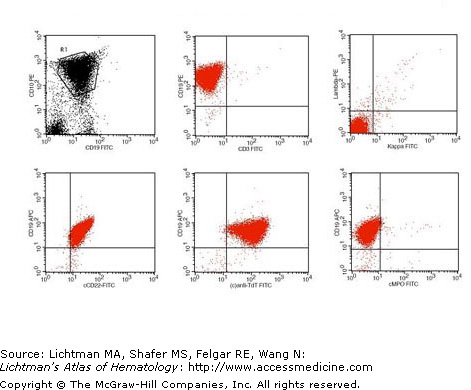
Acute lymphoblastic leukemia, precursor B-cell phenotype. Flow cytometry. Staining pattern of leukocytes of a pediatric precursor B-cell acute lymphoblastic leukemia. Analysis of blood cells shows a prominent CD19+/CD10+ population (upper left plot). Gating for this population based on CD45 vs. side-scatter showed bright CD19 staining and lack of surface CD3 and absence of either surface kappa or lambda expression (upper middle and upper right plots). In the lower row are prototypical cytoplasmic staining findings. In conjunction with surface CD19 staining (y-axis), blasts show co-expression of cytoplasmic CD22 and terminal deoxynucleotidyl transferase (TdT), but lack of myeloperoxidase (MPO) staining using an anti-MPO antibody. Blasts on the blood film were also negative on cytochemistry stain for myeloperoxidase activity.
IX.C.003 Chronic Lymphocytic Leukemia–Small Lymphocytic Lymphoma
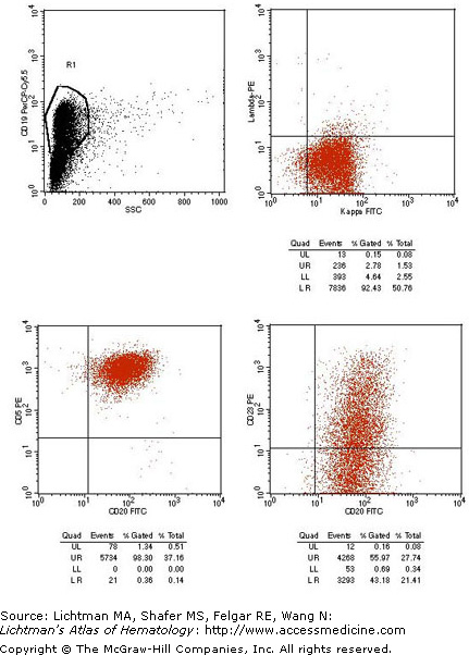
IX.C.003
Related posts:

Stay updated, free articles. Join our Telegram channel

Full access? Get Clinical Tree


