Classification according to etiology [1]
Primary (congenital) lymphedema
Are based on a genetic developmental disorder of lymphatic vessels and/or lymph nodes (aplasia, atresia, hypoplasia, hyperplasia, lymphangioma). They can occur in combination with other dysplasia/syndromes (e.g., Turner syndrome, Noonan syndrome, Klippel-Trenaunay-Weber syndrome) or isolated
Secondary (acquired) lymphedema
Are due to an acquired injury to the lymph vessel system (e.g., post-traumatic, post-inflammatory, artificial, post-reconstructive, or caused by malignant tumors or metastases)
Classification according to clinical stages [2]
Stage 0 (interval stage)
There is no edema, but a pathological scintigraphical finding indicating reduced transport capacity
Stage 1
Is spontaneously reversible, and the swelling may resolve overnight
Stage 2 (spontaneous irreversible stage)
The swelling persists without resolving
Stage 3 (elephantiasis)
There is a monstrous swelling
Classification according to localization [2]
Localized lymphedema
Head, arm, leg, or genital lymphedema or lymphostatic enteropathy
Generalized lymphedema
The whole body can be affected
45.3 Diagnosis
A diagnosis of lymphedema can be based on both basic and advanced diagnostic measures [3].
Basic measures comprise medical history, examination, palpation, and measurement.
Taking a medical history involves asking about malignant disease, surgery, erysipelas infections, soft tissue lesions, venous and arterial disease, medication intake, hereditary factors, and pain—although classic lymphedema is pain-free. If a patient is experiencing pain, this may point to malignant lymphedema.
Examination: Which parts of the body are affected by the swelling? Asymmetry is one of the salient clinical signs of lymphedema. Bilateral swelling normally indicates a systemic cause, e.g., cardiogenic, nephrogenic, hepatogenic, or from medicine intake. Localization of primary lymphedema is mainly peripheral. In secondary lymphedema, proximal localizations are more typical. The presence of skin signs of lymphedema such as deep skin creases, box-shaped teeth, hyperkeratosis, papillomatosis cutis lymphostatica (Fig. 45.1), lymph cysts, or lymph fistulae confirms the diagnosis.
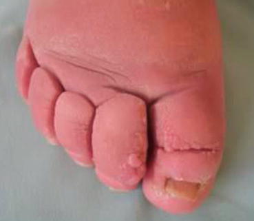

Fig. 45.1
Deep skin creases, box-shaped teeth, and papillomatosis cutis lymphostatica at the first and second toe (picture: ©Apich)
Palpation: Fibrosis can be detected using the skinfolding test. The skinfold between the thumb and index finger is compared with the thickness of the skinfold with the opposite side. If there is a lymphedema, the skinfold is always thicker (Fig. 45.2). A thickened skinfold detected in the region of the second or third toe is referred to as a positive Stemmer sign (Fig. 45.3).
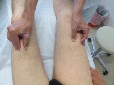
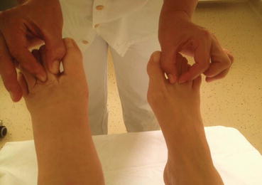

Fig. 45.2
Positive skinfolding test of the right thigh (picture: ©Apich)

Fig. 45.3
Positive Stemmer sign on the right second toe (picture: ©Apich)
The measurement can be done with a measuring tape (Kuhnke method) or with an optoelectronic device (Perometer) [4].
45.3.1 Advanced Diagnostic Measures
If medical history, physical examination, and palpation yield no clinical findings and if it’s still unsure as to whether or not the patient has lymphedema, then diagnostic measures [5] using high-resolution ultrasound, indirect lymphography, scintigraphic quantification of lymphatic function (gold standard), computed tomography (CT), magnetic resonance imaging (MRI), near-infrared fluorescence (NIRF) imaging methods, or bioelectrical impedance analysis should be used.
45.4 Differential Diagnosis
45.4.1 Asymmetrical Edema
These may occur in venous thromboses, post-thrombosis syndrome, post-reconstructive edema, status following supination trauma of the ankle, inflammatory edema attributable to polyarthritis, in Sudeck’s atrophy, a ruptured Baker’s cyst can lead to asymmetric swelling—to name but a few.
45.4.2 Symmetrical Edema
It can be cardiac, nephrogenic, or hepatogenic or can be found in cases of lipoedema or obesity or by taking medication.
Especially elderly patients might have the so-called mixed edema, a combination of lymphedema (asymmetric) and symmetrical edema in generalized systemic diseases.
45.5 Therapy Options for Lymphedema
45.5.1 Conservative Therapy
Complete decongestive therapy (CDT) has the following purposes:
Reducing the current stage of lymphedema to a lower stage
Edema and volume reduction (Fig. 45.4)
Consistency normalization
Improving muscle and joint pump function
Stay updated, free articles. Join our Telegram channel

Full access? Get Clinical Tree


