Figure 28.1 Differential diagnostic scheme for fever and lymphadenopathy.
Important elements of the history should also include the presence or absence of pain, occupational and animal exposures, geographic residence, travel history, sexual and drug use behavior, trauma and presence or absence of systemic symptoms, and/or history of underlying disease. A thorough examination must include the location of lymphadenopathy including an evaluation of all accessible lymph node-bearing areas, the size and consistency of palpated nodes, whether they are discrete or matted and whether tenderness is present and, if so, at what level of severity.
As a general rule, a node larger than 1 cm should be considered abnormal. Stony-hard nodes are usually a sign of malignancy. Very firm, rubbery nodes suggest lymphoma. Softer nodes are the result of infectious or inflammatory conditions and when suppuration is present, these nodes may tend to be fluctuant. The term shotty refers to small nodes that feel like “buckshot” under the skin, as found in the cervical nodes of children with viral illnesses. A group of nodes that feel connected and seem to move as a unit is said to be matted and can be either benign (e.g., tuberculosis [TB], sarcoidosis, lymphogranuloma venereum, and human immunodeficiency virus [HIV]) or malignant (e.g., metastatic carcinoma and lymphoma). Pain and tenderness is usually the result of an inflammatory process or suppuration within the nodes but may also represent hemorrhage into the necrotic center of a malignant node. The presence or absence of tenderness does not reliably differentiate benign from malignant nodes.
Localized lymphadenopathy
Lymphadenopathy is considered localized if no more than two contiguous lymph node groups are involved. Anatomically and clinically, the node-bearing areas are divided into five major groups, namely (1) the head and neck area, (2) the axilla, (3) the inguinal area, (4) the mediastinal-hilar areas, and (5) the retroperitoneal and para-aortic areas. Basic knowledge of the anatomy and areas drained by these lymph nodes can help in narrowing the differential diagnosis (see Table 28.1). Infectious local adenopathy may be acute or chronic. It is usually associated with a primary lesion. At times, the peripheral lesion may be subtle or inapparent. In patients with unexplained localized lymphadenopathy and a reassuring clinical picture, a 3- to 4-week period of observation may be appropriate before considering biopsy. It is important to bear in mind and examine carefully the areas of drainage of each nodal group, as this will often reveal the primary site of infection or other pathology. The lymph nodes of the head and neck are collectively called cervical nodes and occipital and auricular nodes and are more accurately subdivided into several anatomic and clinical areas (see Figure 28.2). The occipital and posterior auricular nodes drain large areas of the scalp and face. Adenopathy of these groups may be associated with primary infectious lesions in these areas (usually from staphylococcal and streptococcal infections) but can be a common feature of acute viral illnesses as well. In children, secondarily infected wounds from insect bites and ringworm (dermatophyte infection) are common causes. The anterior auricular nodes drain the eyelids, palpebral conjunctivae, external auditory meatus, and pinna of the ear. Conjunctivitis and anterior auricular adenopathy (the oculoglandular syndrome) is classically associated with Francisella tularensis via direct inoculation of the conjunctival sac but may also be seen with conjunctival infection from Neisseria gonorrhoeae, Bartonella henselae (cat scratch disease), and epidemic keratoconjunctivitis. The sternocleidomastoid muscle divides the cervical nodes into anterior and posterior sections, each with different drainage areas and resultant clinical importance. The anterior nodes, including the tonsillar, submaxillary, and submental nodes, are most commonly involved as they drain the tonsils and other structures in the pharyngeal area, including the teeth and gums. Enlargement of these nodes should prompt careful inspection of the contents of the mouth. In addition, this group also drains the external structures of the medial face including the lips, chin, cheeks, and medial aspects of the conjunctivae. The posterior cervical nodes are in the occipital triangle of the neck, posterior to the sternocleidomastoid muscle and above the inferior belly of the omohyoid muscle. The drainage of these nodes is more limited and if enlarged, systemic disease should be considered including infectious mononucleosis and mononucleosis-like syndromes, including HIV infection.
| Lymph node area | Anatomic area | Areas drained | Associated conditions/comments |
|---|---|---|---|
| Head and neck | Occipital and posterior auricular | Scalp, face | Local infections with skin pathogens: Staphylococcus, Streptococcus, acute viral illnesses Children: secondarily infected insect, tick, or spider bites, dermatophyte infections (ringworm), viral infections |
| Anterior auricular | Eyelids, palpebral conjunctivae, external auditory meatus, pinna | Conjunctivitis, oculoglandular syndrome (Francisella tularensis, Neisseria gonorrheae, Bartonella henselae), keratoconjunctivitis | |
| Tonsillar, submaxillary, submental | Pharynx, mouth, teeth, lips, tongue, and cheeks | Infections of the head, neck, sinuses, ears, scalp, pharynx, teeth, and oral mucosa | |
| Posterior cervical | Scalp and neck, skin of arms and pectorals, thorax, cervical, and axillary nodes | Mononucleosis, Kikuchi disease, tuberculosis, lymphoma, head and neck malignancies | |
| Axillary | Upper extremity Thoracic wall, breasts, and back | Acute pyogenic infection, cat scratch disease, brucellosis, melanoma, breast malignancy | |
| Inguinal | Lower extremities Abdominal wall Genitalia: penis, scrotum, vulva, vagina, perineum, and perianal region | Pyogenic infections of the lower extremities Sexually transmitted diseases: herpes simplex, syphilis, chancroid, lymphogranuloma venereum Pelvic and perianal malignancy | |
| Mediastinal-hilar | Lungs, trachea, esophagus | Granulomatous disease (infectious and noninfectious) Malignancies | |
| Abdominal Retroperitoneal Para-aortic | Abdominal viscera Retroperitoneal organs: kidneys Pelvic organs | Usually granulomatous disease: Mycobacterium tuberculosis, Mycobacterium avium complex Malignancies: lymphoma |
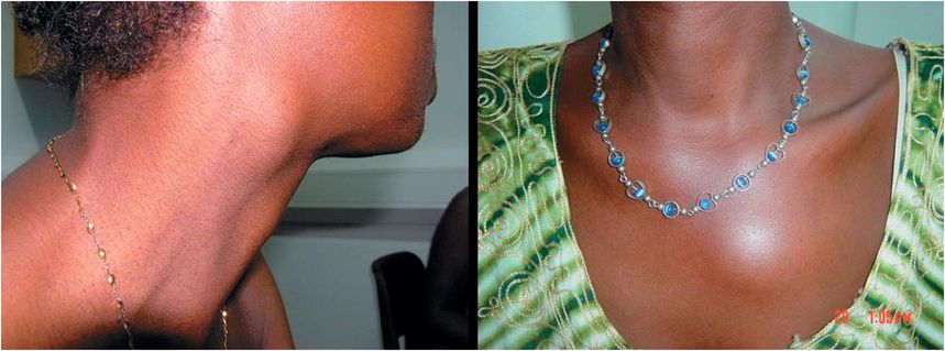
Figure 28.2 These pictures demonstrate palpable lymph nodes in the anterior cervical area (left image) and left parasternal areas (right image).
Kikuchi disease, or histiocytic necrotizing lymphadenitis, is a rare self-limited disorder involving the cervical lymph nodes. This condition was first recognized in Japan and has been predominantly described in females of Asian and Middle Eastern origin, commonly younger than 40 years of age. Clinical manifestations of this entity include fever or flu-like symptoms, rash, localized and sometimes tender cervical lymphadenopathy, elevated erythrocyte sedimentation rate (ESR), and leukopenia. The involved nodes are usually rubbery or firm, discrete, and rarely greater than 2 cm in diameter. Its etiology is still obscure, but it has been associated with Kaposi’s sarcoma-associated herpesvirus (KHSV-HHV-8). This condition does not respond to antibiotics but usually resolves spontaneously in 1 to 2 months. Because of its association with systemic lupus erythematosus (SLE) and Still’s disease, it is also thought to be autoimmune in origin. Recognition of this disease is important because pathologists who are unfamiliar with it commonly mistake it for other conditions that require treatment, such as malignant lymphoma and Kawasaki disease.
The inferior deep cervical nodes lie below the level of the inferior belly of the omohyoid muscle and anteroposterior to the sternocleidomastoid muscle. These nodes receive drainage from the scalp; the superior deep cervical nodes; the axillary nodes; and the nodes of the hilum of the lung, the mediastinum, and abdominal viscera. Infectious and noninfectious entities that involve these structures should be considered when these nodes are involved. Adenopathy in this area is usually subtle and not recognized clinically, but may be detected with the Valsalva maneuver.
Supraclavicular lymphadenopathy has been associated with malignancy in the majority of patients older than 40 years of age. The left supraclavicular (Virchow) node receives lymphatic flow from the thorax and abdomen and may indicate pathology involving the testes, ovaries, kidneys, pancreas, prostate, stomach, or gallbladder. Mediastinal and hilar adenopathies are usually detected only on radiography. These nodes are infrequently involved in acute suppurative disease. Acute suppurative mediastinal lymphadenitis, if present, can be a fulminant process, typically a complication of progressive infections of the upper respiratory tract, or perforation of the esophagus or bronchial tree as a result of trauma or surgery. A useful diagnostic criterion is whether the lymphadenopathy is unilateral or bilateral. Unilateral enlargement most frequently suggests granulomatous disease of both infectious (e.g., Mycobacterium tuberculosis and histoplasmosis) and noninfectious (e.g., sarcoidosis) origin (see Figure 28.3
Related posts:

Stay updated, free articles. Join our Telegram channel

Full access? Get Clinical Tree


