Figure 35.1 Chest radiograph showing a large cavitating lung abscess with air–fluid level in the left lower lobe.
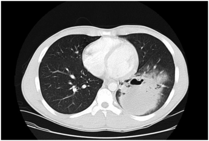
Figure 35.2 Computed tomography scan of the chest further defines the location and extent of the abscess cavity measuring 12 × 9 cm.
The etiologic diagnosis of lung abscess is hampered by contamination of specimens by the normal anaerobic flora of the mouth, prior antimicrobial therapy, and difficulties inherent in culturing and identifying anaerobic bacteria. Although the Gram stain of sputum may be helpful in suggesting an etiologic diagnosis, routine sputum cultures are of no value because all contain anaerobic organisms. Techniques that have been used to obtain uncontaminated lower-airway specimens for anaerobic cultures include transtracheal needle aspiration, transthoracic needle aspiration, and open lung biopsy. Using these techniques, early investigators demonstrated anaerobic bacteria in virtually all untreated patients. These invasive techniques are seldom warranted in the clinical management of patients today. Quantitative cultures of bronchoalveolar lavage or other bronchoscopically obtained lower-airway specimens, such as those obtained with a protected specimen brush, may prove useful in the occasional patient suspected of having lung abscess, but are not needed in most. Transthoracic fine-needle aspiration guided by either ultrasound or computed tomography (CT) has been used increasingly for diagnostic purposes. Although generally safe in experienced hands, serious complications such as pneumothorax and bacterial contamination can result and greatly prolong the recovery period.
Anaerobic organisms most commonly recovered from lung abscesses are listed in Table 35.1. Multiple anaerobic organisms are commonly present along with aerobic or microaerophilic organisms. The viridans streptococci, particularly the Streptococcus milleri group, appear to be significant pathogens. K. pneumoniae was the most common pathogen in a retrospective review of 90 cases of community-acquired lung abscess from Taiwan. K. pneumoniae was recovered from 30 patients (33%), compared to 28 patients with anaerobic organisms. There are likely several explanations for these findings, including prior antibiotic therapy and geographic location. Other than determination of β-lactamase production, susceptibility testing of anaerobic organisms is usually not required.
| Organism |
|---|
| Gram-negative bacilli |
| Pigmented Prevotella spp. Pigmented Porphyromonas spp. Nonpigmented Prevotella spp. Bacteroides fragilis group Fusobacterium nucleatum Fusobacterium spp. |
| Gram-positive cocci |
| Peptostreptoccus spp. Peptococcus spp. |
| Gram-positive bacilli |
| Clostridium perfringens Clostridium spp. Propionibacterium acnes Actinomyces spp. |
Therapy
Most lung abscesses are treated empirically. Table 35.2 provides therapeutic options for intravenous treatment of lung abscess. Selection of agents should be guided by the spectrum of pathogens suspected or isolated from appropriate collected specimens. Historically, penicillin has been the antibiotic of choice because of its good in vitro activity against most anaerobic and microaerophilic bacteria present in the oral cavity. Two randomized clinical trials found that clindamycin is superior to penicillin, with time to resolution of symptoms and failure rate significantly lower in clindamycin-treated patients. Failure of penicillin therapy was associated with the isolation of penicillin-resistant Bacteroides species in one of these studies. Although increasing resistance of anaerobes and gram-positive bacteria to clindamycin has been reported, it is still preferred over penicillin for treatment of lung abscess when anaerobes or microaerophilic streptococci are likely to be predominant pathogens.
Related posts:

Stay updated, free articles. Join our Telegram channel

Full access? Get Clinical Tree


