Distribution of body iron
Iron is contained in haemoglobin, the reticuloendothelial system (as ferritin and haemosiderin), muscle (myoglobin), plasma (bound to transferrin) and cellular enzymes (e.g. cytochromes, catalase) (Fig. 9.1). Reticuloendothelial cells (macrophages) gain iron from the haemoglobin of effete red cells and release it to plasma transferrin which transports iron to bone marrow and other tissues with transferrin receptors (TFRs).
Hepcidin
Hepcidin is a protein synthesized by the liver that controls iron absorption and circulation (Fig. 9.2). It lowers cell levels of ferroportin, the protein that allows iron entry into the portal circulation (Fig. 9.3) from the duodenal enterocytes and into the blood circulation from macrophages. Hepcidin therefore reduces both iron absorption and iron release from macrophages to transferrin. Hepcidin synthesis is controlled by various proteins, e.g. HFE, hemojuvelin (HJV) and the minor transferrin receptor TFR2. Mutation of any of these lowers hepcidin secretion and causes excess iron absorption (see Chapter 10). Inflammation increases hepcidin synthesis through increased levels of IL-6. Increased iron stores stimulate hepcidin synthesis while iron deficiency reduces it. Increased erythropoiesis lowers hepcidin synthesis because of a protein GDF15 released from erythroblasts.
Iron intake, absorption and loss
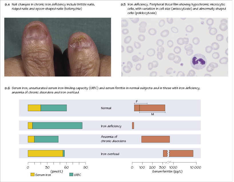
The average Western diet contains 10–15 mg/day of iron, of which 5–10% (about 1 mg) is normally absorbed through the upper small intestine. Absorption is normally adjusted to body needs (increased in iron deficiency and pregnancy, reduced in iron overload). Absorption is regulated by DMT-1 at the villous tip and ferroportin (controlled by hepcidin) at the basolateral surface of the enterocyte (Fig. 9.3). At the luminal surface iron is reduced to the Fe2+ state and on entry to portal plasma reoxidized to Fe3+. It then binds to transferrin. Haem from food is degraded after absorption through the cell surface to release Fe2+. Some iron remains in the enterocyte as ferritin.
Related posts:

Stay updated, free articles. Join our Telegram channel

Full access? Get Clinical Tree


