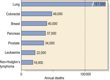
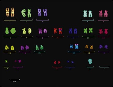
19 Introduction The leukaemias are a heterogeneous group of malignant blood disorders. In this introductory section, general characteristics such as definitions, aetiology and classification are discussed. Each of the more common types of leukaemia is subsequently described in more detail. Definition Leukaemia is a type of cancer caused by the unregulated proliferation of a clone of immature blood cells derived from mutant haematopoietic stem cells. The disease is the result of multiple acquired genetic and epigenetic events which can vary widely between patients. Leukaemic transformation is assumed to occur at or near the level of the leukaemic stem cell prior to definite lineage commitment. The leukaemic cells do not differentiate normally. They may avoid standard mechanisms of cell death (apoptosis) and they may also retain the stem cell signature of self-renewal. This relentless proliferating clone of aberrant cells eventually squeezes out normal cells from the bone marrow causing marrow failure and death. Incidence Leukaemia is not a common disorder but it is a significant cause of death from cancer (Fig 19.1). There is a male preponderance in most types of leukaemia. Geographic variations exist; for instance, chronic lymphocytic leukaemia is the predominant form of leukaemia in the Western world but is much less frequent in Japan, South America and Africa. Fig 19.1 Annual causes of death from malignancy in the year 2011(estimated data from United States). Aetiology As for other malignancies, the evolution of leukaemia is likely to be a multistep process. Thus, accumulated genetic mutations corrupt normal cellular pathways controlling proliferation and differentiation and lead to the production of an autonomous proliferating stem cell clone (‘clonal evolution’). It is easiest to think about the aetiology in terms of these acquired genetic abnormalities and other more general predisposing factors. Genetic abnormalities Cytogenetic analysis and particularly molecular genetic techniques have revealed various acquired non-random chromosomal derangements which play a fundamental role in leukaemogenesis (Fig 19.2). There are a number of different types of possible chromosomal change. Fig 19.2 Fluorescence in situ hybridisation (FISH) study of a complex karyotype (including t(8;16) ) in a patient with acute myeloid leukaemia. Chromosomal translocations One chromosome breaks and donates a fragment to another chromosome which reciprocates by returning a fragment of its own. Such translocations can result in the movement of proto-oncogenes to new sites where they have the capacity to cause leukaemic transformations. The classical example of a balanced translocation is the ‘Philadelphia chromosome’, found in 95% of cases of chronic myeloid leukaemia (CML), where breakages in chromosomes 9 and 22 result in the creation of a new fusion gene (BCR-ABL) which encodes a novel protein with intense tyrosine kinase activity. In a manner incompletely understood, this protein causes deregulated myeloid cell growth. Chromosome deletions and additions Only gold members can continue reading. Log In or Register to continue You may also needAcute myeloid leukaemiaThe myelodysplastic syndromesMyelomaHaematology in the elderlyChronic lymphocytic leukaemiaAplastic anaemiaLaboratory haematology I – Blood and bone marrowOther leukaemias Share this:Click to share on Twitter (Opens in new window)Click to share on Facebook (Opens in new window)Click to share on Google+ (Opens in new window) Related Tags: Haematology An Illustrated Colour Text Jun 12, 2016 | Posted by admin in HEMATOLOGY | Comments Off on Introduction