Immunologic Tolerance


Immunologic Tolerance


Christopher C. Goodnow

Pamela S. Ohashi


INTRODUCTION

How the immune system tolerates molecules that are part of our body yet mounts a destructive response against molecules that are foreign has been a major biologic and medical conundrum for decades. The idea that the repertoire of lymphocytes is made actively tolerant to each individual’s unique set of self-molecules has posed many practical and conceptual challenges. If all lymphocytes that have the potential to respond to self-antigens are silenced or eliminated, will the residual repertoire be sufficient to respond to the enormous array of unknown pathogens? If all self-reactive lymphocytes are actively tolerized, why then do a bewildering array of autoimmune diseases collectively affect more than 5% of people? If we are programmed during development to eliminate tissue-specific lymphocytes, how does immune surveillance against tumors occur? Most importantly, how can we harness the physiologic processes of immunologic tolerance either to prevent transplant rejection and allergy or to promote rejection of tumor cells? In this chapter, we will cover our understanding of the basic mechanisms that are currently known to promote immunologic self-tolerance.


HISTORY

The history of immunologic tolerance illustrates superbly how scientific progress wobbles and sways between two pillars: what people can conceive and what they can test experimentally.


The Discovery of Actively Acquired Tolerance

“Horror autotoxicus” was the insightful term used by Paul Ehrlich early in the 20th century to describe the immune system’s avoidance of producing autoantibodies.1 There were nevertheless much more pressing issues at that time defining the nature of protective antibodies against microbes and the production of vaccines, and little consideration was given to the problem of avoiding autoantibodies.

A key turning point came in 1948 and 1949 in the form of a book and a genetics article synthesizing all that was known about The Production of Antibodies by Burnet and Fenner.2,3 Owen had published in 1945 an extraordinary analysis of antibody production against blood cell alloantigens in cattle.4 He discovered that nonidentical twins had become hemopoietic chimeras in utero and that as adults they avoided making antibodies against the foreign alloantigens that were continuously presented on their sibling’s red blood cells. Traub had earlier observed that mice infected with lymphocytic choriomeningitis virus (LCMV) in utero became lifelong carriers of the virus without producing neutralizing antibodies.5 Burnet and Fenner put these observations together2: “This finding … has the important implication that cells foreign to the host may be tolerated indefinitely provided they are implanted early in embryonic life.”

How the immune system could distinguish self- from foreign antigens was clearly a big problem for Burnet and Fenner’s theory. They proposed that tolerance-inducing antigens would be recognized by antibody on lymphocytes alongside a second receptor for a “self-marker.” It is ironic that Burnet and Fenner, as microbiologists first and foremost, locked onto this idea and did not conceive the reciprocal mechanism: that microbial antigens provoke immunity because they are seen by antibody on lymphocytes together with a second receptor for a “microbe marker.”

The failure to conceive of specific receptors for microbes, other than antibody-like molecules, proved to be a prolonged conceptual handicap for the field of actively acquired tolerance and immunity. It stemmed from a turn-of-thecentury rivalry between the schools of thought spearheaded by 1908 Nobel laureates Ehrlich and Metchnikoff, which artificially divided immunologists into those focused on adaptive immunity, exemplified by Ehrlich’s antibodies with their extraordinary antigen specificity, and students of Metchnikoff’s innate immunity which was viewed by those that followed Ehrlich as “nonspecific.”

In 1963, Claman6 and Talmage7 published experiments and theory that bacterial lipopolysaccharide (LPS) or similar components of acid-fast bacilli used in adjuvants provided a second “nonspecific” signal needed to switch lymphocytes from tolerance to immunity. Citing Talmage’s theoretical work, Claman concluded,


These experiments may nevertheless be regarded in the framework of a theory which considers that the production of antibody requires two stimuli, one specific and (at least) one nonspecific. The specific stimulus (or antigenic determinant) designates which specific antibody will be produced. The nonspecific stimulus (or adjuvant factor) carries the ability to stimulate cell proliferation, and is concerned with the quantity of antibody produced.

In 1975, Moller published further experimental and theoretical evidence for this idea, naming it the “one nonspecific signal” model.8 Their use of the term “nonspecific” to describe the effects of LPS exemplifies the erroneous assumption held by most adaptive immunologists, but not
by those that studied innate immunity, that the adjuvant effects of microbial products like LPS were not mediated by specific receptors. Janeway articulated the need for a collective mea culpa among adaptive immunologists in 1989, describing the attitude to microbial adjuvants as “the immunologist’s dirty little secret,”9 and went on to hypothesize a parallel set of pathogen-associated molecular pattern receptors.10 The question of how LPS, and by extension many microbial adjuvants, were recognized by the vertebrate immune system was answered dramatically with the discovery of precise mutations in the signaling domain of a receptor that looked nothing like antibody, toll-like receptor (TLR) 4, in strains of mice that were specifically unable to respond to LPS.11 Mouse molecular genetics subsequently illuminated specific sensing functions for a diverse family of vertebrate TLRs and other receptors for “microbe markers.”12

Partly because of this conceptual gap, Burnet’s experiments to test the “self-marker” theory that tolerance was specifically acquired in utero yielded negative results. Chickens13 or mice14 exposed to foreign antigens in ovo or in utero were still fully capable of making antibodies against those antigens. However, most of the experiments employed influenza virus and bacterial flagellin that, like LPS, are recognized by antibody-like receptors and by various members of the TLR family, and would have resulted in the induction of immunity rather than tolerance. Although nonmicrobial antigens were also employed, they were not of the type that persisted long-term to maintain tolerance, unlike the allogeneic blood stem cells that had been exchanged in utero between Owen’s twin cattle. True to the Popperian tradition of attempting to falsify one’s hypothesis, Burnet noted that these results “may mean that the whole conception of the development of selfmarker recognition during embryonic life is wrong.”13

Burnet and Fenner’s book had nevertheless triggered Medawar to approach the problem of immunologic tolerance from a different angle, as a transplant scientist seeking to solve the problem of human skin graft rejection in burn patients. He extended Owen’s findings to show that nonidentical twin cattle also accepted skin grafts from each other, representing extraordinary evidence for immunologic tolerance as allogeneic skin grafts remain to this day the most difficult to block from rejection. Medawar’s team then performed the famous experiment of injecting allogeneic blood cells from one inbred mouse strain into newborn mice of another.15 The blood cells would have engrafted to establish lifelong chimerism like the twin cattle and would not have stimulated the TLR system, allowing Medawar to reveal profound tolerance to skin grafts that was extraordinarily specific for the foreign tissue antigens encountered neonatally. In their 1953 paper, Medawar crystallized the concept: “This phenomenon is the exact inverse of actively acquired immunity and we therefore propose to describe it as actively acquired tolerance.”15

These concepts and experimental findings are now employed in clinics around the world, with greatest success in children with primary immune deficiency diseases where tolerance is acquired so completely to semiallogeneic bone marrow transplants that immunosuppressive drugs can be withdrawn several months after transplantation.


The Clonal Selection Theory

While Medawar’s experiments put the phenomenon of actively acquired tolerance on firm foundations, it opened up an even more perplexing problem: how were both tolerance and immunity acquired at the cellular level? Talmage16 and Burnet17,18 independently arrived at the “one cell-one antibody” concept of clonal proliferation to explain acquisition of immunity. Burnet’s PhD student at the time, Gus Nossal, soon obtained the first experimental evidence for that concept using single cell micromanipulation methods with the bacterial geneticist Lederberg.19 Many experiments followed in the 1960s, 1970s, and beyond that affirmed the role of clonal proliferation in acquired immunity. Clonal proliferation itself made the experiments feasible, by increasing the frequency and the specificity of antigen-reactive cells so that they could be accurately enumerated, micromanipulated, and experimentally studied.

The reciprocal mechanism—clonal deletion to explain the acquisition of tolerance—was Burnet’s elegant mirror image concept in The Clonal Selection Theory.18 Lederberg20 extended the concept by pointing out that the timing of antigen encounter, at an immature stage of lymphocyte development, could itself be the “self-marker” that triggered clonal deletion or maturation arrest to prevent the cells from making antibody: “The distinction between the function of an antigen as inhibitor (self-marker) or as inducer of antibody formation is … the time when the antigen is introduced into the potential antibody forming cell.”

Talmage7 and Claman6 proposed a prescient alternative: that antigen alone would trigger exhaustive differentiation of specific lymphocytes without clonal expansion, depleting the pool of lymphocytes left to respond to a second challenge. In their view, antigen plus a second “nonspecific” signal from LPS, other adjuvants, or protein aggregation was necessary for clonal expansion of specific lymphocytes to prevent depletion of the pool of responders when some differentiated into effector cells.

Testing for the disappearance of self-reactive clones in tolerant animals was nevertheless like asking if a needle had been eliminated from a haystack. It was confounded for 30 years by the fundamental technical barrier that antigenbinding lymphocytes are rare and heterogeneous in the preimmune repertoire.


The Experimental and Conceptual Swing against Clonal Deletion in the 1960s and 1970s

Experiments were performed in the 1960s and 1970s to see if lymphocytes capable of binding self-antigens were absent from the circulating repertoire, either by direct antigenbinding measurements, by polyclonally activating cells into antibody secretion in culture, or by immunizing with foreign antigens that resemble self-antigens. Lymphocytes with self-antigen binding antibodies could be found,21 leading many to question the idea of clonal deletion.

We know now that many B cells in the preimmune repertoire bear antibodies that are polyreactive, binding with low and variable affinity to many different antigens including self-antigens.22,23,24,25,26,27,28 The same is true for most circulating
T cells, which are indeed biased to be weakly self-major histocompatibility complex (MHC) reactive. Many of these polyreactive receptors do not pose a risk of autoimmune disease unless made in extraordinary concentrations, and when expressed in transgenic mice some have an affinity for self that falls below that which invokes actively acquired tolerance, as described later. But the assays used at the time could not distinguish between lymphocytes bearing frequent but harmless polyreactive receptors and much rarer cells bearing potentially destructive receptors with higher affinity and specificity, which we know now are usually controlled by active tolerance mechanisms. In other cases, highaffinity receptors were isolated but the assays used confused genuine autoantibodies with antibodies that bound to misfolded, proteolytically cleaved, or posttranslationally modi-fied “altered self.”29

Contemporaneous experiments in the 1970s supported clonal deletion or inactivation of immature lymphocytes, but the necessary experimental contrivances made it hard to shift the growing consensus that self-reactive lymphocytes were not deleted. Cooper neatly bypassed the problem of varying antigen affinity by showing that treatment of developing chicks or neonatal mice with antibodies to the constant region of immunoglobulin (Ig)M completely blocked B-cell development and antibody formation.30 He noted: “Acting at an early stage of differentiation, anti-mu antibody might inactivate or eliminate…cells at the time that they begin to express IgM surface antibody. This concept is analogous to the elimination of ‘forbidden clones’ recognizing self-antigens.”30 Nossal31 and Klinman32 performed sophisticated tissue culture experiments that showed immature B cells exposed to haptenic antigens, but not their mature counterparts, were actively incapacitated from making antibody-secreting progeny, although they could not see the fate of the antigen-specific cells directly.

Set against these findings of clonal deletion or inactivation in immature lymphocytes, Claman,6 Dresser,33 Weigle,34 and others showed that even mature antigenspecific lymphocytes were somehow incapacitated when chronically exposed to nonimmunogenic forms of antigenlike xenogeneic gammaglobulin, provided these persisted in the body, were deaggregated so that they did not provoke inflammatory responses, and were free of the bacterial ligand for TLR 4, LPS.

Miller’s discovery of helper T cells in the 1960s provided an alternative way to reconcile the presence of self-antigen binding B cells with Burnet’s concept of clonal deletion or inactivation. While B cells might not actively acquire tolerance, the repertoire of helper T cells could still be purged of self-reactive clones. This revised view was made poignantly by Miller when he received the inaugural Burnet Medal in 197135: “Specific tolerance can be induced much more readily in T cells than in B cells and, in many cases, tolerance in B cells in vivo is just not demonstrable. It may in fact turn out that tolerance to self-components is a property confined exclusively to the T cell population, tolerance in B cells being merely a laboratory artifice.” Effectively, this allowed the goalposts of clonal deletion to be moved from B cells to T cells. But that solution soon came into doubt as well: the 1974 discovery of MHC restriction by Doherty and Zinkernagel36 indicated that all circulating T cells were inherently self-reactive.


Tolerance by Cell-Cell Interaction Networks and Suppressor T Cells

The inherent self-MHC reactivity of T cells was probably the final trigger that drove many to discard clonal deletion altogether and conceive alternative mechanisms for actively acquired tolerance. By 1983 when Nossal reviewed the field of immunological tolerance,37 he noted that most immunologists had discarded Burnet’s concept of clonal deletion as well as Nossal’s “clonal anergy.” One camp pursued alternative ideas of Jerne, who hypothesized that tolerance was achieved by complex networks of consultation between lymphocytes through idiotype-anti-idiotype interactions.38 It is fair to say that experimental studies have yet to establish the importance of idiotype networks for actively acquired tolerance.

An important alternative school of thought was fostered in the 1970s by Cohn’s two-signal model.39 Its inception hypothesized that antibody responses were only triggered when a newly introduced antigen was recognized simultaneously by two antigen-binding lymphocytes: the B cell and a helper T cell that delivered a second signal to the B cell. In this model, tolerance could still be actively acquired in the B cell by deletion, anergy, or some other mechanism that paralyzed antibody formation if it encountered antigen (signal 1) without receiving signal 2 from the helper T cell. Cohn’s model extended to include T cells the two-signal concepts developed 10 years earlier by Dresser, Claman, and Talmage, that recognition of antigen alone promoted tolerance whereas immunity required a second signal coming directly from microbes through the mitogenic action of LPS and other microbial components.6,7,8,40 This still left the question of how T cells might select between tolerating or rejecting an antigen. Based on transplantation tolerance experiments, Lafferty initiated the conceptual leap that T-cell proliferation and rejection of foreign tissue depended upon recognizing antigen and MHC alongside a second “costimulator” signal unrelated to antigen, on specialized antigenpresenting cells that lay as sentinels in each tissue of the body.41 Steinman discovered dendritic cells as fulfilling this property in the 1970s.42

An intensely popular new idea was that self-reactive helper or cytotoxic T cells could be controlled by suppressor T cells that were actively induced by self-antigens or idiotypes, and were marked by CD8 and a distinctive MHC molecule, I-J.43,44 Thousands of publications appeared in the 1970s and 1980s on suppressor T cells, including more than 300 referring to I-J. However, the experimental tools available at the time to test these ideas were not up to the task, and this period illustrates the potential for even the best minds to be engulfed in mass hallucination when our ideas are not grounded by falsifiable experiments. As molecular genetics revolutionized the field in the 1980s, “suppressor T cell” became a tainted term: Steinmetz and Hood sequenced the MHC and showed there was no
I-J molecule,45,46 and antigen-specific suppressor T-cell clones were found to lack a T-cell receptor (TCR).47 While molecular genetics swept I-J+ CD8+ suppressor T cells away in the 1980s, in 2001 these same powerful experimental methods gave back CD4+ Foxp3+ “regulatory T (Treg) cells” and showed these were critical for tolerance, when Ramsdell and colleagues revealed that deficiency of the Foxp3 transcription factor unleashed lethal autoimmunity in the scurfy mutant mouse and in human immune dysregulation polyendocrinopathy enteropathy X-linked (IPEX) syndrome.48,49,50

Effectively, this turbulent history has laid out a conceptual landscape comprising three general ways to govern tolerance and immunity: 1) “individual choice” where tolerance is acquired by cell-autonomous processes of clonal deletion or inactivation; 2) “committee governance” where cell-cell interactions and additional signals are needed to launch immune reactions; and 3) “governance by police” where tolerance is imposed through the action of dedicated inhibitory cells. As detailed in the following, modern molecular and cellular methods have established important roles in actively acquired self-tolerance for each of these three fundamental concepts.


THYMIC TOLERANCE


T Cells are Positively Selected for Self-Reactivity

The finding that T cells were self-MHC restricted51,52 was soon followed by transplantation studies demonstrating their self-MHC requirement was imprinted during immature T-cell development in the thymus53,54,55: a result that ran entirely contrary to the idea of clonal deletion. The transplantation experiments involved construction of bone marrow chimeras or thymus chimeric mice, where bone marrow of MHC type A × B gave rise to T cells within a mouse or a transplanted thymus of MHC type B or MHC type A. Although controversial, the resulting T cells primarily recognized antigen when it was presented by cells that bore the same MHC type as the thymus in which they had differentiated.53,54,55 These experiments showed T cells were somehow positively selected during their development to recognize self-MHC, triggering intense discussions about whether T cells employed two receptors—one for antigen and one for self-MHC—or if there was one receptor for “altered self.” While we take “altered self” for granted now, it is important to bear in mind that these discoveries took place 7 years before biochemical experiments showed that antigen peptides bind MHC molecules56 and 9 years before the peptidebinding groove of MHC was revealed crystallographically.57

The key event that opened up experiments on thymic tolerance was the identification and cloning of the TCR.58,59,60,61,62,63,64 This led to key experiments that established that there was a single receptor for antigen and MHC,65,66,67,68 and also resulted in the development of two key experimental approaches to analyze the cellular basis of T-cell self-tolerance: antibodies specific for TCR Vβ regions and the ability to generate TCR transgenic mice.69,70,71,72,73 Both of these approaches allowed investigators to follow the fate of T cells with defined specificity during thymocyte development.

The series of experiments that conclusively demonstrated positive selection of self-MHC reactive T cells was the use of TCR transgenic mice. TCRs were cloned from T-cell lines of a defined specificity, and TCR transgenic mice were generated from these receptors. Here, the prediction would be that the rearranged transgenic TCRs would only mature in mice that had the same MHC type as the original donor T cell. Studies using a TCR from a female H2b CD8+ T cell, which was restricted to recognizing H-Y male-specific antigen associated with the H2-Db MHC molecule, clearly demonstrated that CD8+ T cells bearing this TCR matured in female mice expressing H2b but not in mice with other MHC types.74 These studies demonstrated that the transgenic TCR was positively selected and skewed the repertoire to the CD8 lineage during development, as the receptor was specific for the class I MHC molecule.


T Cells are Negatively Selected for Self-Reactivity

One reagent that turned out to be critical in defining T-cell clonal deletion as a mechanism of self-tolerance was the generation of TCR-specific antibodies. The majority of TCR antibodies were specific for Vβ regions. Kappler et al. evaluated the T-cell populations that react with a monoclonal antibody specific for Vβ17. They showed that Vβ17+ T cells react with the class II MHC molecule I-E and are not present in the peripheral lymphoid tissues of mice that express I-E. Importantly, in I-E+ mice, immature CD4+8+ thymocytes were present bearing Vβ17, but these were eliminated from the mature CD4+8- single-positive (SP) thymocyte subset, demonstrating that the self-MHC-specific T cells were deleted during development in the thymus.70 Other studies showed that the infusion of tolerizing agents such as staphylococcal enterotoxin B (SEB) led to clonal deletion of specific cells in the thymus.75 These experiments clearly supported the idea that self-reactive T cells are removed from the repertoire because they encountered their antigen when still immature in the thymus.

Similar studies were done using other Vβ-specific antibodies that correlated the absence of particular Vβ with the presence of self-antigens. One of the most popular models to evaluate tolerance in this way used different strains of mice that expressed minor histocompatibility antigens known as minor lymphocyte stimulatory (Mls) antigens. This group of molecules was defined by Festenstein in the early 1970s76 and later shown to associate with endogenous retroviruses. Mice were characterized as expressing Mls-1a, Mls-1b, or Mls-1c antigens, by the ability of T cells to generate a proliferative response against cells from different strains of mice. Early studies demonstrated that T cells expressing Vβ6 or Vβ8.1 were absent in mouse strains that express Mls-1a.77,78 Collectively, these studies demonstrated that clonal deletion of T cells occurred in the presence of a defined self-antigen.

Conceptually, it is surprising that antigen specificity could be associated with a particular Vβ segment independent of the Dβ, Jβ, and Vα segments of the TCR. Importantly, antigens such as Mls-1a or SEB defined a unique class of antigens dubbed “superantigens” because they were able to stimulate a much higher proportion of T cells than conventional antigens and in a way that did not obey the rules of MHC restriction, although certain MHC types were shown to preferentially present superantigens.79 Research has shown
that superantigens bind the sides of MHC II and TCR Vβ segments in an unconventional way that does not involve the conventional peptide-binding groove of MHC and complementarity determining regions of the TCR.

Although Vβ-specific monoclonal antibodies provided key evidence for clonal deletion, it was essential to demonstrate that this also occurred for self-reactive T cells recognizing conventional peptide antigens bound to self-MHC. The first study to demonstrate clonal deletion of conventional selfreactive T cells employed a TCR transgenic mouse.73 In this model, the TCR α and β transgenes were taken from a CD8+ T-cell clone that was specific for the H-Y male antigen and H-2Db. When female transgenic mice were examined, many T cells with the H-Y specific TCR were positively selected to the CD8 lineage due to the class I H-2Db restriction, as noted previously. In TCR transgenic male mice, however, no T cells developed with the characteristics of the original H-Y T-cell clone bearing high levels of CD8 and the H-Y TCR, although many unusual T cells developed bearing the H-Y TCR and low levels of CD8. In the thymus of male TCR-transgenic mice, there was a drastic reduction in the number of immature CD4+8+ double-positive (DP) thymocytes, implying that thymic deletion occurred at or before this stage. However, follow-up studies established that the loss of DP cells and accumulation of unusual H-Y T cells in male mice was due to premature expression of the TCR alpha chain at the CD4-8-double-negative stage, which caused lineage misdirection at this stage as opposed to clonal deletion.80 When the H-Y TCR alpha chain transgene was controlled so that it was only activated at the DP stage, there was little decrease in the DP population in male mice and negative selection occurred at the CD8+CD4- SP stage of thymocyte development,80 comparable to the findings with superantigens.70 The H-Y studies were reinforced by parallel TCR transgenic experiments with other conventional TCR specificities, providing solid evidence that self-reactive T cells are eliminated from the T-cell repertoire during thymic development.72,81


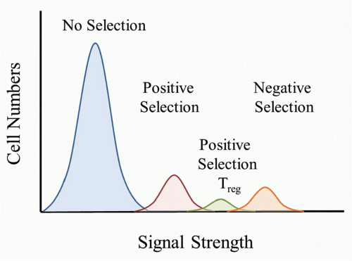
FIG 32.1. The Strength of T-Cell Receptor (TCR) Peptide/Major Histocompatibility Complex (MHC) Interactions Programs Tolerance Mechanisms. The random rearrangements of TCR gene segments lead to a wide range of specificities. Thymocyte fate will be determined by the strength of interaction between the TCR and peptide/MHC complexes that are found in the thymus. The majority of cells will not meet the threshold for positive selection. Stronger TCR interactions lead to the maturation of regulatory T cells or T-cell tolerance resulting in anergy or clonal deletion. The relative proportion of selected T cells is estimated.


Anergy as a Mechanism for Thymocyte Tolerance

Anergy is a term proposed by Nossal to describe lymphocytes that were present but rendered intrinsically unable to respond to stimulation with cognate antigen because of earlier exposure to the same antigen.37 In T cells, evidence for anergy as an alternative mechanism of self-tolerance came from studies using parent into F1 bone marrow chimeras.82 In these studies, Mls-1b bone marrow was transplanted into irradiated Mls-1a Mls-1b hybrid recipients. In this case, where synthesis of the Mls-1a self-superantigen was limited to radioresistant cells in the thymus, Vβ6+ T cells that would normally be deleted in Mls-1a mice were able to mature and emigrate from the thymus but were unresponsive to Mls-1a. This demonstrated that an unresponsive state to self-antigen could be induced in the thymus and suggested that anergy versus deletion were alternative fates for selfreactive thymocytes depending upon the amount of TCR stimulation or the cell type presenting the self-antigen.


T-Cell Affinity/Avidity Defines Thresholds for Positive and Negative Selection

The observation that T cells were positively selected for selfreactivity during thymic development and also negatively selected for self-reactivity drew into sharp focus the central paradox for actively acquired tolerance and immunity. How would a TCR be able to transmit signals for differentiation and survival (positive selection) versus death (negative selection)? Lederberg’s20 and Nossal’s31 earlier concept of a developmental window for deletion could not be invoked here because the alternative fates were both acquired in immature T cells. Over the years, evidence accumulated to support an affinity/avidity model that suggested that T-cell fate was determined by the strength of the signal transmitted by the TCR (Fig. 32.1).83,84,85 The absence of a detectable signal in thymocytes would result in a process called death by neglect. A T cell expressing a receptor that could not engage
self-peptide/MHC complexes (pMHC) would be destined to die because it would be a “useless” TCR to have in the repertoire. TCRs that bound only weakly to self-pMHC complexes in the thymus would send a signal sufficient for the T cell to survive and differentiate to either the CD4 or CD8 lineage. According to this model, a strong signal would be sent if the TCR bound strongly to self-pMHC complexes and would lead to the death of the T cell. It is important to keep in mind that as the thymocyte undergoes positive selection and matures, the TCR level increases, and therefore the T cell would have increased sensitivity to self-pMHC complexes as they progress through thymocyte selection.

Studies to evaluate the affinity/avidity models of thymocyte selection were again dependent upon the development of new technologies. In the 1980s, Smithies and Capecchi reported a way to generate knockout mice, where the expression of a defined gene was disrupted.86,87 This led to a flurry of activity with several groups generating a variety of genedeficient mice. Amongst the early gene knockout mice were animals with a defect in MHC class I protein display due to the absence of the β2-microglobulin subunit of MHC I molecules or the absence of self-peptides available to bind and fold MHC I molecules because of disruption of the transporter associated with antigen processing.88,89 These mice were the basis for a very unique set of experiments in fetal thymic organ cultures, where defined peptides could be added in culture to evaluate their consequences for thymic selection.90,91 When a TCR transgenic model was combined with the class I mutant mice, this provided a novel way to evaluate the consequences of defined peptides on positive selection as well as negative selection.

A series of experiments were done with the OT-1 TCR transgenic mice, that are specific for the ovalbumin (OVA) peptide SIINFEKL and H-2Db.83 The OT-1 TCR transgene was bred onto the transporter associated with antigen processing-deficient background. Fetal thymic organ culture was done using OT-1 transporter associated with antigen processing-1-/- (Tap1-/-) thymic lobes. In this model, no mature OT-1 T cells were generated in the thymus due to the absence of class I expression. However, a series of variant peptides resembling the OVA SIINFEKL peptide could be added to the culture and these would restore class I expression, with the resulting MHC molecules homogeneously complexed with a defined peptide. In this model, the SIINFEKL peptide itself and related peptide sequences that were capable of activating mature OT-1 T cells (agonist peptides) triggered negative selection of immature thymocytes bearing the OT-1 TCR. By contrast, variant peptides that were unable to activate mature OT-1 cells but instead antagonized activation by SIINFEKL itself (antagonist peptide ligands) promoted positive selection of immature thymocytes to accumulate in the cultures as CD8 SP T cells.

At the same time, other studies were done using P14 TCR transgenic mice that express a TCR specific for the LCMVglycoprotein (gp). These studies bred the TCR transgene on a β2-microglobulin-deficient background. Two groups demonstrated that different concentrations of the same agonist peptide promoted positive selection at low concentrations and negative selection at high concentrations.84,85,92 Notably, in the P14 model, antagonist peptide ligands could not support positive selection of the P14 TCR.93 Collectively, these studies provided key evidence that supported an affinity/avidity model for thymocyte selection.

The affinity of TCR binding to MHC molecules bearing the various peptides was subsequently measured by surface plasmon resonance. Using this approach for the OT-1 TCR, Alam et al. demonstrated that the difference between positive selection and negative selection was a threefold difference in affinity for pMHC.94 Interestingly, the agonist peptides (A4Y, L6F) that could support positive selection of the P14 TCR had a similar affinity to the antagonist peptides that supported positive selection of the OT-1 TCR.95 Further studies have also shown that in some models, agonist ligands lead to the differentiation of T cells bearing a CD8αα homodimer and this population of cells have innate-like properties.93,96,97 Collectively, these studies support the concept that a certain affinity/avidity is required to promote positive selection and that increasing the affinity/avidity leads to the induction of negative selection (see Fig. 32.1).

What are the biochemical differences in the signal transmitted by the TCR that leads to survival and differentiation of weak pMHC binding thymocytes but death of strongly self-reactive thymocytes? Several studies demonstrated a critical role for the ERK MAP-kinase signaling pathway for positive selection.98,99 Further in-depth studies to follow TCR signaling events used the OT-1 and P14 models to evaluate differences in peptide-specific interactions that were known to result in positive or negative selection. Interestingly, positive selection was shown to occur as a result of sustained low level ERK signaling, while negative selection resulted a stronger, yet transient peak of ERK activation.100,101,102,103 Accordingly, studies have shown that there was impaired thymocyte positive selection in the absence of ERK1 and ERK2.104,105 Evidence also suggests that calcineurin has an impact on Erk activation and positive selection,106 but not negative selection. An essential upstream activator of ERK, RasGRP1, was also found to be essential for positive but not negative selection of thymocytes.107 Further studies have identified downstream ERK-activated transcription factors that are also important for positive selection.108,109

Recent studies also provided evidence that the outcome of thymocyte positive or negative selection is correlated with a different TCR conformation.110 Further in-depth analyses have observed that signals that induce negative selection lead to the membrane compartmentalization of molecules involved in signal transduction such as ZAP-70, LAT, and ERK1/2. Importantly, similar changes in localization of signaling components were not observed under positively selecting conditions.111 Therefore, the different thymocyte fates of positive or negative selection involve detectable molecular changes on the cell surface and in intracellular compartments.


Regulatory T Cells are Selected by High Affinity/Avidity Interactions

Treg cells were first identified as a critical population that prevented autoimmunity and arose during thymic development. Early studies demonstrated neonatal thymectomy of some
inbred rodent strains led to the development of autoimmunity targeting the gonads and thyroid gland.112,113,114 Further studies using various markers identified a subset of cells, first in the rat and then in the mouse, that had the potential to block the induction of autoimmunity. Mason’s team demonstrated that adoptive transfer of naïve T cells to immunodeficient rats resulted in autoimmune diabetes and inflammatory bowel disease that was blocked if a subset of CD4 T cells with markers of activated or memory T cells was cotransferred.115,116 These findings were extended to mice by Powrie117 and by Sakaguchi, whose group demonstrated that the transfer of CD4+CD25- cells into 6-week-old BALB/c nude mice resulted in the development of autoimmune oophoritis, gastritis, and thyroiditis that was suppressed by the cotransfer of CD4+ CD25+ cells.118 While these experiments suggested the existence of a new type of suppressor T cell (dubbed a Treg cell to avoid confusion with the CD8+ suppressor T cells of the 1980s), their interpretation was uncertain: suppression by cells with markers of activated or memory T cells could simply reflect better control of microbial flora in the immunodeficient recipients, so that there was less stimulus for inflammatory bowel disease and autoimmunity.

The critical turning point establishing the existence of Tregs came from the identification of the transcription factor, FoxP3, as a specific marker of mouse Tregs that was required for CD4+25+ T cell differentiation in the thymus. Foxp3 was first identified by genetic mapping of an autoimmune mutation in the scurfy mouse strain.48 That led to the identification of FOXP3 mutations as the cause of a human inflammatory disorder known as IPEX syndrome.49,50,119 Foxp3 was necessary and sufficient for Treg differentiation, and thymic-derived Foxp3+ “natural Tregs” were shown to be critical to suppress autoimmunity, allergy, and inflammation of mucosal and cutaneous barriers.120,121,122,123,124 However, it should be noted that the marker FoxP3 is not exclusively linked to Treg cells in humans.

Caton’s group was the first to show that natural Treg differentiation was an alternative fate to clonal deletion for CD4 thymocytes with a strongly self-reactive TCR, further complicating the question of how alternative cell fates are determined in developing thymocytes.125 The strength of TCR signal has again been invoked as an instructive in-fluence, supported by additional studies that indicate that Tregs bear TCRs that bind to self-pMHC more strongly than TCRs that promote positive selection of helper T cells 126-129 (see Fig. 32.1). However, other studies argue that Foxp3 expression may be stochastic and protect strongly self-reactive CD4 cells from deletion.130


MOLECULAR PATHWAYS INVOLVED IN THYMIC CLONAL DELETION

The precise molecular pathway that leads to the death of self-reactive T cells remains unknown. Evaluating the role of a given signaling molecule in positive and negative selection often points to a role for that molecule in both events, supporting the idea that TCR signals are critical for determining T-cell fate.131,132,133,134,135 However, in general, the role of molecules involved in regulating negative selection has met with controversy, although interesting candidates are beginning to emerge. Some of the uncertainty is likely due to the models that were used to study negative selection. In the early literature, mice were often treated with anti-CD3 antibodies to induce death of T cells. However, because the majority of T cells are activated, this leads to a stress-induced death that is dependent upon glucocorticoids,136 and therefore is not a good model to study negative selection.


Controversial Role for Apoptotic Pathways in Negative Selection

Two major mechanisms that regulate apoptotic cell death are known as the intrinsic and extrinsic pathways and are regulated by Bcl2 family members and tumor necrosis factor receptor (TNFR) family members, respectively. Because many members of the TNFR family possess a “death domain” that can interact with other death domain-containing proteins, this family became the focus for studies investigating the mechanisms of death during thymocyte negative selection. TNFR, Fas, CD30, Trail, DR3, as well as the downstream effector molecules have all been investigated, and the results remain controversial.137,138,139,140,141,142,143,144,145,146,147,148,149,150,151


The Role for ERK and Nur77 in Negative Selection

Several approaches have been taken to address the question of whether the ERK pathway is essential for negative selection. Experiments using dominant negative forms of RAS or MEK, which limit Erk activation, supported the idea that ERK was important for positive selection but not negative selection.98 Other studies using pharmacologic inhibitors for the Erk pathway demonstrated that Erk played a role in both positive and negative selection,101,152,153 whereas yet other studies supported the idea that Erk did not influence negative selection.154 It is possible that the discrepancies in these studies are related to the strength of the signal required for selection of the TCR in the different models that were being studied. For example, because positive selection requires sustained signaling that results from weak TCR interactions, these signals would be more likely to be perturbed by transgenic forms of dominant negative signaling molecules, because this system would still have the endogenous molecules that could provide some signals.

Studies using mice that were deficient in both ERK1 and ERK2 demonstrated that thymic clonal deletion could still occur by following deletion of the OT-1 TCR transgenic T cells using various models.155 However, there are many ERK family members and it is therefore possible that only certain members of the ERK family are critical or that the various ERK family members can compensate for each other. Studies using dominant negative or constitutively activated MEK5, which activates the ERK5 pathway, was able to modulate clonal deletion of thymocytes, but had no role in positive selection.156 Importantly, ERK5 activity correlated with the induction of Nur77, which has been previously implicated in thymocyte negative selection.157,158,159 In several other models, Nur77 is also upregulated during thymocyte
negative selection, thereby supporting the importance of this molecule.160 Therefore, studies support a role for ERK5 and Nur77 in thymocyte clonal deletion.


Bim Links Negative Selection and Autoimmunity

If a particular molecule is essential for the death of selfreactive T cells, one prediction would be that the absence of this molecule may lead to spontaneous autoimmune disease. This prediction would not hold true if that particular molecule was also essential for T-cell differentiation or activation. One such candidate that was considered is the BH3-only-containing protein Bim. Bim is a proapoptotic molecule that gives rise to three alternatively spliced forms: Bims, BimL, and BimEL. In general, Bim is thought to promote apoptosis by binding and sequestering survival molecules Bcl-2, Bcl-XL, and other members of that family. Initial studies reported that Bim-deficient mice spontaneously develop autoimmunity, and that Bim was critical for thymocyte negative selection.161,162

Recent studies, however, question the role of Bim in negative selection and autoimmunity. Baldwin’s group evaluated negative selection using the HYcd4 TCR transgenic model.163 They demonstrated that Bim played a role in apoptosis of thymocytes but not in self-antigen-specific negative selection. Other studies used an elegant TCR transgenic model to accurately follow the fate of Bim-deficient thymocytes. Kovalovsky et al. demonstrated that self-reactive Bim-/-T cells undergoing negative selection were arrested at a CD4loCD8lo immature stage of development.160 Death of these cells was dependent on an alternative pathway that required the thymic microenvironment. By contrast, evidence from unpublished work has shown that Bim deficiency almost completely abolished negative selection in 3A9 TCR transgenic mice crossed with hen egg lysozyme transgenic mice.163a In these animals, the negatively selecting antigen was expressed selectively in Aire+ thymic medullary epithelial cells by the insulin promoter, and in their Bim+/+ counterparts negative selection was induced at the early CD4 SP stage. Thus, the relative dependence on Bim may vary depending on the stage of thymocyte development when self-antigen is encountered, the amount of self-antigen presented in the thymus, or the nature of the antigen-presenting cells that induce negative selection. Further studies using Bim-/- mice have shown that these mice have impaired induction of experimental autoimmune encephalomyelitis. Unexpectedly, Ludwinski et al. also showed that Bim-deficient CD4+ T cells have a strikingly reduced ability to produce cytokines, particularly interleukin (IL)-6 and interferon-γ.164. This adds further complexity as to the mechanism of spontaneous autoimmune disease in Bim-deficient mice.161

Many insights have been uncovered by evaluating both animal models for autoimmunity and clinical studies. Several groups have shown that the nonobese diabetic (NOD) mouse strain that spontaneously develop diabetes has impaired negative selection as one of several factors that may contribute to the onset of disease.165,166 Interestingly, recent studies have evaluated the genes that are expressed in stimulated NOD thymocytes versus wild-type thymocytes. Interestingly, Bim and Nur77 are among the genes that are not upregulated in the NOD background, lending potential support for these molecules in tolerance induction.167,168,169


MINK Links Jnk and Bim in Negative Selection

There is compelling evidence published by Cantor’s group that a molecule known as MINK plays a role in negative selection but not positive selection.170 They showed that MINK expression is increased 20-fold in DP thymocytes compared to the double negative subset. Expression of MINK was also shown to be very low in SP thymocytes as well as peripheral T cells. McCarty et al. studied the role of MINK in thymocyte selection by generating chimeric mice with bone marrow that was infected with a lentivirus that expressed green fluorescent protein as well as a small hairpin ribonucleic acid that reduced MINK expression. Three models of thymocyte negative selection supported a role for MINK. These include the superantigen Mtv-9-induced Vβ5 deletion in the C57Bl/6 and BALB/c backgrounds, the male-specific H-Y TCR transgenic model, as well as peptide-induced deletion using OT-II transgenic mice. Interestingly, they provided evidence that MINK acts via c-jun N-terminal kinase activation and the upregulation of the proapoptotic molecules Bim and BimEL.

Several groups have also evaluated the role of the c-jun N-terminal kinase/SAPK or p38 MAPK pathways in negative selection. Using knockout animals or pharmacologic inhibitors, evidence suggests that both of these pathways play a role in the death of thymocytes.154,171,172 However, further experiments are necessary to solidify a role for Jnk in thymocyte deletion. Importantly, Jnk activation has been linked with Bim expression in neuronal cell types, further strengthening the possibility that Jnk may be involved in thymocyte deletion.173,174


EXPRESSION OF SELF-ANTIGENS IN THE THYMUS

Negative selection in the thymus provides a key mechanism to eliminate self-reactive cells early in development. However, this mechanism was thought to be incapable of establishing self-tolerance to the numerous self-antigens that are expressed only in specific organs, such as insulin or elastase in the endocrine and exocrine pancreas, respectively. Pioneering work by Hanahan identified cells in the thymic medulla that he dubbed “peripheral antigen-expressing cells” that were able to express organ-specific genes like insulin and elastase, and induced T-cell tolerance to a transgenic self-antigen controlled by the insulin promoter.175,176 Several years later, work by Derbinski et al. showed that medullary thymic epithelial cells were able to express a wide array of self-antigens.177,178

At the same time as Hanahan’s definition of peripheral antigen-expressing cells, two human molecular genetics teams discovered that mutations in a novel gene, dubbed autoimmune regulator (AIRE), were responsible for a devastating recessive syndrome known as autoimmune polyendocrinopathy-candidiasis ectodermal dystrophy
(APECED) or autoimmune polyendocrinopathy syndrometype 1 (APS-1).179,180 APS-1 is remarkable because of the progressive development of multiple organ-specific autoimmune diseases, most frequently autoimmune adrenocortical failure and hypoparathyroidism, but also autoimmune thyroid disease, diabetes, vitiligo, alopecia, ovarian failure, hypogonadism, liver disease, pernicious anemia, and exocrine pancreatic insuffi-ciency.181 Almost all patients develop mucocutaneous Candida albicans infections because they produce neutralizing autoantibodies to IL-17, a cytokine that is essential for immune control of Candida.182,183 While this experiment of nature established that AIRE underpinned a critical mechanism for self-tolerance to a wide range of discrete organs, the AIRE gene and protein provided few clues to its mechanism of action: it contained motifs that suggested a role in transcription regulation,179,180 it was mostly expressed in the thymus and not in the organs that were affected by autoimmunity,180 and in the thymus the protein was primarily expressed in medullary thymic epithelial cells where it was concentrated in nuclear dots.184

Aire-deficient mice were generated and were also found to develop spontaneous organ-specific autoimmune diseases, albeit not an identical spectrum to humans.185,186 No overt abnormalities in T-cell development or T-cell subsets were apparent in Aire-knockout mice. Consistent with the predominant Aire expression in medullary thymic epithelial cells, transplantation studies established that spontaneous autoimmunity resulted even when AIRE was only absent from thymic epithelium and not when it was absent from lymphoid cells.186 The key turning point connecting Hanahan’s peripheral antigen-expressing cells with the human autoimmune disease came when Aire-deficient mice were analyzed for expression of organ-specific genes in the thymus186 and for thymic negative selection of organ-specific T cells,187 which revealed these processes were selectively and severely disrupted. AIRE has been shown to be an unusual transcription factor that promotes ectopic expression of a variety of organ-specific genes in thymic medullary epithelial cells by relieving the tendency for ribonucleic acid polymerase to stall shortly after the transcription start site.188

These studies supported the idea that human APS-1 results from failure of thymic negative selection to organ-specific genes and the development of a T-cell repertoire that is poised to develop organ-specific autoimmunity. To further test this hypothesis, Anderson’s group identified an autoantigen that was specifically expressed in the retina of the eye, interphotoreceptor retinoid-binding protein (IRBP), as a target of autoantibodies in AIRE-deficient mice that had developed autoimmune uveitis.189 By crossing Aire-/- mice with IRBP gene knockout mice, they showed that IRBP expression in the eye was essential for spontaneous development of autoimmune eye disease in Aire-/- mice but not for other organ-specific autoimmune diseases in the same animals. IRBP messenger ribonucleic acid (mRNA) expression in the thymus was abolished in AIRE-deficient mice, and selective deficiency of IRBP in thymic stromal cells was sufficient to recapitulate the eye specific autoimmunity that occurs in AIRE-deficient mice.189 With a similar rationale, Trucco’s group generated mice where insulin was deleted from thymic epithelial cells while maintaining expression in the pancreas.190 Importantly, diabetes spontaneously developed in 3-week-old animals. Anderson’s group has gone on to identify other genes that are regulated by AIRE, that are also targets of autoimmunity in mice and humans.191

An important question concerns the possibility that organ-specific autoimmunity caused by AIRE deficiency might reflect failure to induce organ-specific Treg cells, as opposed to the failure to delete organ-specific helper and cytotoxic T cells. Anderson et al.192 addressed this question definitively by performing elegant double-thymus transplant experiments in nude mice, which lack normal thymic epithelium. When the mice were engrafted with one Aire-deficient thymus and one Aire-sufficient thymus, half of the circulating T-cell repertoire would have been selected in the thymus that lacked expression of organ-specific genes, while the other half was formed in the thymus that expressed organ-specific genes. If a deficiency of organ-specific Tregs was responsible for the organ-specific autoimmunity in Aire-deficient mice, this would have been complemented by the production of organ-specific Tregs from the wild-type thymus in the doubly transplanted mice. By contrast, the wild-type thymus would have no impact if autoimmunity resulted from self-reactive effector T cells that escaped deletion in the Aire-deficient thymus. Consistent with the latter, autoimmunity developed with the same organ-specific pattern in the doubly transplanted mice. The same conclusion was reached when equal mixtures of T cells from Aire-deficient and wild-type mice were cotransferred into immunodeficient Rag1-knockout mice.192 Further evidence that AIRE mediates self-tolerance through mechanisms that do not involve Treg formation comes from comparing Foxp3-deficient mice with normal AIRE or lacking AIRE, because the latter have markedly accelerated autoimmunity.193 Likewise, analysis of mice with a T-cell repertoire constrained to junctional diversity between a single TCR Vα and Jα found that the great majority of Treg-specific TCRs were selected independently of AIRE.194

Collectively, these studies demonstrate that thymic clonal deletion is an important mechanism for actively acquired self-tolerance in man and mouse. This conclusion is reinforced by the finding that human susceptibility to type 1 diabetes correlates with genetic variants in the insulin promoter that selectively diminish insulin gene expression in the thymus.195,196 Similarly, a genetic variant in the promoter of the CHRNA gene that diminishes its transcription in the thymus contributes to susceptibility to forming autoantibodies against the acetylcholine receptor chain encoded by this gene and development of myasthenia gravis.197


PERIPHERAL T-CELL TOLERANCE

Powerful mechanisms exist to regulate mature T-cell responses against self-antigens after they have emigrated from the thymus: a process known as peripheral tolerance. One line of evidence for this conclusion comes from an experiment of nature: most organs and antigens remain unaffected by autoimmunity in AIRE-deficient humans and mice despite the failure of thymic tolerance to antigens in almost every tissue. Experimental evidence supporting this conclusion traces back to the demonstration that adult
mice could be rendered tolerant to adjuvant-free, deaggregated gammaglobulin from other species,6,33,34 and this tolerant state would persist for months provided the animals had been thymectomized to prevent generation of fresh T cells.198 However, unlike in the thymus, where the engagement of strong TCR-pMHC interactions leads to clonal deletion or anergy, strong pMHC signals in mature T cells normally leads to the development of an effective immune response. Therefore, with mature T cells we are left with the puzzle of how a strong TCR interaction results in tolerance versus activation. Solving this puzzle holds the most practical potential for overcoming the major clinical problems of transplant rejection and tumor immunology and for restoring tolerance in people with autoimmunity or allergy.

As summarized in the section “History” at the start of this chapter, early work began to lay out two-signal concepts for tolerance or immunity by demonstrating that bacterial LPS, adjuvants, or protein aggregation were required together with antigen for inducing immunity and not tolerance in animal models, although the adjuvant effect was not viewed as a specific sensing pathway.6,40,199 In the early 1970s, Gery and Waksman discovered that LPS-stimulated macrophages produced a soluble activity that augmented T-cell proliferation, dubbed “lymphocyte activating factor.”200 “Monokines” with other names such as leukocyte pyrogen201 and mitogenic protein202 were subsequently observed to have similar T-cell-augmenting activity and shown to represent the same small protein, renamed IL-1.203 Independently, experiments inducing tolerance to allografts led Lafferty to conclude that T cells need to receive not only an antigen stimulus but also a specific “costimulus” from a dedicated antigen-presenting leukocyte for induction of immunity and not tolerance.41,204 It nevertheless became apparent that a cell-bound T-cell costimulator that was not IL-1 also existed,205,206 leading to the discovery of the T-cell costimulatory receptor, CD28, and its membrane bound ligands CD80/B7-1 and CD86/B7-2.12,207,208,209,210

The study of T-cell costimulation by IL-1 languished after the discovery of CD28 and its ligands, and only recently has IL-1 been demonstrated to serve as a powerful additional signal that is necessary—in conjunction with CD80 and CD86—for survival and accumulation of large numbers of daughter cells when CD4 T cells are triggered into division by antigen in vivo.211 IL-12 and interferon alpha and beta, made by antigen-presenting cells in response to bacteria and viruses, similarly provide critical signals for CD8 cells that have received TCR and CD28 stimulation, promoting the survival and accumulation of progeny over successive cell divisions and their acquisition of cytolytic effector functions including interferon gamma and granzyme B expression.212,213,214,215,216,217,218,219,220 Interferon alpha and beta have also been shown to promote survival of activated CD4 cells.221

Much of the early work on T-cell costimulators focused on macrophages as a principal source,206 but parallel studies by Steinman led to the recognition that another myeloid cell, the dendritic cell (DC), was the key antigen-presenting cell required to induce CD4 and CD8 immunity.222,223 Furthermore, if the DC had not been stimulated by adjuvants to “mature,” then antigens encountered on the “immature” DC induced T-cell tolerance, while antigens encountered on mature DCs induced immunity.224,225,226 DC maturation was shown to be triggered by pathogen-derived signals, such as TLR signals, and that maturation included the upregulation of a variety of key costimulatory molecules on the DC, including CD80/B7-1 and CD86/B7-2 ligands for CD28,12,207,208,209,210 as well as the production of IL-1, IL-12, or other cytokines. The theory governing peripheral T-cell tolerance versus immunity in its simplest form often reports signal 1 as the TCR-derived antigen-specific signal, and signal 2 that stems from interactions with mature DCs has been typically thought of as CD28/B7 interactions. However, it is clear that multiple receptor ligand interactions as well as cytokine signals are critical to promote T cell-mediated immunity.

In this section, we will introduce the basic mechanisms of peripheral T-cell tolerance, which include clonal deletion and anergy. Tolerance is also maintained by two other mechanisms. One is known as T-cell ignorance, which is the inability of the T cells to detect sequestered self-antigens. In addition, potentially autoreactive cell function is held in check by Treg cells that arise either in the thymus or may also be induced from the mature CD4 T cell population. It is also clear that CD8 Treg cells exist; however, much less work has been done with this population.


Evidence for T Cell Anergy

The first direct evidence for T-cell clonal anergy came from tissue culture experiments with human T-cell clones that recognized specific influenza virus peptides and MHC II molecules.227 When these T cells were cultured for more than 3 hours (optimally 18 hours) with high concentrations of the relevant peptide in the absence of stimulatory antigen-presenting cells (an unfractionated mixture that included monocytes, DCs, and B cells), the T cells became profoundly refractory to restimulation of proliferation with antigen presented by stimulatory antigen-presenting cells yet retained a normal proliferative response to IL-2. This selective tolerance to antigen persisted despite culturing the anergic T cells for more than 168 hours in the presence of IL-2. Human T-cell clones express MHC II molecules, so that in the presence of high concentrations of peptide the T cells could present p/MHC II complexes to each other228 in such a way that a high proportion of surface CD3 molecules were initially modulated from the cell surface.229 Whether the long-term unresponsive cells regained normal CD3 expression was not examined in this study.

The concept of T cell anergy resulting from antigen recognition in the absence of a specific costimulatory signal from specialized antigen-presenting cells was extended in experiments where cloned mouse T cells were cultured with the relevant peptide antigen presented on chemically fixed antigen-presenting cells230,231 or on synthetic lipid bilayers containing only MHC II molecules.232 Like the human T-cell clones, this also resulted in a persistent state of antigen tolerance that blocked the proliferation response of T cells when subsequently exposed to antigen on unfixed antigen-presenting cells, yet left untouched the proliferative response to IL-2.
This antigen-tolerant state, dubbed T cell clonal anergy by Schwartz and coworkers, was not accompanied by long-lasting downregulation of surface CD3 and TCR. The missing costimulatory signal from the antigen-presenting cells could not be replaced by exogenous IL-1 or IL-2,232,233,234 but was later shown could be replaced by stimulatory antibodies against CD28.235 The tolerance-inducing signal from antigen on fixed antigen-presenting cells could be mimicked by selectively elevating intracellular calcium and required calcium-activation of calcineurin and induction of protein synthesis,233,234 leading to the concept that TCR-induced activation of calcium, calcineurin, and the transcription factor NFAT induced the expression of a lasting tolerance program in mature T cells if it was not accompanied by costimulatory signals.205,236

To extend these tissue culture experiments to a more physiologic milieu, an essential requirement for studying peripheral T-cell tolerance was the ability to follow a T cell with a defined TCR specificity in vivo. Early studies using monoclonal antibodies specific for Vβ regions allowed one to evaluate peripheral T-cell tolerance induction to superantigens such as Mls-1a and others. Pioneering studies performed by Rammensee et al. provided evidence for the induction of T-cell anergy in vivo. Mls-1a cells were given to Mls-1b mice and by following Vβ6+ Mls-1a-specific T cells, it was shown that the remaining Vβ6+ T cells were unresponsive to the Mls-1a antigen.237 A multitude of studies using superantigens or conventional antigens supported these findings that mature T cells could be present in a functionally tolerant state after encountering a tolerogenic antigen in vivo.212,213,214,215,238,239,240,241,242,243,244,245,246,247,248,249,250,251,252,253,254,255,256,257,258,259,260,261,262,263,264,265,266,267,268 Examples of these studies are summarized in Table 32.1.


Tolerance by Anergy, Abortive Proliferation, and Clonal Deletion of Peripheral T Cells

Almost all the in vivo examples of peripherally induced T-cell clonal anergy cited previously are embedded in a more dynamic process that includes T-cell division, death, and differentiation. A large body of evidence, examples of which are summarized in Table 32.1, demonstrates that TCR stimulation of mature T cells without CD28 or microbial adjuvant costimulation in vivo does not immediately induce anergy but frequently induces several rounds of cell division. Tolerance nevertheless follows because the burst of T-cell proliferation is aborted by apoptosis, and because the daughter cells fail to differentiate into fully fledged helper or cytotoxic effector cells.


Peripheral Tolerance to Exogenous Superantigens

In a pivotal set of studies analyzing peripheral T-cell tolerance in vivo, mice were thymectomized to prevent new T-cell production and then infused with Mls-1a+ spleen cells. Vβ6+ Mls-1a reactive T cells proliferated in response to the antigen, but subsequently were eliminated from the T-cell repertoire presumably by exhaustive differentiation into short-lived effector cells269 —a fate analogous to an original two-signal model for acquisition of tolerance articulated by Talmage nearly three decades earlier.7

These findings of abortive proliferation were reinforced and extended by analyses of tolerance in mice that had been injected with a bacterial superantigen, SEB.242,243,244,245,246,247,248,249 SEB engages TCRs bearing the Vβ8 element. Twenty-four hours after SEB injection into BALB/c mice, flow cytometric analysis of deoxyribonucleic acid (DNA) content in Vβ8+ CD4 T cells revealed that 30% were in S, G2, or M phase of cell cycle.245 The same fraction were induced into cell division in CD28-knockout mice, establishing that CD28 costimulation is unnecessary to initiate T-cell proliferation at least in the context of an unconventionally potent superantigen stimulus.249 CD28 deficiency, nevertheless, had a large effect: it prevented normal levels of CD25 from being induced and caused a rapid drop in the fraction of dividing Vβ8+ CD4 T cells and in their accumulation 48 hours and 72 hours after SEB injection. This effect of CD28 in promoting the survival of dividing daughter cells can at least partly be explained through induction of the prosurvival protein, Bcl-XL.270

Despite evidence for CD28 costimulation during acquisition of tolerance to SEB, when carboxyfluorescein diacetate succinimidyl ester (CFSE)-labeled Vβ8+ CD4 cells were tracked, the fraction that had divided peaked on day 3 and remained steady on day 4, with most of the cells dividing only three to five times and many displaying the apoptosis marker, annexin V, after their fourth or fifth division.248 Continuous labeling with BrdU from the time of SEB injection revealed that almost all of the Vβ8+ CD4 T cells that were induced to divide had disappeared by 14 days.246 Approximately 45% of Vβ8+ CD4 T cells never entered division, despite almost all responding by CD69 induction, and it was these “nondividers” that persisted at later times in an anergic state characterized by diminished proliferation or IL-2 production to restimulation with SEB in vitro. It is conceivable that the “nondividers” to SEB either received a weaker TCR stimulus by chance, by the nature of the antigen-presenting cell they encountered, or because of their particular TCRβ junction sequence or TCRα chain sequence.

Deletion of most of the divided T cells between days 4 and 7 during the response to SEB appears to reflect combined actions of the intrinsic (Bcl2-regulated) and extrinsic (Fas– or Tnfr1 -regulated) pathways for apoptosis. Thus, several studies found the decrease in Vβ8+ CD4 T cells was either slower or less marked in mice homozygous for the Faslpr mutation.247,271,272 Hildeman et al.,273 however, found deletion was unaffected in mice with defective Fas-ligand or with a triple deficiency of Fas, Tnfr1, and Tnfr2 but was inhibited in Bcl2-transgenic mice or in mice lacking Bim, the proapoptotic inhibitor of Bcl-2. Strasser et al.274 found that complete blockade of peripheral deletion occurred only when both pathways were blocked in Faslpr/lpr Bcl2-transgenic mice, resulting in a dramatic increase in Vβ8+ CD4 T cells 7 days after SEB treatment.


Peripheral Tolerance to Exogenous Peptide Antigens in T-Cell Receptor-Transgenic Mice

Abortive T-cell proliferation and anergy in response to conventional protein antigens has been demonstrated by tracking CD8 or CD4 cells with defined TCR specificities in TCR-transgenic mice (see Table 32.1). One of the first of


these studies was by Kyburz et al.,254 who compared the fate of TCR-transgenic CD8 cells recognizing a dominant peptide from LCMV presented by the MHC class I molecule, H2Db, after injecting thymectomized TCR-transgenic mice with LCMV peptide in incomplete Freund’s adjuvant (which provides no microbial costimulus but creates a reservoir of peptide). The specific CD8 T cells became large, increased in number, and developed cytotoxic T-lymphocyte (CTL) activity transiently, peaking on day 2 when many appeared apoptotic, with a rapid loss of blast cells and ex vivo CTL activity by day 3 and a drop in overall numbers below starting levels on day 5 and decreasing further on days 10 and 20. This abortive activation was independent of CD28, although CD28 deficiency made the response even more transient.256 The cells present after day 2 appeared functionally tolerant, as they could not be induced to proliferate or form CTLs in vitro from this time onwards, even when their subsequent deletion was abolished by transgenic expression of either Bcl-2 or Bcl-XL.255 Deletion of the activated T cells in this model thus resulted from activation of the intrinsic pathway of apoptosis, implying that the peptide-activated T-cell blasts received insufficient external signals for cell growth and metabolism, whereas the extrinsic apoptosis pathways triggered by Fas or TNFR1 had no measurable role.257,275


TABLE 32.1 Examples of Experimental Studies Demonstrating Abortive Proliferation, With or Without Anergy, Induced in Mature T Cells by Tolerogenic Antigens in Vivo


Tolerizing Antigen


TCR Population Followed in Vivo


Evidence of Abortive Proliferation


Evidence of Functionally Tolerant Cells


Pathways Involved


Citation


Exogenous Mls-a spleen cells


Vb6+ CD4 cells in thymectomized mice


Increased frequency day 4, 30% of starting frequency by day 22


Poor thymidine incorporation


269


Exogenous SEB


Vb8+ CD4 cells in Balb/c mice


Increased frequency day 4, 75% of starting frequency by day 7


Poor thymidine incorporation and IL-2 on day 7


242,243


Exogenous SEB


Vb8+ CD4 cells in Balb/c or C57BL/6 mice


Increased DNA content day 1, increased frequency day 4, CFSE dilution, BrDU incorporation, frequency below starting by day 7, many CFSE low cells divided four to five times are Annexin V+ day, many subdiploid DNA content day 7


Poor thymidine incorporation and IL-2 protein and mRNA for IL-2, IL-3, IFNg starting day 3


Deletion partly inhibited by Fas-lpr


244,245,246,247,248


Exogenous SEB


Vb8+ CD4 cells in Balb/c mice


Increased DNA content: 30% in cycle day 1 regardless of CD28-/-, sustained day 2 and increased frequency day 4 in CD28+/+ but aborted by day 2 in CD28-/- and frequencies dropped, decline in frequency of CD28-/- on day 3 and 4


Poor thymidine incorporation


CD28-/- diminished survival and sustained division


249


Exogenous SEB


Vb8+ CD4 cells in C3H/HeJ mice


Decreased numbers in wild-type controls on day 7


Deletion fully inhibited in Fas-lpr + Bcl2-transgenic mice resulting in 300% increased numbers day 7. Partial effect of single mutants.


274


Exogenous SEB


Vb8+ CD4 cells in MRL mice


Decline in frequency days 4 to 6


Deletion partly inhibited by Fas-lpr


272


Exogenous SEB


Vb8+ CD4 cells in MRL mice


Decline in frequency days 7 to 14


Deletion partly inhibited by Fas-lpr


271


Exogenous SEB


Vb8+ CD4 cells in C57BL/6 mice


Decline in frequency days 7 to 15


Deletion blocked in Bim-/- and Bcl2-transgenic mice, but not in B6-gld or B6-lpr/lpr Tnfr1-/- Tnfr2-/- mice


273


Exogenous SEB


Vb8+ CD4 cells in C57BL/10 or 129/Sv mice


Increased death of cells isolated 2 days after SEB and placed in culture


In vitro apoptosis suppressed by IFN alpha or beta


221


Exogenous SEA


Vb3+ CD4 cells in Balb/c mice


Increased frequency relative to starting on day 4, decline in frequency relative to day 4 on days 5 to 7


Deletion prevented by LPS independently of CD80-86 blockade but partly reversed by TNFa blockade


280


Exogenous cytochrome c peptide in PBS (I-Ek)


2B4 TCR-Tg CD4 cells in TCR-transgenic mice


Increased numbers 200% to 300% of starting in MRL-lpr/lpr day 7; decreased numbers 30% of starting in MRL+/+ controls day 7


Deletion blocked by Fas-lpr mutation


137


Exogenous OVA peptide in PBS IV (I-Ed)


DO11.10 TCR-Tg CD4 cells transferred to Balb/c


DNA content, BrdU incorporation, and increased numbers days 2 to 3, numbers dropped by day 5 and below untreated controls by day 17


Poor proliferation in vitro or after rechallenge in vivo with peptide/CFA or peptide/IFA; failure to form follicular T cells in vivo, lower surface TCR, diminished IL-2 mRNA, and protein


Complete Freund adjuvant sustains numbers day 5, requiring CD28: reversed by CD80 and CD86 blockade and enhanced by CTLA4 blockade


250,251,252


Exogenous OVA in PBS SC (I-Ed)


DO11.10 TCR-Tg CD4 cells transferred to Balb/c


Increased numbers peaking day 3, declining days 5 to 12, LPS enhanced numbers at all times and increased IL2 production per cell 8 to 12 hours after OVA


CD28-/- diminished IL-2 production and the increase in numbers day 3, and abolished the effect of LPS. Accumulation of T cells not decreased by IL-2 deficiency but enhanced.


281


Exogenous OVA in IFA SC


DO11.10 TCR-Tg CD4 cells transferred to Balb/c


CFSE dilution on day 3, number of cells day 6


CD80 and CD86 blockade diminished induction of CD25, fraction dividing, and number of divisions, but had little effect on IL-2 production as a function of cell division. BclXL transgene prevented the diminution of cell numbers by CTLA4-Ig on day 6.


282,283


Exogenous OVA or cytochrome c in PBS IV (I-Ad, I-Ek)


OT-II or 5CC7 TCR-Tg CD4 cells transferred to B6 or B10.A


CFSE dilution on day 7 but low accumulation of divided cells


Concurrent IL-1a or IL-1b increased accumulation of divided cells massively, 10- to 50-fold more than LPS alone. Blocked cell autonomously by IL1R deficiency in half the OT-II cells.


211


Endogenous or exogenous lysozyme presented by anergic or activated B cells or via other APCs (I-Ak)


3A9 TCR-Tg CD4 cells transferred to B10. Br mice


Induction of CD69 and in vivo B cell killing via Fas, CFSE dilution, peak numbers of divided cells day 3 declined by day 5


Activation and CD86 induction on B cells did not affect proliferation or loss of divided cells on day 5, whereas lysozyme in CFA promoted large accumulation of divided cells


284


Exogenous lysozyme peptide coupled to anti-DEC205 antibody (I-Ak)


3A9 TCR-Tg CD4 cells transferred to B10. Br mice


Increased numbers of CFSE diluted cells on day 3, decline in numbers of divided CFSE-low cells by days 7 and 20


Agonistic antibody to CD40 prevents deletion of CFSE low cells on day 7


226


Exogenous LCMV peptide in IFA (Db)


P14 TCR-Tg CD8 cells in thymectomized TCR-transgenic mice


Transient cell enlargement, increased numbers, ex vivo CTL activity days 1 and 2 with peptide, most with subdiploid DNA content day 2 after peptide, decrease frequency relative to starting by day 5 completely blocked by Bcl2 or BclXl transgenes. Slower but sustained and greater increase after LCMV infection.


Loss of in vitro proliferation and CTL activity from day 2, loss of CD69 induction after in vivo peptide rechallenge, inability to control subsequent challenge with LCMV


Deletion blocked by Bcl2 and BclX transgenes but not by Fas&TNFR1 double deficiency. Persisting cells anergic. Initial proliferation unaffected by CD28-/- but unable to sustain ex vivo CTL activity unless peptide administered every 12 hours.


254,255,256,257


Exogenous OVA peptide in PBS IV (Kb)


OT-1 TCR-Tg CD8 cells transferred to C57BL/6


CFSE dilution on day 2 or 3 but little accumulation of divided cells with lower peptide dose and no CTL activity unless ive IL-12 or LPS concurrently.


Little increase in numbers and no CTL activity on rechallenge with peptide+LPS day 40, unless given IL-12 initially


IL-12 as “signal 3” for survival of daughter cells and acquisition of effector function. Sustained exposure to antigen, b7 and IL-12 needed for 1 to 2 days. IFN alpha had similar activity in vitro.


212,213,214,215


Exogenous heart graft expressing ovalbumin (Kb and I-Ab)


OT-1 TCR-Tg CD8 cells and OT-II TCR-Tg CD4 cells transferred to C57BL/6


In absence of helper cells, OT-1 CFSE dilution on day 2 or 5 not accompanied by increase in numbers or acquisition of ex vivo or in vivo CTL activity due to low granzyme B and IFN gamma


Helper cells increased OT-1 CD25 induction, numbers accumulating on day 5, and CTL activity, IFN gamma and granzyme B. Helper effect blocked by IL-12Rb-/- in OT-1 cells, or CD40L-/- in OT-II cells, and mimicked by IL-12 treatment.


216


Exogenous influenza peptide in PBS IP (Dd)


F5 TCR-Tg CD8 cells in thymectomized TCRtransgenic mice


Increased frequency and CTL activity day 4, low frequency after 5 weeks of peptide injections


IL2-/- did not block proliferation in vivo days 2 to 4, but prevented acquisition of ex vivo CTL activity


515,516


Exogenous influenza HA peptide in PBS IP (Kd)


Clone 4 TCR-Tg CD8 cells transferred to Balb/c mice


Higher fraction diluting CFSE on day 4 with high-dose peptide, and more divisions but no CTL activity or IFN gamma expression


CD8 cells on day 4 after highdose peptide have poor ERK phosphorylation in response to peptide in vitro


Propose that low, repeated stimulation with antigen triggers proliferation followed by deletion, whereas high repeated stimulation desensitizes dividing cells and diminishes deletion


258,259


Endogenous H-Y antigen ubiquitously expressed (Db)


T1.70 TCR-Tg CD8 cells transferred to C57BL/6 nude


Increased number day 5, decline in numbers day 9 and 60


Poor proliferation in vitro days 5 onwards, unable to increase frequency upon secondary transfer to fresh male nude mice


Deletion blocked by Bcl2 transgene but not by Fas-lpr mutation


240,253,286


Endogenous H2-Kb from metallothionein or albumin promoters


DES TCR-Tg CD8 cells in TCR-Tg mice or transferred to B10.A


CD69 induction, BrdU incorporation, CFSE dilution, increased numbers peaking 7 days if LN activation, transient hepatitis, and ALT elevation. Almost complete loss of cells by 30 days after transfer, failure to accumulate cells after emigration from thymus in thymus grafted mice


No evidence for anergy: normal CFSE measured in vitro proliferation of Bim-deficient cells from LN of Alb-Kb on day 15


When antigen presented only by hepatocytes induced almost normal initial CFSE dilution on day 2.5, but poor CD25 or ICAM1 induction, high Bim, and Bim-dependent failure to accumulate in large numbers in LNs or acquire CTL activity ex vivo or based on liver ALT


287,288,289,290


Endogenous LCMV gp33 from H2Kb promoter (Db)


P14 TCR-Tg CD8 cells transferred to C57BL/6


CFSE dilution on day 2 but no detectable expansion or CTL activity unless concurrently give LCMV, VSV, Listeria, LPS or poly I:C within 1 to 3 days of T-cell transfer


LCMV-expanded cells day 35 had lower surface TCR, could not be stimulated to proliferate or form CTLs in vitro


268


Endogenous OVA from insulin promoter presented by DCs (Kb)


OT-1 TCR-Tg CD8 cells transferred to C57BL6.H2-bm1 chimeric mice with H2b marrow


CD69 induction, CFSE dilution, diabetes induction if antigen also presented directly by beta cells, decline in numbers after 8 weeks or after thymectomy


No. Persistent cells when deletion blocked by Bim or Bcl2 proliferated normally in vitro.


Deletion not blocked by Fas-lpr but completely blocked by Bim-/- or Bcl2 transgene resulting in accumulation. Deletion also blocked by OT-II helper T cells.


292,293,294


Endogenous OVA from keratin5 promoter presented by mature Langerhans and DCs (Kb)


OT-1 TCR-Tg CD8 cells transferred to C57BL/6 mice.


CFSE dilution on day 3, decline in numbers after 6 weeks


High CD86 on mature Langerhans and DCs insufficient to prevent deletion


517


Endogenous OVA from keratin14 promoter presented by mature Langerhans cells, or from actin or H2k promoter systemically


OT-1 TCR-Tg CD8 cells transferred to C57BL/6 mice.


CFSE dilution but little accumulation of OT-1 cells in systemic expressors or in K14-OVA × actin-OVA double transgenics. CTL activity acquired and 100- to 1000-fold expansion of OT-1 cells days 4 to 6 in K14-OVA mice or B6 mice infected with OVA-vaccinia virus, declining to very low numbers by day 21


Normal expansion upon in vivo rechallenge with OVA peptide + LPS on day 21 in K14-OVA animals, but suppressed in Actin-OVA recipients


High CD86 (+ signal 3 cytokines?) on mature Langerhans sufficient to promote accumulation and acquisition of CTL activity


260


Endogenous HSV peptide from insulin promoter presented by CD8+ DCs (Kb)


gBT1 TCR-Tg CD8 cells transferred to C57BL/6 mice


CFSE dilution on day 3, elimination by 7 weeks


Same CD8 + DCs induce tolerance and priming of HSV T cells


518


Endogenous influenza HA peptide from insulin promoter (Kd)


Clone 4 TCR-Tg CD8 cells transferred to Balb/c mice


CFSE dilution day 3 in fraction of cells (transgene dose-dependent) but only slight increase in numbers day 4 and no CTL activity or IFN gamma expression, unaffected by CD80 and CD86 blockade. Numbers fell by day 8.


Ten to fourteen days after transfer of low numbers of T cells, influenza infection no longer precipitated diabetes. Divided cells sorted on day 4 and parked in non-transgenic recipient for 4 weeks expand normally following infection.


Bim-deficiency or Bcl2-transgene increased numbers of divided cells day 4, whereas Faslpr or TNFR1-/- did not. Concurrent influenza infection at time of transfer precipitated diabetes, greatly increased Cl4 number, CD25 and IFN gamma expression and CTL activity day 4, inhibited by CD80 and CD86 blockade.


259, 296,297,298


Endogenous influenza HA peptide from insulin promoter (Kd and I-Ad)


Clone 4 TCR-Tg CD8 cells plus HNT TCR-Tg CD4 cells transferred to Balb/c mice


CFSE dilution in fraction of CD4 and CD8 cells, no enhancement of CD8 cell division or accumulation or CTL activity by naïve helper cells. In vitro activated CD4 cells increased accumulation of CD8 cells and IFNg and CTL activity, precipitating diabetes.


CD80 and CD86 blockade negated effects of activated CD4 cells. Agonistic antibody to CD40 did not mimic helper cells despite inducing CD86 on DCs unless accompanied by exogenous IL-12 as “signal 3.”


217


Endogenous influenza HA peptide at low levels from H2K promoter in hemopoietic cells (Dd)


F5 TCR-Tg CD8 cells in RAG1-/- TCR-Tg mice


Induction of CD44 and in vivo CTL activity, but normal numbers


Increased CD5, some with lower TCR, poor in vitro proliferation, IL-2 or IFN gamma production but normal CTL activity


Evidence for elimination of antigen-presenting DCs and macrophages


261


Endogenous SV40 T-antigen expressed by insulin promoter (I-Ak)


TCR1 TCR-Tg CD4 cells in TCR-Tg mice


CD69 and CD44 induction in draining lymph nodes, BrdU incorporation in vivo over 12 days, insulitis at 1 to 3 weeks of age. Decline in frequency and numbers 4 to 12 weeks of age, rapid decline after thymectomy.


Poor proliferation to peptide in vitro


262


Endogenous influenza HA peptide from C3(I) systemic promoter at low or high levels (I-Ed)


6.5 TCR-Tg CD4 cells transferred to Balb/c mice


CFSE dilution on day 5, increased numbers on day 9 in HA-high strain but not in HA-low strain


Poor proliferation and IL-2 secretion in vitro


263,264


Endogenous lysozyme from insulin promoter (I-Ak)


3A9 TCR-Tg CD4 cells transferred to MRL+/+ mice


CFSE dilution in ˜20% cells in pancreatic node, one to three divisions after 10 days, increased to ˜70% by CTLA4-deficiency but no increase in numbers and no diabetes.


Six days after transfer cells have poor proliferation to HELpeptide in vitro


Coadministration of lysozyme in PBS or alum increases CTLA4-/- numbers in pancreatic node sixfold, and precipitates diabetes. Lysozyme in CFA has similar effect for CTLA4+/+ cells.


266,267


ALT, alanine transaminase; APC, antigen-presenting cell; CD, cluster of differentiation; CFA, complete Freund’s adjuvant; CFSE, carboxyfluorescein diacetate succinimidyl ester; CTL, cytotoxic T-lymphocyte; DC, dendritic cell; DNA, deoxyribonucleic acid; HA, hemagglutinin; HEL, hen egg lysozyme; HSV, IFA, incomplete Freund’s adjuvant; IFN, interferon; IL, interleukin; LCMV, lymphocytic choriomeningitis virus; LN, lymph node; LPS, lipopolysaccharide; MHC, major histocompatibility complex; mRNA, messenger ribonucleic acid; OVA, ovalbumin; PBS, Phosphate Buffered Saline; SEB, staphylococcal enterotoxin B; TCR, T-cell receptor; Tg, transgenic; TNF, tumor necrosis factor; VSV, vesicular stomatitis virus.


Color codes denote studies involving the following: yellow, exogenous superantigens; orange, exogenous conventional protein antigens; purple, endogenous conventional protein antigens; blue, MHC II-restricted CD4 T cells; green, MHC I-restricted CD8 T cells; red, in vivo evidence of functionally tolerant (“anergic”) T cells.

Only gold members can continue reading. Log In or Register to continue

Stay updated, free articles. Join our Telegram channel

Aug 29, 2016 | Posted by in IMMUNOLOGY | Comments Off on Immunologic Tolerance

Full access? Get Clinical Tree

Get Clinical Tree app for offline access