Immunologic Mechanisms of Allergic Disorders


Immunologic Mechanisms of Allergic Disorders


Marsha Wills-Karp

Ian Lewkowich


INTRODUCTION

This chapter will provide an overview of our current understanding of diseases gathered under the rubric of “allergy.” These diverse diseases (anaphylaxis, asthma, allergic rhinitis, atopic dermatitis, food allergy) are united at least superficially by the facts that a) these conditions all result from the expression of harmful immune responses; b) the implicated immune responses are all associated with the generation of immunoglobulin (Ig)E (whether or not IgE is integral to pathogenesis); and c) the antigens driving such immune responses are not derived from infectious pathogens. The basic cellular and molecular mechanisms that underlie the pathogenesis of allergic disorders, as well as the environmental and genetic substrates for their generation, will be closely considered. Although the study of allergic diseases has focused on the adaptive immune response in the last decade, in the period since the last edition of this book, the focus has shifted to dissecting the role of innate immune mechanisms in regulating susceptibility to the development of aberrant immune responses in allergic diseases. Thus, this edition will reflect this change in perspective when possible. Although much of our current understanding of mechanism in allergic disease has derived from the study of animal models, mechanistic data on human disease will be discussed wherever possible. The chapter finishes with a brief survey of the clinical and therapeutic characteristics of the major human allergic disorders. Readers are referred to clinically oriented texts for a fuller discussion of such issues.


HISTORICAL PERSPECTIVE

The term allergy was coined in 1906 by the astute pediatrician Clemens von Pirquet, who argued that antigenic stimuli led to two distinct categories or patterns of response: immunity and allergy.1 The former, an old concept, referred to those responses leading to protection from infectious challenge. The latter, a novel theoretical construct, referred to “altered reactivity” that itself led to host damage. This idea of allergy, that is, the notion that the immune response can itself be a cause of disease, was a powerful conceptual advance that led to novel insights into the pathogenesis of a variety of diseases. Quite naturally, this concept of allergy initially included autoimmune diseases in addition to those conditions that find classification as allergic diseases today. As noted previously, current usage largely restricts allergy to diseases caused by the subset of harmful immune responses (to pathogen-unrelated antigens) that is associated with the generation of IgE. There is some artificiality to this. Very similar patterns of immune response can drive pathology in response to infectious pathogens such as tissue helminths. Further, IgE may be more a marker of an underlying pattern of immune response than a mechanistic participant in the immunopathogenesis of at least some subtypes of allergic disease. As long as these caveats are kept in mind, however, this concept of allergic disease long enshrined by clinical subspecialists has considerable theoretical and practical utility.

The trail leading to the specific identification of IgE began with demonstration by Prausnitz and Kuster in 1921 that hypersensitivity to an antigen could be passively transferred in serum from one individual to another.2 The instigating antigens (allergens in contemporary parlance) were known as atopens, and the mysterious plasma factor that conferred sensitivity was called atopic reagin. It was not until 1966 that Teruko and Kimishige Ishizaka demonstrated that reaginic activity was carried by a novel class of Ig, IgE.3,4,5 The word atopy has since come to denote the propensity for developing allergic reactions to common environmental antigens (allergens), a propensity defined operationally by elevations in serum levels of IgE reactive with, or by skin test reactivity to, such antigens. Definitions of other key terms are given in Table 45.1.


General Features of Atopic Disorders

Allergic disorders are categorized by the anatomic site where disease is manifested: atopic dermatitis (skin), atopic rhinitis (nasal passages), atopic asthma (lung), food allergy (gut), and anaphylaxis (systemic) (Table 45.2). These clinical entities all involve a similar allergic effector cascade, at least superficially, with differences in presentation likely reflecting variation in the physiochemical characteristics of the allergen, the site of initial sensitization to the allergen, the route and dose of allergen exposure, and the programmed response of resident cells (eg, epithelial cells) to injury and inflammation. Anaphylaxis aside, there is often a stereotypical sequence in the development of allergic manifestations of disease in patients with atopy, with early expression of food sensitivities or atopic dermatitis and the subsequent development of either atopic rhinitis or asthma. Many individuals will have all three of the latter clinical entities, which form the “atopic triad.”

Atopic disorders represent a major health problem worldwide, affecting 5% to 30% of the population. The incidence of atopic diseases, including asthma, atopic rhinitis, and atopic dermatitis, has increased dramatically in recent
years in westernized countries.6,7,8 In such countries, 30% of the population manifest some form of atopic disease at some time in their lives. Interestingly, the low baseline level of atopic diseases in developing countries has not changed over the same time period, suggesting that factors associated with the westernized lifestyle predispose to atopic disease. The widespread prevalence and morbidity of atopic diseases imposes a heavy burden on society.


TABLE 45.1 Definitions of Key Terms


Term


Definition


Allergen


An environmental antigen that typically elicits allergic responses in susceptible individuals. These antigens ordinarily have little or no intrinsic toxicity.


Allergy


Clinically adverse reactions to environmental antigens reflecting acquired immune responses that are marked phenotypically, by the presence of allergen-specific IgE, along with mast cell and eosinophil recruitment and/or activation. CD4+ T cells that produce a Th2 profile of cytokines (IL-4, IL-5, and IL-13) are thought to be central to the development of allergic responses.


Atopy


The propensity for developing immediate hypersensitivity reactions to common environmental allergens, defined operationally by elevations in serum levels of IgE reactive with allergens or by skin-test reactivity to allergens.


Allergic diseases


The group of clinical disorders (such as allergic asthma, allergic rhinitis [hay fever], and atopic dermatitis) in which IgE-associated immune responses, typically directed against otherwise innocuous environmental allergens, are thought to have a pathogenic role.


CD, cluster of differentiation; Ig, immunoglobullin; IL, interleukin.


The defining feature of atopy is the production of IgE in response to exposure (via muocosa or the skin) to a variety of ubiquitous, and otherwise innocuous, antigens. Such IgE production is a tightly regulated process, part of a complex network of cellular and molecular events necessary for the development of the allergic response. Initiation of this response appears to occur with presentation of the allergen by antigen-presenting cells (APCs) residing in the mucosa to cluster of differentiation (CD)4+ T cells in the draining lymph nodes (a process referred to as sensitization). In atopic individuals, responding allergen-specific T cells polarize to a Th2 pattern of production, with the elaboration of cytokines such as interleukin (IL)-4, IL-13, IL-5, and IL-9 (vide infra). Although T cells from nonatopic individuals clearly recognize these same environmental antigens, the expansion and differentiation of such T cells does not involve Th2-deviation. Instead, it is thought that nonatopic individuals develop “tolerance” to these innocuous environmental antigens. The mechanisms controlling allergen-associated Th2 polarization in atopic individuals or tolerance in nonaffected individuals are not completely understood. It appears likely that genetic and environmental factors impacting on the antigen-presenting process play a key role.


TABLE 45.2 Major Features of Allergic Immune Responses


1.


Responses are elicited by certain groups of environmental allergens such as foods, drugs, and proteins derived from pollens, insects (house dust mite), and animal dander.


2.


In susceptible individuals, allergens are sensed by a variety of pattern recognition receptors in the mucosal epithelium resulting in the elaboration of cytokines (TSLP, IL-15, IL-33) and chemokines that recruit, activate, and instruct antigenpresenting cells residing in the mucosa of the skin, gastrointestinal tract, or respiratory tract to drive naïve T cells to undergo differentiation to a Th2 cytokine producing pattern.


3.


Elaboration of Th2 cytokines (IL-4, IL-5, IL-3, IL-9) initiates the allergic cascade via their combined ability to regulate IgE production, FcεRI expression, mast cell phenotype, and development, recruitment, and activation of eosinophils.


4.


Under the control of Th2 cell-derived signals (IL-4, IL-13, and CD40L), B cells undergo class switching to production of the IgE subclass.


5.


Upon reexposure to the offending allergen, acute responses occurring within minutes of allergen exposure result from release of preformed mediators (histamine, tryptase) from FcεRI-bearing cells via the cross-linking of allergen and IgE on their surface. Cells activated during the acute phase also release cytokines and mediators that perpetuate the Th2-driven response.


6.


Late-phase responses are due to the combined effects of inflammatory cells (eosinophils and T cells) recruited to the tissues within 6 to 24 hours after the initial allergen exposure.


7.


Repeated allergen exposures in the context of an already inflamed tissue results in structural changes (remodeling) such as smooth muscle thickening, tissue fibrosis, and mucus cell hyperplasia.


CD, cluster of differentiation; Ig, immunoglobulin; IL, interleukin; TSLP, thymic stromal lymphopoietin.


The elaboration of Th2 cytokines sets into motion a complex series of events leading to IgE production; the development, recruitment, and activation of effector cells such as mast cells, basophils, eosinophils, and effector T cells; and a variety of downstream effector cascades. Once an atopic individual is sensitized, the manifestations of allergy are readily induced upon reexposure to the allergen (the elicitation phase). Although the effector phases of IgE-associated atopic disorders generally appear as a continuum, it is useful to define three temporal patterns: 1) acute reactions (developing within seconds to minutes of allergen exposure), 2) delayed or late reactions (developing hours after allergen exposure), and 3) chronic reactions (developing over days to years). Acute reactions result from cross-linking of high-affinity FcεR (FcεRI) on the surface of mast cells/basophils, induced by the interaction of allergen with cell-bound IgE. Such cross-linking results in the release of vasoactive mediators, chemotactic factors, and cytokines that initiate the so-called allergic cascade. This early reaction may resolve within minutes but is often followed by late-phase responses that begin 3 to 6 hours after antigen challenge and
may persist for days in the absence of therapy. The pathophysiologic consequences of chronic reactions are associated with the migration of eosinophils and lymphocytes from the blood into affected tissues.


ALLERGENS


Definition and General Characteristics of Allergens

Allergens are, by definition, antigens that can elicit specific IgE responses in genetically susceptible individuals. The list of structures that have been identified as allergens represents a tiny subset of the antigenic universe to which humans are routinely exposed. Allergens are generally subdivided by route of exposure and source. Such allergens include aeroallergens (pollens, mold spores, animal dander, fecal material excreted by mites and cockroaches), food allergens, stinging insects, pharmaceuticals, and latex.


Allergen Classification

Purified allergens are named in accordance with guidelines published in 1994 by the World Health Organization International Union of Immunologic Societies Allergen Nomenclature Sub-Committee, based on their source and the order in which they were discovered.9 The names incorporate the first three letters of the genus and the first letter of the species from which the allergen is derived, plus an Arabic numeral that is used to denote structurally homologous allergens from the same species. For example, the two major species of dust mite (Dermatophagoides pteronyssinus and Dermatophagoides farinae) are designated as Der p (Der p 1, Der p 2) and Der f (Der f 1, Der f 2). Other major allergens include Fel d 1 from the cat, Bet v 1, from birch pollen, Amb a 1 from ragweed pollen, Phl p 1 from the pollen of timothy grass, and Bla g 2 from cockroach.


Specific Allergens


Aeroallergens

Aeroallergens are airborne proteins or glycoproteins derived from a variety of different sources, including pollinating trees and grasses, mold spores, animal dander (cat, dog, and rodent), and particulates secreted by dust mites and cockroaches. Factors that affect the growth or accumulation of these latter organisms (high humidity, well-insulated homes, fitted carpets) increase the levels of these allergens in the indoor environment. Exposure to such indoor allergens is also dependent on a variety of geographical, climatic, and socioeconomic factors. Interestingly, whereas indoor allergens are more closely associated with development of asthma, outdoor allergens (eg, ragweed pollen) appear to be more important to the development of allergic rhinitis. The mechanisms underlying such associations remain obscure. Speculation has focussed on the physiochemical nature (size, chemical structure) and pattern of exposure (acute versus chronic).


Food Allergens

Although hundreds of different foods are ingested, only a small number account for the vast majority of food allergy. The most common foods responsible for childhood food allergy are milk, egg, peanut, soy, and wheat. Responses to food allergens are relatively common in children under the age of 2 but usually disappear as the child ages. In contrast, in adult food allergy, the most common offending foods are peanuts, tree nuts, fish, and shellfish. Most food allergens have been found to be water-soluble glycoproteins ranging in size from 10 to 40 kD that are heat and acid stable and resistant to proteolytic degradation. Exceptions to this are fruit and vegetable allergens. Reactions to food allergens can be fatal, with one of the most severe food reactions occurring in response to peanut allergens.


Latex Allergens

A new class of antigens associated with immediate hypersensitivity reactions to latex rubber has been identified in the last few years. Latex allergy is frequently seen in health care workers, rubber industry workers, and subjects undergoing multiple surgical procedures in early infancy.10,11 Symptoms manifest as contact urticaria, rhinoconjunctivitis, asthma, and mucosal swelling. However, severe reactions and death have occurred upon exposure of patients to latex balloons on the rectal mucosa, especially in children with spina bifida. Multiple individual latex allergens have been identified, eight of which have received an international nomenclature designation. These include Hev b1, rubber elongation factor; Hev b2, B-1,3-glucanase; Hev b3, homologous to Hev b1; Hev b4, a microhelix component; Hev b6, prohevein/hevein; and Hev b7, a patatin-like protein.12 It has recently been appreciated that individuals with allergies to certain fruits such as banana, avocado, kiwi, and chestnut develop clinical symptoms upon initial contact with latex.13 This phenomenon has been coined “the latex-fruit syndrome.” It is thought to occur as a result of the presence of IgE reactive to enzymes such as β-glucanase and chitinases that are present in both fruits and rubber. Interestingly, mammalian chitinases have been identified as inducible by Th2 cytokines such as IL-13.


Pharmaceuticals

Adverse drug reactions are relatively common clinical problems. Most conventional pharmaceutical agents are relatively low-molecular-weight compounds that become allergens only after their haptenization to endogenous proteins. The penicillins are classic instigators of allergic reactions. Penicillin is associated with a relatively high incidence of allergic reactions because of the chemical reactivity of penicillin and its metabolites. Although penicillin itself is the major allergen, its metabolic products, penicilloate and penilloate, are minor allergens but are responsible for a disproportionate share of severe, life-threatening reactions. Moreover, the drug is often administered parenterally, which greatly increases the probability that an adverse IgE-associated response will be fatal. Other agents, such as quaternary ammonium compounds (neuromuscular blocking agents) and sulfonamides (antibiotics) are relatively common stimuli of allergic reactions.


Insect Venom Allergens

Stinging insect hypersensitivity develops in both nonatopic and atopic individuals. Individuals are sensitized when
relatively high levels of proteins (approximately 50 µg) in venom are injected subcutaneously during a sting. The venom-associated allergens of several vespids (yellow jacket, wasp, fire-ants, and white faced hornet) are cross-reactive and include antigen 5, phospholipase, and hyaluronidase. The honeybee venom contains distinct allergens, including two major ones, phospholipase A2 and hyaluronidase, and a less important one, melittin. Many of these allergens have proteolytic activity.


Biological Properties of Allergens

The major allergens are a diverse group of proteins in which no one biologic property appears to be dominant. Allergens constitute a diverse range of molecules that derive from a variety of environmental sources, such as plants (trees, grasses), fungi (Alternaria alternata), arthropods (mites, cockroaches), and other mammals (cats, dogs, cows). As they are derived from complex living organisms, they serve a broad range of functions in their respective hosts, from structural to enzymatic. For example, the common house dust mite allergens include several cysteine proteases (Der p 1, Der p 3), serine proteases (Der p 3, Der p 6, Der p 9), chitinases (Der p 15, Der p 18), lipid-binding molecules (Der p 2), and structural molecules such as tropomyosin (Der p 10). Some are species specific; others are molecules with broad biochemical homology that are found in many species. The fact that there does not appear to be a common strucutural motif or conformational sequence associated with allergenic potential leaves open the possibility that proteins with allergenic potential exhibit a necessary commonality of biologic function. Indeed, it has been recently proposed that allergens are linked by their ability to activate the innate immune system at mucosal surfaces, triggering the activation of structural cells and initiating the influx of innate immune cells that subsequently promote Th2-polarized adaptive immune responses.

Recent studies suggest that allergens may activate the innate immune response either through their intrinsic enzymatic activity or through activation of pattern recognition receptors (PRRs) on mucosal epithelial cells or APCs directly. Allergens from diverse sources have enzymatic activity that may bias the immune response toward a Th2 phenotype. Several allergens have cysteine or serine protease activity, including diverse allergens from arthopods (eg, house dust mites14,15), German cockroaches,16 fungi,17 mammals (eg, Felis domesticus18), and plants (eg, pollens from ragweed19). In addition, many forms of occupational allergy are associated with encounters with proteolytic enzymes such as those used in the manufacture of detergents (alkaline detergents)20 or in the food industry (papain).21 Moreover, the potent peanut allergen, Ara h 2, is homologous to and functions as a trypsin inhibitor.22 Other allergens, such as ragweed pollen, have been shown to contain intrinsic nicotinamide adenine dinucleotide phosphateoxidases. Pollen-derived nicotinamide adenine dinucleotide phosphate-oxidases have been shown to rapidly increase the levels of reactive oxygen species (ROS) in lung epithelium as well as the amount of oxidized glutathione and 4-hydroxynonenal in airway-lining fluid.23 These oxidases, as well as products of oxidative stress (such as oxidized glutathione and 4-hydroxynonenal) generated by these enzymes, are thought to play a pivotal role in the development of lung inflammation.24

Alternatively, it has recently been recognized with the discovery of PRRs that allergens and their soluble components may contain pathogen-associated molecular patterns (PAMPs) that actively interact with innate recognition systems present in the mucosal layer of various tissues. Specifically, it has long been appreciated that allergens contain substances such as endotoxin, chitin, or β-glycans, which are recognized by PRRs (toll-like receptor [TLR]4, TLR2, dectin-1, mannose receptor) on cells lining mucosal surfaces such as the epithelium or on underlying dendritic cells (DCs). Activation of these PRRs both directly and indirectly provides signals required for productive DC:T cell interactions at mucosal surfaces. The ability to provide these danger signals to DCs likely underlies the unique ability of allergens to initiate allergic responses. The best example of this comes from the recent observation that the house dust mite allergen Der p 2 has been shown to be a molecular and functional mimic of the TLR4 receptor adapter molecule MD-225 and to drive allergic inflammation through activation of the TLR4 complex directly in the absence of MD-2. Several other members of the MD-2-like lipid-binding family are major allergens, suggesting generality for these findings.

Many allergens also contain carbohydrate moieties that can stimulate the C-type lectin receptors that recognize complex carbohydrates. For example, the peanut allergen Ara h 126 and the dust mite allergen Der p 127 have both been shown to bind to C-type lectin receptors on DCs, thereby enhancing their uptake and activation of a Th2 immune response. Evidence for the importance of these biologic properties to allergenicity will be discussed further in the following. Nonetheless, although many of these allergens can potentially create a microenvironment conducive to Th2-cell differentiation and expansion, normal individuals do not mount Th2 responses when exposed to these allergens, which suggests that despite the nature of these antigens, other factors are necessary for the development of allergic outcomes in susceptible individuals.


TYPE 2 POLARIZED IMMUNE RESPONSES IN ATOPY

As the primary orchestrator of specific immune responses to foreign antigens, the T-lymphocyte has been implicated in the pathogenesis of allergic diseases. Several lines of evidence support a causal role for T-lymphocytes in allergic disorders. Increased numbers of T-lymphocytes are found in the bronchial mucosa, nasal mucosa, and skin of patients with allergic asthma, rhinitis, and dermatitis, respectively, when compared with nonatopic controls.28,29,30 In asthma and allergic rhinitis, CD4+ T cells predominate. In atopic dermatitis, however, excess CD4+ and CD8+ T populations are both present in skin lesions.30 Further, there is a generalized increase in T-cell activation in allergic individuals both at the site of disease and systemically. Experimental data support
a generalized increase in T-cell activation in all disorders of the atopic triad, with increased expression of the IL-2 receptor, class II histocompatibility antigens (human leukocyte antigen [HLA]-DR), and very late activation antigen-1.28,30 These activated T cells have the capacity to rapidly expand in response to specific stimuli, and through the release of a variety of cytokines they recruit and activate other immune cells (B cells, CD8 T cells, macrophages, mast cells, neutrophils, eosinophils, basophils) thereby initiating a complex series of events resulting in the symptoms of allergic diseases. Following initial activation, CD4+ T cells remain in lymphoid tissues as memory cells. These memory cells retain the ability to respond to specific antigens upon reexposure throughout the lifetime of the individual.

As has been covered in detail in other sections of this text, functional subsets of CD4+ T cells have been distinguished at both clonal and population levels by the unique profiles of cytokines that they produce.31,32 The differential presence of these cytokine phenotypes in a variety of allergic and infectious diseases both in mice and in humans has provided descriptive power and theoretical insight into disease pathogenesis.33,34 Th1 cells producing tumor necrosis factor (TNF)-β, and interferon (IFN)γ, are critical in the development of cell-mediated immunity, macrophage activation, and the production of complement fixing antibody isotypes. Th2 cells producing IL-4, IL-13, IL-5, IL-9, and IL-6 are important in the stimulation of IgE production, mucosal mastocytosis, eosinophilia, and macrophage deactivation. Another subpopulation of T cells, referred to as regulatory T (Treg) cells,35 have immunosuppressive functions and cytokine profiles distinct from either Th1 or Th2 cells. These cells are thought to play an important role in limiting immune responses to self- or exogenous antigens by preventing the activation and function of nonregulatory effector T cells through through cell-to-cell interactions or through elaboration of IL-10 and/or transforming growth factor (TGF)-β.

Until recently, the development of allergies has been thought to be due solely to an imbalance between allergenspecific Th1 and Th2 cells with a skew toward Th2 immune responses. Several lines of evidence suggest that allergic diseases may arise as a result of an imbalance between allergen-specific Tregs and Th2 cells, resulting in a loss of tolerance mediated via Tregs. Whether this imbalance occurs due to overzealous Th2 immune responses, to impaired Treg responses, or a combination of both is an open question. Recent evidence also suggests a role for the Th17 subset, which uniquely produces IL-17A and IL-17F.36 This subset has been generally shown to be important in neutrophil development, activation, and recruitment, and the induction of inflammatory mediators from a variety of tissue resident cells such as epithelial cells. In the context of allergic disorders, Th17 cells are likely to enhance Th2-dependent immune responses. Evidence for each will be discussed subsequently.


Type 2 Polarized Immune Responses in Atopy Disorders

Several lines of evidence support the involvement of Th2 cytokines in the pathogenesis of allergic disorders. Firstly, T cells at the site of disease (bronchoalveolar lavage, bronchial biopsies, nasal biopsies) in allergic individuals (allergic asthma, atopic rhinitis) express elevated levels of messenger ribonucleic acid (mRNA) for IL-4, IL-13, granulocyte macrophage-colony stimulating factor (GM-CSF), and IL-5.37,38,39,40,41 In atopic dermatitis patients, elevated Th2 cytokines (IL-4, IL-13, IL-5) and their receptors (IL-4R, IL-5R) are found in skin lesions in acute disease,38 while cytokine patterns in chronic lesions are mixed, with both Th2 cytokines (IL-5 and IL-13) and Th1 cytokines (IFNγ) being expressed.39 Secondly, it has been shown that successful therapeutic treatment of these disorders is associated with a reduction in the Th2 cytokine pattern. For example, both steroid treatment and immunotherapeutic regimes result in reductions in Th2 cytokine levels in the nasal mucosa40 of patients with allergic rhinitis and in bronchoalveolar lavage (BAL) of asthmatic patients.41

Although considerable descriptive evidence suggests that CD4+ T-lymphocytes and Th2 cytokines are important in the pathogenesis of atopic disorders in humans, definitive proof is, of course, difficult to obtain. As a result, experimental animal models have been extremely useful in mechanistic delineation of the role of CD4+ T cells and T cell-derived cytokines in the pathogenesis of allergic disorders. Murine models of antigen-driven asthma have consistently revealed a causal role for CD4+ T cells in the development of the signs of allergic airway disease.42,43 In these models, sensitization with various allergens (ovalbumin, house dust mite, ragweed, aspergillius) by either intraperitoneal injections or airway installation, followed by direct airway challenge, induces a phenotype closely resembling that observed in human asthmatics. Specifically, allergen sensitization and challenge results in airway hyperresponsiveness (AHR), eosinophilic inflammation, elevations in allergen-specific IgE levels, and mucus hypersecretion. Regardless of mouse strains or exposure protocols, an absolute requirement for CD4+ T cells for the development of allergic responses is clear in such models. A lack of CD4+ T cells, achieved either by antibody depletion42 or gene targeting,43 is associated with prevention of the development of allergen-induced airway responses. Furthermore, adoptive transfer of Th2 clones into the mouse lung is sufficient for the development of allergic airway symptoms.44 On the other hand, transfer of Th1 clones44 or administration of agents such as IL-12 and IFNγ that inhibit Th2 cytokine production and stimulate Th1 pathways prevent the development of allergen-induced AHR and eosinophilic inflammation in murine models.45,46,47 Conversely, mice deficient in T-bet, a transcription factor important in IFNγ secretion, spontaneously develop Th2-mediated allergic airway responses.48 Several studies have shown evidence of association between polymorphisms in the T-bet gene and asthma phenotypes in humans.49

The involvement of each of the specific Th2 cytokines in atopic airway responses has been demonstrated in studies in which IL-4, IL-5, IL-13, and IL-9 have been manipulated through either antibody blockade50,51 or gene targeting.52,53,54 Similar roles for CD4+ T cells and Th2 cytokines have been demonstrated in experimental mouse models of each of the atopic disorders including atopic rhinitis,55,56 food allergy,57
and atopic dermatitis.58 Collectively, the Th2 cytokines orchestrate the elicitation of the allergic response via their ability to regulate IgE production and recruitment and activation of various effector cells (eg, mast cells, eosinophils). In particular, IL-4, through its critical role in Th2 differentiation, has been shown to be essential in the initiation of allergic airway responses.59,60 IL-5 clearly plays a role in eosinophil development, recruitment, and activation at the site of Th2-inflammatory responses,61 whereas IL-9 appears to be an important regulator of mast cell activation.62 IL-13 has been shown to have a singular role in the effector phase of the allergic response.63,64 Specifically, it is sufficient to induce many of the manifestations of allergic disease including airway inflammation, AHR, and mucus cell hypersecretion in allergic airway diseases.


ROLE OF NATURAL KILLER T CELLS IN ALLERGIC DISEASE

Although allergic diseases have clearly been associated with polarized Th2 cytokine production in tissues, the source of these Th2 cytokines has been recently debated. Specifically, studies have implicated invariant T-cell receptor (TCR)+ CD1d-restricted CD4+ natural killer (NK) T cells as one of the major sources of Th2 cytokines.65,66,67 Akbari et al.65 demonstrated that NKT-deficient mice do not develop AHR in response to systemic ovalbumin priming in the presence of adjuvants and subsequent allergen aerosol challenge. The resistance of these mice to the development of allergen-specific AHR can be overcome by adoptive transfer of tetramer purified NKT cells producing IL-4 and IL-13 or by rIL-13 treatment. In support of a role for NKT cells, Morishima et al.68 have shown that priming of mice with ovalbumin plus the NKT cell ligand α-GalCer overcomes the tolerance that is normally seen with airway exposure to ovalbumin (OVA) alone. In this model, the development of Th2-mediated AHR is CD1d+ dependent but it does not occur in mice deficient in major histocompatibility complex (MHC) class II, suggesting that NKT cells are necessary but not sufficient to induce Th2-mediated responses. Interestingly, a single administration of α-GalCer to the mouse airway induces AHR, suggesting that NKT cells may serve as effector cells in the absence of conventional Th2 responses through their ability to produce Th2 cytokines such as IL-13. The effect of NKT-cell activation appears to be dependent upon the timing and context of activation as other studies have shown that activation of NKT cells with α-GalCer + OVA in OVA-primed mice actually inhibits AHR, and Th2 cytokine production in a NKT cell and IFNγ-dependent manner.69 In support of a role for NKT cells in human asthma, Akbari et al.70 demonstrated that a large fraction of CD4+CD3+ cells in the lungs of allergic asthmatic individuals are not MHC class II-restricted, but rather NKT cells. Similarly, Sen et al.71 found that Vα24+ invariant NKT cells in the blood of patients with asthma selectively expressed CCR9, and that large numbers of CCR9+ and Vα24+ cells were present in bronchial-biopsy samples from patients with asthma, but not from control subjects. They also showed that conventional CD3+α/β T cells could be polarized to a Th2 phenotype by cell-to-cell contact with Vα24+ invariant NKT cells, with enhanced expression of CCR9, from patients with asthma. The induction of Th2 cytokine production requires CCL25 and CCR9 to activate adjacent membrane signaling by CD226, a leukocyte-adhesion molecule, that is expressed on monocytes. CD226 appears to be critical for activating Vα24+ invariant NKT cells for the induction of a Th2 bias in conventional T cells. DCs and epithelial cells are major sources of CCL25. They also showed that the numbers of CCR9+Vα24+ NKT cells in the peripheral blood of symptomatic patients with asthma decreased after steroid treatment and when asthma was clinically silent. Taken together, these studies suggest that NKT cells play a prominent role in the development of some Th2 immune allergic responses; however, the exact nature of their role is dependent upon both the timing and dose of the activating ligand. Thus, NKT cells can function as an adjuvant when iNKT cells are activated during adminstration of a protein antigen, can direct induction of AHR when they are activated in the absence of other signals, or can function to prevent the development of AHR when they are activated by strong NKT cell activating agents once inflammation is established. Recent studies demonstrate that house dust extracts collected from different homes contain antigens capable of activating both mouse and human iNKT cells. These extracts enhanced OVA-induced AHR in an iNKT-dependent manner, suggesting that they have adjuvant properties. Interestingly, administration of these extracts together with OVA augmented the synthesis of cytokines from both Va14i NKT cells and conventional CD4+ T cells in the lung.72 These results suggest a scenario in which NKT cells and conventional CD4+ T cells responding to their respective glycolipid and peptide antigens work in concert to mediate the development of allergic responses. Moreover, human CD1d-restricted T cells recognize lipids in pollens.73 Interestingly, iNKT cells do not appear to be required for the development of another allergic disorder, namely atopic dermatitis.74 Further studies will be required to determine the extent of the contribution of NKT cells in various allergic disorders and to define the specific endogenous or exogenous glycoplipid antigens that are activating NKT cells in allergic patients.


TH17 CELLS IN ALLERGIC DISEASE

Although allergic diseases are typically thought to be Th2-dominated, recent studies suggest that certain forms of disease may also be associated with elevations in the cytokines IL-17A, IL-17F, IL-21, and IL-22 produced by the newly described Th17 subset.75 These cytokines are strongly associated with neutrophil responses and protection from bacterial or fungal pathogens. Evidence for a pathogenic role for Th17 cells in severe forms of asthma include the observations that elevated levels of IL-17A in serum, sputum, or bronchial biopsy specimens,76,77,78 and polymorphisms in the IL17A or IL17F promoters79,80 are significant risk factors for the development of severe asthma. Similarly, single nucleotide polymorphisms (SNPs) in the gene that encodes the p40 subunit of IL-23 (also shared by IL-12) are associated with
severe asthma.81,82 Interestingly, exposure to many known triggers of asthma exacerbations, including organic dust, inhaled particulate matter, and ozone, have been shown to trigger release of Th17 cytokines.83,84,85

Evidence from animal studies also supports a role for Th17 cells in more severe allergic disease. A number of studies demonstrate that mice lacking IL-17A (either through genetic manipulation, following anti-IL-17A treatment or pharmacologic intervention) display reduced airway inflammation and AHR.86,87,88,89 Similarly, IL-23-deficient mice have decreased airway inflammation and Th2 immune responses.90 Moreover, mouse strains that are genetically predisposed to mount a mixed Th2/Th17 response (A/J mice) demonstrate increased IL-23 production and more severe AHR than those that mount a pure Th2 response (C3H/HeJ mice).76,91 Blockade of IL-17A reduced severity of AHR in A/J mice, while exogenous IL-17A exacerbated AHR in C3H/HeJ mice.76 Similarly, simultaneous transfer of both Th2- and Th17-cytokine-producing T cells induces a significantly more intense AHR.92,93,94

Conflicting reports do exist however, with some reports arguing for either no role for IL-17A in promoting the development of allergic asthma, or a protective role for this cytokine.95,96 While the reasons for these discrepancies remain unclear, a recent report by Besnard et al.97 suggest that the pathogenicity of IL-17A may in turn be regulated by an additional cytokine, IL-22. In the presence of IL-22, the authors demonstrate that IL-17A is pathogenic, while in its absence, IL-17A appears to play a protective role. Thus, the presence of IL-22, a cytokine coproduced by Th17 cells, may regulate the pathogenicity of IL-17A. It is interesting that recent evidence suggests that different “subsets” of Th17 cells develop in the presence of different extrinsic signals,98,99 suggesting the possibility that different Th17 cell subsets may play protective or pathogenic roles based on which signals they are exposed to during development.

While the role of Th17 cells has been studied most extensively in allergic asthma, there is also evidence that IL-17A plays a role in other allergic diseases. Increased IL-17A mRNA expression is associated with the presence of nasal polyps in patients with both chronic rhinosinusitis and allergic rhinitis but not in those with allergic rhinitis alone.100,101 These findings suggest that Th17 cells may be involved in particular subtypes of allergic rhinitis. In atopic dermatitis, increased IL-17A expression is found in biopsies of acute skin lesions, and there is a correlation between the number of Th17 cells found in the peripheral blood and the severity of acute atopic dermatitis (AD) lesions.102,103 Filaggrin-deficient mice also display increased IL-17A expression in skin lesions after epicutaneous exposure to an allergen.104 Interestingly, the association between IL-17A and AD lesions does not extend to chronic lesions, as very little IL-17A expression is detected in chronic lesions in patients with AD patients.105,106 Rather, chronic AD lesions appear to be associated with infiltration of “Th22” cells—a subset of T cells that produce high levels of IL-22, but limited IL-4, IL-17A, or IFNγ.106,107 Thus, the interplay of IL-17A and IL-22 may also play an important role in regulating pathogenicity of Th17 cells in AD.

While the mechanisms through which Th17 cells enhance the severity of allergic diseases remain unclear, a number of potential mechanisms have been proposed. The ability of IL-17A to regulate disease severity may be related to its capacity to support neutrophil accumulation, as asthma and allergic rhinitis in some patients are associated with neutrophilia.108,109 Although the role of neutrophils in these diseases is not entirely clear, mice lacking neutrophils have been demonstrated to develop less severe allergen-induced AHR. However, enhancing neutrophil recruitment was not sufficient to trigger enhanced disease.110 IL-17A also directly induces the production of the mucins Muc5AC and Muc5B by tracheal epithelial cells,111 and overexpression of IL-17A by pulmonary epithelial cells is accompanied by increased mucus staining,112 suggesting that IL-17A may be involved in mucus hypersecretion or goblet cell hyperplasia. Finally, of relevance for all models of allergic disease, IL-17A has also been shown to directly enhance IL-13-driven signaling both in vitro and in vivo,76,113 suggesting that the presence of Th17 cytokines may serve to exacerbate Th2-driven immunopathology.

It has been recently postulated that Th17-derived cytokines may directly contribute to the steroid resistance observed in individuals with severe asthma. McKinley et al.92 have reported that while in vitro culture of Th2 cells in the presence of the steroid dexamethasone completely abrogated Th2 cytokine production, culture of Th17 cells with dexamethasone failed to suppress their cytokine-producing capacity. Similarly, transfer of Th2 cells into naïve mice induced the development of steroid-sensitive AHR whereas transfer of Th17 cells triggered AHR that was resistant to steroid treatment. While the mechanisms of IL-17-driven steroid resistance are unclear, a recent report showed that IL-17-driven inhibition of histone deacetylase 2 activity was responsible for promoting steroid-insensitive production of IL-8 by human bronchial epithelial cells, suggesting that epigenetic mechanisms may be at play.114


IMPAIRED TREGULATORY CELL FUNCTION IN ATOPIC INDIVIDUALS

It has recently been hypothesized that under noninflammatory conditions, the outcome of immune responses to innocuous environmental allergens is the development of immunologic tolerance. Moreover, it is thought that a loss of tolerance results in Th2-biased immune responses at mucosal surfaces. Although the specific immunologic events that mediate tolerance in this setting are not well understood, recent studies have suggested that Treg cells protect against the development of allergic disease and that their function is impaired in genetically susceptible individuals. As is described in depth in other chapters within this text, Tregs are cells that inhibit the development and function of other nonregulatory T cells. To date, two major categories of Treg cells have been described. The first is the naturally occurring, thymically derived CD4+CD25+ Treg cells that express high levels of the transcription factor Foxp3, which is essential for their development and function. The other major category is the antigen-specific Treg, which can be induced in vitro and in vivo under particular conditions
of antigen stimulation. These antigen-specific Tregs secrete anti-inflammatory cytokines such as IL-10 and TGF-β and regulate immune responses and inflammatory pathologies. Antigen-induced Tregs that secrete IL-10 are often referred to as IL-10-Treg cells, or Tr1 cells, whereas those that secrete TGF-β have been referred to as Th3 cells.

Several lines of evidence from both human and animal studies have suggested that alterations in Treg populations and function may contribute to susceptibility to allergic disease. In animal models, several studies show that adoptive transfer of CD4+CD25+ Tregs can reverse established airway inflammation through an IL-10-dependent process. Conversely, reduced expression of membrane-bound TGF-β exacerbates airway pathology in an asthma model.115 Furthermore, environmental exposures to agents that induce Treg cell expansion have been shown to ameliorate the development of asthma. In particular, exposure of mice early in life to lipopolysaccharide (LPS) resulted in the expansion of T cells expressing CD25 and IL-10 concomitant with reduced allergic manifestations upon sensitization and challenge as compared to those mice that were not exposed to LPS.116 Treatment of mice with mycobacterium-induced allergen-specific Treg cells producing IL-10 and TGF-β protected against airway inflammation.117 Similarly, heat-killed Listeria monocytogenes treatment induced allergen-specific Treg, producing IFN-γ and IL-10, which protected against food allergy in dogs.118

Although fewer conclusive studies have been conducted in humans, several support the hypothesis that impaired Treg function may contribute to development of Th2-dependent pathologies in humans. For example, the frequency of allergen-specific IL-10-secreting cells was significantly decreased in allergic patients as compared to nonatopic individuals.119 Whether the IL-10 is derived from Tregs or activated effector T cells is unknown. In addition to a relative lack of Tregs in atopic individuals, Ling et al.120 have shown that CD4+CD25+ cells from atopic individuals have impaired ability to suppress effector T-cell cytokine production. Another interesting observation regarding the role of Tregs in susceptibility to atopy is the finding that children who outgrew milk allergy had higher numbers of CD4+CD25+ cells in their blood.121 Furthermore, therapies shown to be beneficial in the treatment of allergy and asthma, such as allergen immunotherapy or glucocorticoid therapy, have been shown to increase the induction of allergen-specific IL-10-secreting Treg cells, concomitant with a reduction in allergen-specific Th2 responses.122

Although the mechanisms by which Tregs regulate the development of allergic inflammation are not completely understood, they are thought to prevent allergic symptoms by suppression of Th2 cell effector function through the elaboration of the suppressive cytokines, IL-10 and TGF-β. However, recent studies now support the concept that Tregs may alter sensitization and ongoing Th2 immune responses via regulating airway DC function. Mice lacking the transcription factor RunX3, which is involved in downstream TGF-β signaling, spontaneously develop symptoms of asthma concomitant with increased numbers of lung DCs displaying a mature phenotype with increased expression of MHC II, OX40 ligand, and CCR7.123 In further support of a role for Treg cell regulation of DCs, it has been shown that in mice (C3H/HeJ) that are resistant to house dust mite-induced asthma and ARH, Treg depletion with the CD25-depleting antibody similarly led to increased numbers of pulmonary myeloid DCs with increased expression of MHC II, CD80, and CD86 and an increased capacity to stimulate T-cell proliferation and Th2 cytokine production.124 In contrast, Tregs from normally susceptible A/J mice do not suppress inflammation and AHR. These data suggest that resistance to allergen-driven AHR is mediated in part by CD4+CD25+ Treg suppression of DC activation and that the absence of this regulatory pathway contributes to susceptibility.

The lack of allergen-specific tolerance in allergic individuals has been hypothesized to be related to improved hygiene in industrialized countries, possibly due to reduced infections, alterations in commensal microflora in the intestinal tract and/or reduced TLR signaling. This will be discussed in more detail in the following sections. Taken together, these studies suggest that Tregs may induce tolerance to or provide protection against inhaled allergens in healthy individuals and that an imbalance between allergen-specific Tregs and Th2 cytokine-producing cells may underlie susceptibility to the development of atopic diseases.


Determinants of Susceptibility to Type 2 Immune Responses in Atopic Individuals


Overview

Very little is known conclusively about the underlying causes of the aberrant expansion of Th2 cytokine-producing cells in atopic humans. However, the skewed Th2 immune responses in atopic disorders may be due to either overzealous Th2 immune responses, impaired ability to generate either Th1 or T regulatory responses, a lack of exposure to Th1- and Treg-promoting agents, or a combination of each of these. This balance is influenced by a number of genetic, environmental, and epigenetic factors that control both the innate and adapative immune responses to allergens at mucosal surfaces. There is supportive data for each of these determinants in atopic diseases.


Genetic Influences on Allergen Sensitization

There is substantial evidence suggesting that the development of atopic diseases is genetically controlled.125,126,127,128,129 Familial aggregation and twin studies have confirmed a fundamental contribution of genetic factors to the development of atopy and specific clinical atopic phenotypes.125 Specifically, individuals with a first-degree relative with atopy are at a significantly greater risk of developing atopy. Moreover, individuals with two “atopic” parents are at a greater risk of developing an allergic disease than those with only one atopic parent. Further support for a genetic basis of atopy is the fact that there is a greater concordance for atopic disease between monozygotic twins compared to dizygotic twins.126 The concordance is not 100%, suggesting that atopy is a polygenic disorder with strong environmental influences. Despite the
complex genetic nature of atopic disorders, multiple studies have reported evidence of linkage of atopic related traits with five primary chromosomal regions including 1) the Th2 cytokine gene cluster on 5q31-q33 including the IL-4, IL-13, CD14, SPINK5, LTC4S, and CYFIP2 genes; 2) the HLA region on 6p21 including both classical (HLA-DRB1, HLADQB1, HLA-G) and nonclassical HLA genes (TNF, LTA); 3) the region that contains the high-affinity IgE receptor gene (FcεRIb) on 11q13; 4) a large region on chromosome 12q14 that spans several candidate genes (Stat6, IFNγ, stem cell factor, nitric oxide synthase 1); and 5) a region of 16q containing the IL4RA.127 Disease-specific genes have also been identified through candidate gene approaches and genome-wide association studies: asthma (IL33, IL18R1, IL2RB, SMAD3, ORMDL3, HLA-DAQ),128 AD (the epithelial barrier gene, filaggrin),129 and eosinophilic eosphagitis (thymic stromal lymphopoietin [TSLP]).130


Environmental Influences on Development of Atopic Disease

Despite the clear heritable component of allergies, the incomplete concordance of disease in twin studies and the rapid shift in the rate at which the incidence of atopic disorders is rising suggests a recent change in environmental influences on this process. Changes in exposures to allergens, infectious agents or their byproducts, and/or pollutants have all been suspected. It is likely that multiple environmental factors influence the development of allergic diseases, and that there may be complex interactions between these individual factors (Fig. 45.1).


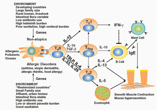
FIG. 45.1. Allergic Diseases are Associated with Aberrant Adaptive Immune Responses to Innocuous Environmental Antigens. The development of allergic responses to harmless antigens is influenced both by environmental factors and the genetic background of the individual. Under conditions in which microbial exposure is minimal such as in developed countries, T cells differentiate along a Th2 pattern with elaboration of interleukin (IL)-4, IL-13, IL-5, and IL-9. In contrast, microbial exposure in early life protects against the development of harmful Th2 immune responses via the induction of regulatory T cells.


Timing of Allergen Exposure

The spectrum of antigens an atopic individual is sensitized to is dependent on their environment in early life. This tenet is based on the positive correlations observed between allergen exposures present during the month of birth and the development of sensitization to the same allergens later in childhood (eg, birch, grass, and dust mite allergens).131,132,133 For example, Scandinavian babies born in late winter or early spring are more likely to develop IgE antibody to birch pollen, which is prevalent during the spring, than those born at other times of the year.131 In addition, avoidance or withholding certain allergenic foods during the first few months of life tends to prevent sensitization and subsequent allergic responses to these particular foods.

It has recently been hypothesized that exposure to environmental antigens occurs both prenatally and postnatally. In this regard, several independent studies have provided support for the postulate that prenatal events may influence susceptibility to allergic diseases.134,135 For example, it has been shown that cord blood mononuclear cells respond to inhalant and food allergens, suggesting that initial priming
of allergen-specific T-cell responses may occur before birth.134,135 It has been demonstrated that the responding cord blood T cells were indeed of fetal origin, as they did not respond to common vaccine antigens such as tetanus toxoid, which the majority of adults in the study population expressed active immunity against.134 These findings suggest that transfer of substances from mother to child (eg, allergens, antibodies, toxins, hormones, or other immune mediators via the placenta) may occur during gestation, which might prime or sensitize the developing infant to environmental antigens. In addition, factors present in the in utero environment may serve to influence the nature of the immune response of the fetus to allergens that cross the placenta. Indeed, mediators such as PGE2 and high levels of progesterone, which has been shown to alter the Th1/Th2 balance by either suppressing Th1 cytokine production or inducing a Th2 pattern, are present during pregnancy. Th2 skewing is thought to take place to suppress IFNγ production that is toxic to the fetus. Accordingly, exogenous antigens that leak across the placenta are likely to be presented to the fetal immune system within a milieu conducive to selection for Th2 immunity. This contention is supported by studies showing that cord blood cells from both allergic and noallergic infants both produce high levels of Th2 cytokines and low IFNγ levels.135,136 Although immune responses in neonates are skewed toward a Th2 phenotype, studies of human infants indicate that this Th2 skew gradually diminishes during the first 2 years of life in nonallergic individuals.137 In allergic infants, the reverse occurs, with the strength of neonatal Th2 responses increasing over a similar period. This has been supported by Holt and colleagues,138 who showed that although the levels of IgE vary considerably during early childhood, atopic subjects eventually lock into a stable pattern of increasing antibody production and Th2-polarized cellular immunity that is associated with stable expression of the IL-4 receptor in allergen-specific Th2 memory cells, which is absent during infancy. The persistence of this neonatal bias and the failure to produce Th1 or Treg-type responses may be an important feature of the atopic disease state.

The redirection of Th2 responses is thought to occur simultaneously with childhood bacterial or viral infections in early life. This relationship has been capsulated in the “hygiene hypothesis,” which argues that early childhood infections inhibit the tendency to develop allergic disease.139,140 Several epidemiologic studies have provided support for this hypothesis by demonstrating an inverse relationship between farm living,141 pet ownership,142 or drinking unprocessed cow’s milk143 in early life and atopy. Although the general implication of these studies is that protection from allergy seems to be associated with an increase in microbial exposure, the link has remained fairly indirect.

The complex interface with the microbial world provided by the gastrointestinal tract might well be important to the interrelationship between infection and allergy. The endogenous flora of the gut provides a wealth of stimuli for the developing immune system at birth. There seem to be both quantitative and qualitative differences in early childhood patterns of bacterial colonization between the developed and developing world. For example, a study of Swedish and Pakistani infants indicates that intestinal colonization with aerobic gram-negative bacteria tends to occur later in developed than developing countries.144 Once they are colonized, infants in developed countries tend to carry the same enterobacterial strains, whereas infants in developing countries are often colonized serially with different strains. Differences in intestinal flora have also been found between allergic and nonallergic children in Europe.145,146 Allergic children seem to be colonized less often with lactobacilli in Sweden and Estonia than nonallergic controls. This idea is consistent with the observation that oral tolerance cannot be induced in germ-free mice.147 Furthermore, elimination of commensal intestinal microflora with broadspectrum antibiotics also prevented oral tolerance from developing and resulted in susceptibility to intestinal inflammation. These observations suggest that TLR signaling by commensal bacteria under normal steady-state conditions is required for maintenance of intestinal epithelial homeostatsis, and possibly for the induction of some forms of Tregs. The intestine has been shown to affect immune responses at other mucosal surfaces including the respiratory tract and lacrimal, salivary, and mammary glands through secretory IgA antibodies.148,149 A recent study suggests that not only are there differences in gut flora between individuals living in developed versus developing countries, but that allergic asthmatic individuals display a different spectrum of bacterial species in their lungs than do healthy control individuals.150 In both compartments, a shift from protective bacteriodes to clostridial species has been observed. Recent studies demonstrating the role of a specific clostridial species, segmented filamentous bacteria, in the generation of Th17 responses further support a role for an altered microbiome in the pathogenesis of allergic disorders.151 Further studies on the influence of the microbiome are likely to provide tremendous insight into the mechanisms underlying the hygiene hypothesis.

Further support for the hygiene hypothesis is the striking association between polymorphisms in genes in the TLR pathways and atopic phenotypes. Specifically, an association has been consistently reported between a polymorphism in the CD14 gene, increased levels of sCD14, and decreased levels of IgE, reflecting the importance of bacterial LPS in downregulating Th2 responses.152 Moreover, a polymorphism in the coding region of the TLR4 gene (A/G 896) resulting in reduced cell surface expression of TLR4 and subsequent disruption of LPS-mediated signaling, has been associated with atopy in children.153 However, the data in asthma is conflicting with some studies showing an association between asthma and TLR4 polymorphisms153 while others do not.154 Interestingly, inheritance of both the TLR4 SNP (A/G+896) and the IL-4 SNP (-590) confers greater risk of asthma pathogenesis in females.155 Similarly, a polymorphism in the TLR2 gene (TLR2-16934) was shown to be a major susceptibility gene for children living on farms.156 Clearly these innate immune pathways play a critical role in determining the inflammatory reactions of the airways and the outcome of T-cell responses to inhaled allergens.


Although the hygiene hypothesis is likely oversimplified, it is theoretically possible that APCs at mucosal surfaces in atopic individuals receive less innate immune stimulation or stimulation of a different type in the form of microbial stimuli (LPS, peptidoglycans, mycobacterial antigens) and therefore they fail to redirect weak Th2 responses into protective Th1 responses or Treg responses. This hypothesis may provide a plausible explanation for the rising prevalence of these disorders in developed countries.

Exposure to other environmental agents such as diesel particles, ozone, secondhand tobacco smoke, and viruses (respiratory syncytial virus [RSV], rhinoviruses) can also enhance sensitization to allergens in young children. Numerous epidemiologic studies illustrate clear associations between exposures to these agents and enhanced antigenic sensitization and worsening of disease.157,158 Nasal challenge with diesel alone increased IgE production in both atopic and nonatopic individuals, suggesting that indeed diesel may be a sensitizer.157 When evaluated together with allergen, diesel exposure of ragweed-sensitive subjects resulted in a significant increase in allergen specific IgE with an increase in Th2 cytokine production. Ozone can alter both immediate and late-phase responses of asthmatics and allergic rhinitics to inhaled allergen.158 Epidemiologic studies have shown that exposure to environmental tobacco smoke in childhood is associated with increased skin test reactivity, serum IgE, and prevalence of eosinophilia.159,160 Recently, it has been shown that controlled chamber exposures of ragweed-sensitive subjects to environmental tobacco smoke resulted in enhanced production of allergen-specific IgE, Th2 cytokines, and decreased IFNγ levels in nasal lavage fluids.161 Although there is controversy regarding whether RSV is an inducer of allergic asthma or rather a predictor of aberrant immune responses, severe RSV infections are strongly associated with asthma.162 Similarly, rhinoviral infections are strongly associated with exacerbations in atopic airway disease.163 In summary, the complex interplay between genetic and environmental factors likely governs susceptibility to the development of atopic disorders.


Epigenetic Influences on Development of Atopy

The inheritance of atopic disorders appears to be preferentially associated with the atopic status of the mother. As asthma risk alleles are inherited from both parents, the preferential link of atopy with the atopic status of the mother suggests that factors present in the in utero environment contribute to subsequent risk of atopy in the offspring164 (Fig. 45.2). These modifications in gene expression in response to environmental cues can result from modifications of deoxyribonucleic acid (DNA; eg, by methylation) or of proteins that intimately associate with DNA (eg, acetylation, methylation, or phosphorylation of histones). Such differences could result in differential “prenatal programming” of immune cells in the fetus depending on the asthma status of the mother or from exposure to asthma medications taken by asthmatic mothers during pregnancy.

The first evidence that fetal genotype interacts with “maternal asthma” to determine risk for asthma in the child was provided by a positional cloning study that identified HLA-G as an asthma susceptibility gene.165 In that study, the -964G allele was associated with asthma only in families with an affected mother, and the -964A allele was associated with asthma in families with an unaffected mother. Subsequent studies implicated a SNP (+3142C/G; rs1063320) that resides in the 3′ untranslated region within a target site for three micro ribonucleic acids (miRNAs).166 In the presence of any of these miRNAs, expression of the G allele is suppressed whereas expression levels are unchanged for the C allele. The GG genotype, which is suppressed in the presence of the miRNAs, was associated with significant protection from asthma
among children of mothers with asthma but with modest risk for asthma among children of mothers without asthma. The mechanism through which the “maternal asthma-fetal HLA-G genotype” interaction influences subsequent risk for asthma in the child is still unknown, but modulation of expression via miRNA targeting could be involved. Because asthma in the mother is such a strong predictor of asthma in her children, it is likely that other, as yet undiscovered, genes also play a role in modulating this effect.


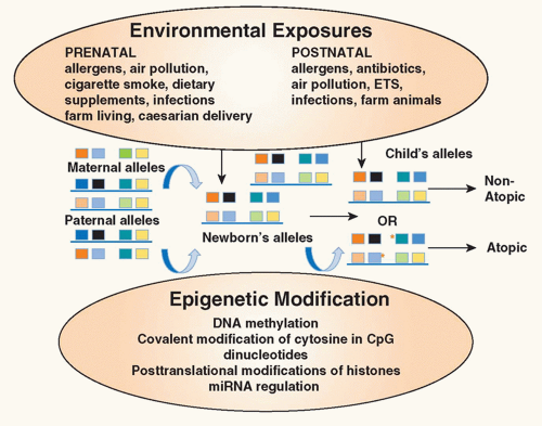
FIG. 45.2. Environmental Epigenetics and Atopy. Exposure of the mother to environmental factors (environmental tobacco smoke, air pollution, allergens, antibiotics, dietary supplements) during pregnancy may induce epigenetic changes (deoxyribonucleic acid methylation, covalent modification of cytosine in cytosin-phosphatidyl-guanosin dinucleotides, posttranslational modifications of histones, micro ribonucleic acid regulation) in gene expression in the offspring providing an explanation for the heretofore unexplained maternal influence on the inheritance of atopic diseases. In addition, exposure of the infant in early childhood to environmental factors may also alter expression of genes important in allergic inflammation. Collectively, these influences contribute to the risk of the child developing atopic diseases. Red asterisks refer to sites of epigenetic changes. Adapted from Miller and Ho.164

Recent evidence suggests that prenatal and postnatal exposures to the key environmental triggers of allergic disease described previously (ie, allergens, microbial infections, tobacco smoke, other pollutants, dietary supplements) can induce epigenetic changes in gene expression and alter disease risk through activating or silencing immune-related genes with subsequent effects on immune programming. For example, Fedulov and Kobzik et al.167 demonstrated that DNA from splenic DCs harvested from pups born to mothers previously sensitized and challenged to OVA was hypermethylated. Moreover, adoptive transfer of these DCs from pups of allergic mothers into offspring from nonatopic mothers transferred increased susceptibility to allergic disease to recipients despite there being no differences in DNA sequences. The specific genes conferring this susceptibility remain to be determined.

There is also strong evidence that the association of maternal smoking in pregnancy with asthma risk may be due to epigenetic changes.164 Indeed, altered DNA methylation patterns have been observed in several genes in buccal cells from children exposed in utero to tobacco smoke.168 Likewise, exposure to diesel exhaust partially augmented the production of IgE following allergen sensitization through hypermethylation of IFNγ cytosin-phosphatidyl-guanosins (CpGs) and hypomethylation of CpGs in the IL-4 locus.169 Further evidence for epigenetic contributions to disease risk is provided by the observation in mouse models of asthma that a maternal diet rich in methyl donors (folate) can increase susceptibility to allergic disease in offspring that is mediated through increased DNA methylation.170 Studies in humans examining the role of folate have been conflicting, with one study showing no association,171 whereas another showed an association between folate supplementation in late pregnancy and a physician’s diagnosis of asthma in the offspring.172

Several lines of evidence suggest that the protective effects of maternal exposure to farm environments, which increase microbial burden prenatally, may occur through epigenetic changes. Specifically, studies have shown that nonpathogenic microbial strains isolated from farm environments can induce epigenetic effects when administered to pregnant animals and protect the offspring from experimental asthma.173 The protective effects were IFNγ-dependent and associated with protection against the loss of histone 4 acetylation of the IFNγ gene in the CD4+ T cells of the offspring. In contrast, a decrease in H4 acetylase was observed in the IL-4 promoter. Moreover, pharmacologic inhibition of H4 acetylation in offspring abolished the protection. Further examination of epigenetic changes in allergy will undoubtedly enhance our understanding of the ontogeny of allergic diseases.


FACTORS REGULATING TYPE 2 IMMUNE RESPONSES IN ALLERGIC INDIVIDUALS

Differentiation of naïve CD4+ T cells into the various subsets requires several signals derived from either APCs or accessory cells. These include presentation of antigen in the context of MHC II, signals conveyed through costimulatory ligand-receptor interactions, and signals conveyed through the actions of specific cytokines. Although the factors driving Th1, Th17, and Treg subsets are well defined, the factors critical for the initiation of the type-2 response have long eluded investigators. In contrast to the steps involved in the differentiation of other T subsets, traditional antigen loaded APCs themselves are not sufficient to drive the early IL-4 required for Th2 differentiation or for full development of Th2 effector functions. Recently, however, several critical observations have led to a more detailed understanding of the type 2 response. First, three type 2-inducing cytokines, TSLP, IL-25, and IL-33, were identified as being necessary for the upregulation of type 2 effector cytokines, mirroring the role of IL-12 in the type 1 response. Secondly, recent evidence suggests that these type 2-promoting cytokines are produced by resident cells in mucosal tissues in response to innate immune receptor stimulation, not by the APCs themselves. Thirdly, exciting evidence suggests that type 2 effector cytokines can be produced by the newly described lineage negative innate lymphoid cell (ILC) populations in response to the type 2-promoting cytokines (IL-25, IL-33). Thus the overall emerging scenario is one in which allergens uniquely interact with mucosal epithelial cells and resident cells to produce these Th2-promoting cytokines through stimulation of various PRRs or danger associated molecular patterns expressed on resident mucosal cells. These cytokines (IL-33, IL-25) can themselves drive type 2 effector cytokines in innate cells (mast cells, basophils, ILCs) independently of APCs. In parallel, they also license traditional APCs to present allergenic peptides to conventional naïve T cells in draining lymph nodes, driving Th2 differentiation and T-cell memory development. The relevance of each of these steps to the development of allergic diseases will be discussed in the following sections (Fig. 45.3).


Innate Immune Responses to Environmental Antigens

Allergens interact with innate recognition systems present on a diversity of cells at the mucosal surface of the respiratory tract, skin, and gastrointestinal tract, including epithelial cells and phagocytic cells (DCs, macrophages). As the primary interface with the environment, the epithelial cell layer represents the front-line defense against injury by pathogens or environmental irritants. Emerging evidence suggests that epithelial cells play a critical role in both antigen recognition and danger recognition. Allergenic particles and their associated soluble components interact directly with epithelial cells through a variety of diverse mechanisms including 1) binding of PRRs including various members of the TLR family, the C-type lectin receptor (CLR) family, and nucleotide oligomerization domain (NOD) receptor family; 2) the activation of protease-activated receptors (PARs); and
3) activation of various danger sensing pathways (extracellular nucleotides, uric acid).


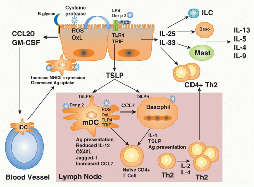
FIG. 45.3. Schematic of Events Leading to the Initiation of Th2-Mediated Immune Responses. Upon encounter with allergen, epithelial cells of the respiratory tract, the intestinal tract, and the skin release mediators that recruit (CCL20), activate (granulocyte macrophage-colony stimulating factor [GM-CSF]), and/or drive Th2 cell differentiation (thymic stromal lymphopoietin [TSLP], interleukin [IL]-25, IL-33). IL-25 and IL-33 recruit and activate a variety of innate immune cells such as basophils, mast cells, and innate lymphoid cells, which can produce the protypical type 2 cytokines IL-4, IL-5, and IL-13 driving the allergic phenotype. The elevations in IL-25 and IL-33 can perpetuate the allergic response by further type 2 cytokine production from basophils, eosinphils, or by initiating conventional Th2-cell expansion through activating dendritic cells (DCs) during encounters with allergens. In parallel, immature myeloid DCs are recruited to mucosal surfaces following the release of chemokines (ie, CCL20) and cytokines (GM-CSF) by epithelial cells in response to inflammatory stimuli such as allergens, pollutants, irritants, and/or viral infections. Once in the tissues, DCs mature and express costimulatory molecules (ie, CD86, OX40L, Jagged-1) under the influence of cytokines (TSLP, IL-33, IL-25) derived from surrounding tissues through stimulation of pattern recognition receptors (likely TLR4, -2; C-type lectins) expressed on their surface. In atopic individuals, it has been postulated that myeloid DCs appear to be the predominant population of DCs taking up antigen and driving the differentiation of naïve T cells toward a Th2 profile. They then migrate to the draining lymph node to stimulate naïve T-cell differentiation and proliferation. Proteasecontaining allergens induce the production of TSLP by epithelial cells through a series of steps including generation of reactive oxygen species, production of oxidized lipids, which in turn stimulate TLR4-TRIF signaling pathways leading to TSLP expression. These same pathways induce the production of the chemokine CCL7 by DCs, which recruits basophils to the lymph node. In the lymph node, they can also directly stimulate basophils to produce TSLP and IL-4. Basophils and DCs drive Th2 cell differentiation under the influence of various cytokines such as TSLP, IL-4, and IL-25. These Th2 cells proliferate in response to IL-2 and IL-4 in an autocrine fashion. Activated Th2 cells in turn migrate back to the site of antigen stimulation under the influence of Th2 selective chemokines (CCL17, CCL12). Once in the tissues, Th2 cells secrete a profile of Th2 cytokines (IL-4, IL-5, IL-13, IL-9) that initiate a cascade of downstream pathways that collectively lead to the development of the allergic phenotype. Not shown on the figure is the fact that through their effects on the airway epithelium, Th2 cytokines may further amplify Th2 immune responses through the induction of Th2-selective chemokines (CCL17, CCL12) and/or IL-25 and IL-33 initiating a positive feedback loop. baso, basophil; iDC, immature dendritic cell; ILC, innate lymphoid cell; mDC, myeloid dendritic cell; ROS, reactive oxygen species.

Activation of epithelial cells through these diverse pathways can induce the release of cytokines (TSLP, IL-25, IL-33), chemokines, and growth factors that both facilitate entry of antigens into the mucosa; recruit APCs; and provide instructive signals to antigen APCs. As the absence of PRR activation and instruction of APCs results in the development of tolerance to exogenous antigens, alterations in these innate immune responses occurring at the epithelial-environmental interface may be important determinants of susceptibility or resistance to the development of allergic diseases (Fig. 45.4).


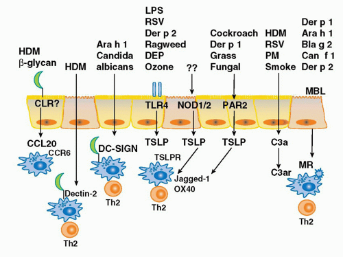
FIG. 45.4. Innate Immune Pathways Activated by Allergens. Allergenic peptides are recognized by several traditional pattern receptors including toll-like receptors, C-type lectin receptors, and nucleotide oligomerization domain receptors. In addition, other classes of allergens initiate Th2-mediated immune responses through the activation of protease-activated receptors and the cleavage of complement pathways. Activation of these innate immune pathways on mucosal epithelial cells leads to the production of chemokines which recruit immature demdritic cells (DCs), the production of Th2-promoting cytokines such as thymic stromal lymphopoietin, the enhancement of DC internalization of allergens, and the upregulation of expression of costimulatory molecules associated with Th2 differentiation such as OX40L and jagged-1. DEP, diesel exhaust particles; HDM, house dust mite; MBL, mannan-binding lectin; MR, mannose receptor; PM, particulate matter. Der p 1 and Der p 2 are house dust mite-derived allergens; Ara h 1 is the major peanut allergen; Blag 2 is the major cockroach allergen; Can f 1 is the major canine allergen.


Role of Toll-Like Receptor Stimulation in Allergic Inflammation

The most well-studied family of PRRs in allergic inflammation is the TLR family. Epidemiologic studies have consistently reported an inverse correlation between high levels of bacterial products such as LPS in the ambient environment during very early life and the subsequent development of atopy and allergic disease.174,175 It has been postulated that such exposures drive counterregulatory immune responses in the developing immune system.176 On the other hand, controlled human challenge studies have shown that LPS exposure of sensitized individuals can exacerbate existing disease.177 Although the mechanisms underlying this apparent paradox are not entirely clear, the complexity of the responses to TLR agonists may be due to several factors including the array of TLR receptors activated by complex allergens (TLR9 versus TLR4), their relative abundance, and the timing of exposure during the life of the individual. For example, TLR9 stimulation clearly prevents and inhibits the development of experimental allergic inflammation at all doses,178 whereas TLR2 and TLR4 pathway stimulation has been shown to both drive179,180 and inhibit180,181 the development of Th2-mediated allergic inflammation in experimental mouse models. Bottomly and colleagues180 have shed some light on this complexity, demonstrating that the impact of TLR4 stimulation on allergic inflammation is highly dependent upon the dose of TLR4 agonist. Specifically, they showed that although airway sensitization with the normally tolerizing antigen, OVA along with 1 ng of LPS (LPS-depleted) was reported to induce tolerance, sensitization in the presence of “low-dose” (0.1 µg) LPS promoted TLR4-dependent, Th2 inflammation, and sensitization in the presence of “high-dose” (100 µg) LPS led to a Th1 response. Although these studies provided a plausible explanation for the LPS dose effects observed in epidemiologic studies, they did not explain how stimulation through the same receptor could result in two distinct biologic outcomes. To address this issue, Tan and colleagues182 examined allergic responses in a series of bone marrow chimeric mice expressing TLR4 in specific compartments. They show that strong (high-dose LPS) TLR4 signaling always results in a Th1 response despite the fact that high LPS stimulation of mice expressing TLR4 only in the stromal compartment drives Th2 responses, as a result of the dominant influence of the hematopoietic cell compartment under these conditions. Surprisingly, they found that at very low LPS levels, mice expressing TLR4 only in the stromal compartment did not mount Th2 or Th1 immune responses. However, when mice that had competent TLR4 signaling in both the stromal and hematopoietic compartments were exposed to low levels of LPS+OVA, they mounted Th2 immune responses, suggesting that once a threshold level of TLR4 stimulation is reached in the stromal compartment, Th2 responses ensue. The authors propose that the ability of stromal cells (presumably epithelial cells) to drive Th2 responses is likely through their ability to secrete TSLP and to promote the maturation of Th2-inducing DCs that express the Notchligand Jagged-1, but not the Th1-inducing ligand, Delta-4. As Jagged-1 has been shown to induce the expression of the critical Th2 transcription factor, GATA3 in CD4+ T cells through binding Notch ICD, these findings may explain the association between low-level activation of TLR4 pathways and Th2-cell differentiation.183 In contrast to Tan’s findings, another group184 showed that stromal cell TLR4 signaling was sufficient to drive Th2 immune responses when mice were exposed to dust mite extracts containing low levels of LPS, suggesting that the dust mite extracts might contain endogenous TLR4 agonists that shift the dose response of the stromal compartment to TLR4 stimulation into the Th2-inducing range.

A recent study has provided a compelling mechanism by which endogenous components of dust mites may drive TLR4 signaling. Based on the recent discovery of a structural homology between Der p 2, one of the major house dust mite allergens, and MD-2, a member of the lipid-recognition (ML) domain family of proteins, which is the LPS-binding member
of the TLR4 signaling complex, Trompette and colleagues25 asked the question whether Der p 2 and MD-2 exhibited functional homology as well. Indeed, they reported that Der p 2 facilitates TLR4 signaling through direct interactions with the TLR4 complex, reconstituting LPS-promoted TLR4 signaling in the absence of MD-2. They also demonstrated that Der p 2 could facilitate LPS signaling in primary APCs, with or without MD-2 being present. Finally, they reported that in vivo delivery of Der p 2 drives the development of experimental allergic asthma in a TLR4-dependent manner, retaining this property in mice with a genetic deletion of MD-2. These data suggest the possibility that Der p 2-mediated activation of TLR4 signaling in the airway epithelium under conditions of low bacterial product exposure—those associated with increasing rates of aeroallergy in the urban, developed world— may shift the LPS/TLR4 response curve from the tolerizing into the Th2-inducing range. Der p 2 exposure may serve to facilitate TLR4 signaling by airway epithelial cells as they have been reported to express TLR4, but little or no MD-2, in the basal state.185

The fact that the major dust mite allergen, Der p 2, is a molecular mimic of an endogenously expressed mammalian lipid binding family member has several important implications for our understanding of allergenicity. As numerous other members of the MD-2-like lipid binding family are major allergens, the activation of innate immune pathways via lipid binding is likely to be a common feature of allergens. Indeed, the recently solved structures of several allergens including Der p 5 and Der p 7 suggest that they possess the propensity to bind hydrophobic compounds.186,187 Of note, Der p 7 has been shown to resemble the LPS binding protein and to bind to the lipopeptide polymyxin B from gram-positive bacteria. More broadly, a wide range of allergens are lipid binding proteins (ie, lipid transfer proteins [peach allergen Pru p 3], steroid-like molecules [cat allergen Fel d 1], lipocalins [horse allergen Equ c 1, mouse allergen Mus m 1]). Further studies are clearly needed to define the lipids naturally bound by these allergens, the receptors activated by such lipids, and the precise pathways of innate and adaptive immune responses driven by such activation.

Despite our rudimentary understanding of the role of PAMPs in allergic responses, one may speculate that either differences in exposures to microbial products or polymorphisms in genes of the TLR pathway may both be important determinants of the risk of developing allergic disease through altering the phenotype and function of DCs. Along these lines, as discussed previously, genetic studies show that polymorphisms of different components of the TLR pathways (CD14, TLR4, TLR2) may partly explain susceptibility to atopy.152,153,154,155,156


Role of Carbohydrate Recognition in Th2 Immunity

Just as the mammalian immune system has evolved mechanisms to recognize bacterial proteins in association with PAMPs that induce appropriate Th1 responses, recent studies suggest an important role for complex carbohydrates in driving Th2 immune responses to allergens. In particular, fucosylated glucans are a diverse class of naturally occurring glucose polymers that are widely expressed in the cell walls of fungi, helminths, pollens, and certain bacteria, but they are not found in mammalian cells. Evidence is emerging that these carbohydrates drive strong Th2-biased immune responses through their interaction(s) with a large array of CLRs.

The C-type lectin family of soluble and transmembrane receptors demonstrate unique specificity for carbohydrate residues via distinct clustering of carbohydrate recognition domains.188 Transmembrane members of the CLR family include collectins, the mannose receptor family, selectins, dectin 1 and 2, and DC-specific intercellular adhesion molecule-3-grabbing nonintegrin. Ligand engagement of these receptors is involved in processes ranging from cellular trafficking to cell signaling and pathogen recognition mediated through FcRγ and Syk activation.188

Several studies suggest the involvement of various C-type lectins in regulating allergic inflammation. For example, it has been reported that β-glucan structures present in the peanut glycoallergen Ara h 1 have Th2-inducing characteristics; native, but not deglycosylated, Ara h 1 has been shown to activate human monocyte-derived DCs and induce IL-4- and IL-13-secreting Th2 cells.26 The Th2-promoting actions of Ara h 1 were mediated through binding of DC-specific intercellular adhesion molecule-3-grabbing nonintegrin and activation of ERK/MAPK signaling in DCs. Consistent with a role for lectins in promoting Th2 immune responses, a recent study showed that the CLR dectin-2 was required for the development of house dust mite-induced allergic inflammation.189 Consistent with a major role for CLRs in initiation of Th2 immune responses, β-glucans have been shown to mediate several aspects of DC function including antigen uptake, recruitment and activation of DCs, and instruction for Th2 differentiation. For example, blockade of the mannose receptor, an endocytic CLR, significantly reduced Der p 1 uptake by DCs.190 Moreover, recent studies have shown that β-glucans contained in house dust mite extracts and in molds may initiate immune responses through the induction of the chemokine, CCL20, which recruits immature DCs. The induction of CCL20 was shown to occur through β-glucan and Syk-dependent signaling pathways.191 Although the study of the role of carbohydrates as Th2-inducing PAMPs is only in its infancy, the evidence thus far suggest that carbohydrate moieties contained in common allergens function as strong Th2 inducers through activation of variety of CLRs on DCs.

Allergens containing lectins may also activate the lectin pathway of complement pathway activation, a highly conserved component of innate immunity. The complement system is a sophisticated network of soluble and membranebound proteins, which serve as both immune sensors and immune activators. Like other PRR pathways, the complement system can be activated by “hardwired” PRRs that have elvolved to recognize both endogenous and exogenous “danger” motifs. Lectin recognition receptors in the complement system include: mannan-binding lectin, and ficolins. Recognition of pathogen-associated carbohydrate residues by the soluble mannan-binding lectin triggers the serine protease-mediated cleavage of complement components C4 and C2 resulting in the generation of C3 convertase that carries out the rest of the complement cascade. Recognition by mannan-binding lectin is an integral part of the first line
of defense against a large number of microbial pathogens including bacteria, fungi, protozoa, and viruses. Indeed, it has recently been shown that the major cat allergen, Fel d 1, is a novel ligand of the cysteine-rich domain of the mannose receptor and the development of allergic inflammation induced by Fel d 1 was attenuated in mannose receptor-deficient mice.192 Likewise the mannose receptor has been shown to mediate the internalization of a diverse range of allergens (Der p 1, Der p 2, Can f 1, Bla g 2, Ara h 1) into monocyte-derived DCs through their carbohydrate moieties.193 Alterations in these pathways may lead to enhanced recognition of allergens as sequence variations in the MRC1 gene have recently been associated with the development of asthma in two independent and ethnically diverse populations (Japanese and African American).194


Nucleotide Oligomerization Domain Receptors in Allergic Inflammation

Although NOD receptors have not been as well studied as other PRRs, it has recently been shown that NOD1 (which binds cell wall peptidoglycans of gram-negative bacteria) and NOD2 signaling in stromal cells drives Th2 immune responses through the TSLP-dependent induction of OX40L.195 Studies demonstrating that polymorphisms in the intracellular NOD1 protein were associated with atopic eczema and asthma196 suggest that alterations in these pathways may play an important role in susceptibility to some atopic disorders.


Proteases in Allergic Inflammation

Several of the common allergens (house mite-derived, Der p 1, and Der p 3; Fed d 1 from domestic cats; cockroach allergens; fungal allergens) possess intrinsic protease activity. While the link between protease activity and type 2 inflammation is well documented, the mechanistic basis for these observations has been unclear. Allergens containing protease activity, such as Der p 1, a cysteine protease, have been demonstrated to produce changes in the barrier function of the epithelium, probably by disrupting the epithelial tight junctions by degrading the tight junction proteins ZO-1 and desmoplakin.197 Disruption of the tight junctions may both facilitate access of allergens to the underlying cells including DCs and may also directly initiate inflammatory cascades (modulate the function or immune and structural cells) in DCs and/or airway epithelium. For example, Der p1 cleaves several important immunomodulatory molecules including CD25, CD23, and various complement components.198,199 As a result of cleavage of CD25, peripheral blood T cells show markedly diminished proliferation and IFNγ secretion in response to potent stimulation by anti-CD3 mAb. These findings suggest that Der p1 decreases the growth and expansion of antigen-specific Th1 cells augmenting expansion of the antigen-specific Th2 cells that favor a proallergic response. Der p 1 cleavage of CD23 on murine B cells that would normally serve to inhibit IgE synthesis would further potentiate allergic responses by disrupting an important negative regulator of IgE production.199

Although the cellular mechanisms by which protease activity induces cytokine release remain unclear, members of a recently identified G protein-coupled family of cell surface receptors, designated PARs, have been implicated. Indeed, allergens such as the house dust mite serine proteases, Der p 3, Der p 9, cockroach extracts, and pollens activate PARs. Asokananthan et al.200 have shown that endogenous peptidases caused the release of cytokines through the activation of PARs on the respiratory epithelium, and that all four members of the PAR family were expressed on respiratory epithelial cells. In vivo experiments in experimental allergen models support the contention that activation of PAR pathways is associated with enhanced allergic responses. Specifically, PAR-2 overexpression enhanced OVA-induced eosinophilia and bronchial hyperresponsiveness, whereas PAR-2 deficiency was associated with reduced allergendriven eosinophilia and IgE production.201 Although the exact mechanisms by which PAR-2 mediates Th2 induction is unknown, PAR-2 was shown to mediate, in part, the induction of TSLP from airway epithelial cells in vitro in response to the protease activity of the common environmental fungus Alternaria alternata, as well as in response to the cysteine protease papain.202


Role of Danger-Associated Molecular Patterns in Regulation of Th2 Immunity

Several sensors of cellular damage have recently been implicated in the development of allergic inflammation. First, Eisenbarth and colleagues203 showed that the adjuvant alum promotes humoral responses by activating the nucleotide binding domain-leucine rich repeat (LRR)-containing receptor (NLR) inflammasome. Moreover, it has been shown that allergen stimulation of airway epithelial cells results in an upregulation of extracellular adenotriphosphate levels that trigger the migration of myeloid DCs (mDCs) into the airways and mediate the development of allergic inflammation in vivo.204 Idzko and colleagues205 have recently shown that these adenotriphosphate-triggered effects on airway inflammation are regulated via the P2Y(2)R. Another sign of cellular damage is the release of the potent antioxidant uric acid. Recent studies show that administration of uric acid crystals together with protein antigen was sufficient to promote Th2 cell immunity and the features of asthma.206 Surprisingly, the adjuvant effects of uric acid did not require the inflammasome (Nlrp3, Pycard) or the IL-1 (Myd88, IL-1r) axis, but promoted Th2 cell immunity by activating DCs through spleen tyrosine kinase (syk) and PI3-kinase δ signaling. Recently, a member of the trefoil factor family (TFF2), which has previously been shown to be a sensor of damage to the gut epithelium, has been shown to regulate Th2 immune responses to both allergens and helminth infections via induction of IL-33 by the epithelium and alveolar macrophages.207

These studies demonstrated that in contrast to Th1-inducing factors, which directly license DCs through PRRs, allergens license antigen-loaded DCs and accessory cells through a variety of innate signals derived from the mucosal epithelium to induce Th2 immunity. Thus, licensing of APCs in trans by signals emanating from the stromal compartment appears to be a unique feature of Th2 immune responses. As will be discussed subsequently, this concept is entirely consistent with studies showing that the primary cytokines driving Th2 differentiation are stromal cell (epithelial cell)-derived.


Cytokine Regulation of Th2 Differentiation in Allergic Disease

One of the most important variables in instruction of T-cell differentiation comes from the local cytokine milieu at the time of antigen presentation. Recently, Th2 cell-polarizing factors have been finally identified, these include TSLP, IL-25, and IL-33 (Fig. 45.5). Alterations in either the production of or responsiveness to these signals could lead to the development of aberrant Th2 immune responses in atopic individuals. Indeed, there is evidence to support each of these possibilities.


Thymic Stromal Lymphopoietin Initation of Th2 Immune Responses

TSLP is an IL-7-like cytokine originally characterized by its ability to promote the activation of B cells and DCs.208 The TSLP receptor is comprised of the IL-7Rα chain and the unique TSLPR chain. TSLP is expressed by epithelial cells with the highest levels being found in lung- and skin-derived epithelial cells. TSLP has been shown to activate DCs such that they acquire the ability to prime naïve T cells for the production of Th2 cytokines, while downregulating IFNγ and IL-10. In support of a role for TSLP in Th2 responses, TSLP levels have been shown to be elevated in the airways of asthmatic individuals209 and in animal models210,211 of atopic disease. Overexpression of TSLP in the lungs of mice has been shown to lead to the development of Th2 immune responses, whereas TSLPR knockout mice develop strong Th1 responses to allergen and are protected against the development of allergen-driven asthmatic responses.210,211 Finally, overexpression of TSLP in the skin of mice results in an atopic eczema-like phenotype.212 In humans, the gene encoding TSLP is located on chromosome 5q22 in a strong linkage region for asthma. Polymorphisms in TSLP have been associated both with asthma213 and eosinophilic eosophagitis.130 Although the IL-7Rα gene is located on chromosome 5p13, it remains to be determined whether polymorphisms in the IL-7Rα receptor influence TSLP signaling. Although the precise mechanisms underlying the ability of TSLP to prime DCs is not known, one mechanism that has been postulated is that TSLP induces DCs to express OX40 ligand (OX40L) and decrease IL-12 production.214 In support of this contention, TSLP-induced OX40L on DCs was required for triggering naive CD4+ T cells to produce IL-4, -5, and -13.215


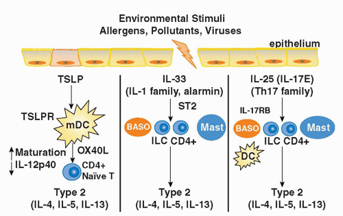
FIG. 45.5. Members of Three Distinct Cytokine Families Promote Th2 Differentiation. Thymic stromal lymphopoietin (TSLP), an interleukin (IL)-7-like cytokine released from mucosal epithelial cells, binds its receptor TSLPR on dendritic cells and induces the expression of OX40L and the suppression of IL-12 production, thus driving the differentiation of naïve T cells toward a Th2 phenotype. IL-33 is an IL-1 family member that binds its unique receptor ST2 on a variety of innate immune cells including basophils, innate lymphoid cells, conventional CD4+ T cells, and mast cells driving the production of type 2 cytokines. IL-25, a member of the IL-17 cytokine family, binds its receptor IL-17RB on a variety of innate immune cells including basophils, innate lymphoid cells, mast cells, and conventional CD4+ T cells driving their production of type 2 cytokines. IL-25 and IL-33 can also stimulate type 2 cytokine production indirectly by activating dendritic cells to promote Th2 differentiation.


Interleukin-25 in Allergic Inflammation

IL-25 is a novel member of the IL-17 cytokine family (IL-17E) that promotes CD4+ T-helper 2 lymphocyte-like (Th type-2) inflammatory responses. IL-25 is produced by epithelial cells, activated human basophils, eosinophils, and mast cells. Recombinant IL-25 administration to mice has been shown to induce IL-4, IL-5, and IL-13 production, and systemic Th2-cell responses, characterized by elevated IgE production, eosinophilia, and remodeling in several organs including the lungs, gastrointestinal tract, and skin of naïve mice.216 Interestingly, IL-25 can induce these Th2-mediated immune responses in mice lacking T and B cells, suggesting that IL-25 acts upon another cell type besides T cells to drive IL-13 production.217 A search for the cellular targets of IL-25 has led to the discovery of a family of ILCs that produce type 2 cytokines (ILC2 cells).217 These ILC2 cells belong to a heterogeneous family of innate cells that do not express surface markers of adaptive immunity (non-T, non-B cells) and are not antigen restricted like their adaptive counterparts. However, as they express CD45 and are dependent on traditional T-cell growth factor signaling pathways such as those mediated through the common γ-chain and IL-7 receptor α-chain (CD127), they have been called ILCs. The population of ILC2s that produce type 2 cytokines in response to IL-25 and IL-33 are lineage-negative (CD4-CD127+IL-1RL1+IL-17RB+, RORγt-). Several ILC2 cells have been discovered in the mouse by independent groups and have been designated natural helper cells,218 multipotent progenitor type 2 cells,219 nuocytes,217 and innate helper type 2 cells.220 Recently, it has been shown that adoptive transfer of wild-type nuocytes producing IL-13, but not IL-13-/-nuocytes, into IL-13-deficient mice was sufficient to induce allergic inflammation.221 Although there is much to learn about these populations and the mechanisms regulating their function, increased IL-25 and IL-25R expression has been detected in patients with asthma, suggesting that overproduction of IL-25 may be associated with aberrant type 2 immune responses.222


Interleukin-33 and Nuocytes in Atopy

IL-33 is a member of the IL-1 family that stimulates type 2 cytokine production through binding its receptor composed of the IL-33 specific receptor chain, IL-1RL1 (ST2) and the
IL-1 receptor accessory protein (IL-1RAcP),223 both of which are widely expressed, particularly by innate immune cells (mast cells, basophils, DCs), Th2 cells, and innate lymphoid populations described previously.217,223 IL-33 is thought to function as an “alarmin” released from stromal cells (epithelial cells, endothelial cells, and airway smooth muscle) following cellular injury to alert the immune system to tissue damage or stress. A role for IL-33 in type 2-cytokine dependent immune responses in vivo is supported by the finding that administration of IL-33 to naive mice induces eosinophilia, increased serum IgE levels, IL-5 and IL-13 production, and mucus cell changes in the respiratory and gastrointestinal tracts.224 Moreover, blocking IL-33 with an ST2 fusion protein decreases eosinophilic airway inflammation, concomitant with a decrease in IL-4 and IL-5 expression.225 Notably, the levels of soluble ST2 protein and IL-33 mRNA protein are increased in sera and tissues from patients with asthma.226,227 Genome-wide association studies have identified polymorphisms in the genes encoding Il1rl1 and Il33 in patients with asthma, hypereosinophilic diseases, and allergic rhinitis, suggesting an association with atopic disorders in general.228,229,230

Although the exact mechanisms by which IL-33 induces type 2 cytokine production are unknown, it has been shown to induce Th2 cytokines from a number of innate immune cells including basophils, mast cells, and the newly described ILC populations. In support of a role for the activation of ILCs by IL-33, it has recently been shown that administration of rIL-33 induces AHR and eosinophilic inflammation in mice independently of adaptive immune cells.224 Interestingly, it was initially reported that OVA-induced AHR was not dependent on ILCs,231 whereas another study suggest that fungal antigen-induced allergic inflammation was dependent upon IL-33 and innate lymphoid populations.232 Collectively, these results suggested that antigens, which contain PAMPs that activate or injure the epithelium, recruit and/or activate ILCs. Consistent with this hypothesis, it has recently been shown that IL-13-producing ILCs are important in protection of the mucosal epithelium following viral infections, despite the fact that the type 2 cytokines do not play a role in clearance of the virus.233 In support of a role for ILCs in allergic disorders, Mjosberg et al.234 recently reported that a lineage-negative CD127(+)CD161(+) ILC population that responded to both IL-25 and IL-33 by producing IL-13 was enrichment in nasal polyps of patients with chronic rhinosinusitis. Although much remains to be learned about the role of ILC2 cells in type 2-mediated immunity, these new studies raise the possibility that expansion of these unique ILCs in response to exposure to allergens, infectious agents, or environmental toxins may unwittingly lead to the establishment of type 2-mediated diseases.

Only gold members can continue reading. Log In or Register to continue

Stay updated, free articles. Join our Telegram channel

Aug 29, 2016 | Posted by in IMMUNOLOGY | Comments Off on Immunologic Mechanisms of Allergic Disorders

Full access? Get Clinical Tree

Get Clinical Tree app for offline access