Immunity to Viruses


Immunity to Viruses


Hildegund C.J. Ertl


INTRODUCTION

Veni, vidi, vici—I came, I saw, I conquered. If viruses could talk or see, this would be their hendiatris. Although viruses are minute particles composed only of a genome surrounded by a few proteins, they have a fiendish way of wreaking havoc not only on humans, as everyone who ever had the flu knows, but also on animals, plants, and even bacteria. Each year, more people die of viral infections than of natural disasters such as hurricanes, earthquakes, and tsunamis combined, or even manmade tragedies such as war. For example, it is estimated that variola major, the causative agent for smallpox, killed nearly half of the population of Native Americans after the virus was introduced into the western hemisphere by European colonizers. The Spanish Flu caused by an H1N1 influenza A virus caused the death of 50 to 100 million humans between 1918 and 1919, which is well in excess of the 16 million casualties of World War I. Human immunodeficiency virus (HIV)-1 has killed more than 25 million humans since 1981 and continues to spread, threatening the already frail economic structures of the most afflicted countries in sub-Saharan Africa, where more than 30% of the adult population carries the virus.

Not only do viruses cause acute or chronic infections with potentially fatal outcome, but they also contribute to other diseases. Viruses are associated with 20% of cancers, they have been implicated in the pathogenesis of human arteriosclerosis and autoimmune diseases, and they are linked to an overall reduction in life expectancy.

How can something so small be so deadly? Viruses, which range in size from 10 to 300 nm in diameter with genomes of minimally 2 kilobases to over 1.2 megabases, are unable to propagate themselves, but require a host cell to replicate. Once a cell becomes infected, viruses hijack its transcription and translation machinery to promote their own replication. The physiologic functions of the infected cell are disrupted as it is being turned into a virus production facility. However, the cell fights back as soon as it senses the virus. This fight initially takes place intrinsically within the infected cells but then rapidly spreads extrinsically once the immune system has been alerted. In turn, many viruses mount defenses against the attack from their host by encoding proteins that actively subvert innate and adaptive immune responses. In acute virus infections, the fight between virus and host literally lasts until the death of one of the adversaries. In chronic infections, a truce is eventually reached where virus and host coexist, generally at the expense of the well-being of the latter.

Fewer than 200 viruses are known to cause disease in humans. Over the last 50 years, on average two new species of human viruses have been discovered annually; one can expect that this number will continue to rise.1 Where does this ever-increasing number of viruses come from? We are not certain about the origin of viruses, although we know from ancient texts as well as more modern data-driven genomic analyses that viruses have been around for a very long time.2 Whether they originated from cells, concomitantly with cells, or even primordially from some genetic soup remains debated.3 No matter how they evolved, their lack of common genes argues for a polyphyletic rather than monophyletic evolution. The constant discovery of new viruses may simply reflect improvements of detection technologies that traditionally were based on cell culture and that are now being replaced with high-throughput genomics. Notwithstanding, many of the newly discovered viruses seem to evolve from animal reservoirs through mutations that allow for an extension of their host range. Of importance is that viral genomes have far higher mutation rates than, for example, mammalian genomes as they fail to correct errors during replication; such errors most commonly lead to loss of viral fitness but occasionally benefit the virus in its quest for replication. One example of a stable host range altering mutation that caught global attention was that of a coronavirus, which caused the outbreak of severe acute respiratory syndrome (SARS) in 2003 and 2004. This virus, termed SARS-CoV, which in its wild-type form infects civets, a cat-like carnivore, mutated and became infectious to humans and then within this host rapidly underwent further positive selection.4 Other viruses such as pathogenic H5N1 avian influenza viruses have been isolated since the late 1990s repeatedly from humans, who commonly died as sequela of the infection. Pathogenic H5N1 viruses, which were, and by some still are, feared to evolve into pandemic viruses, have thus far failed to mutate to achieve sustained human-to-human transmission, while concomitantly, another influenza A virus arose from a triple reassortment between viruses that naturally infect humans, avians, and swine, and caused the 2007 influenza pandemic. Considering that our knowledge of animal viruses remains limited, the sudden emergence of new and potentially deadly viruses from other species continues to threaten global health.

While most other deadly disease can be treated with drugs such as antibiotics to resolve bacterial infections or can be prevented by lifestyle choices, our arsenal to combat viral infections remains limited. Vaccines, which can be effective
in preventing viral infections, and even achieved the eradication of small poxvirus, are only available for 15 viruses. Drugs to specifically treat infections are only effective for some viruses such as HIV-1, herpes, hepatitis B and C, and influenza viruses. Our main defense thus remains the immune system. Over eons, it has evolved to sense viruses as pathogens, to produce factors that stop viral replication, and to develop lymphocytes that destroy those cells that serve as viral production factories. Like an elephant, the immune system never forgets. Nevertheless, this defense like that in every war, even if victorious, comes at a price: a runny nose at best and death due to insufficient or overwhelming responses at worst.


BASICS IN VIROLOGY


Virus Classification

According to the International Committee of Taxometry of Viruses, all viruses are classified into order, family, subfamily, genus, and species. Names of serotypes, genotypes, strains, variants, or isolates of virus species or artificial viruses are not ruled by the International Committee of Taxometry of Viruses. A species is defined as a polythetic class of viruses that constitutes a replicating lineage and occupies a particular ecologic niche. A genus defines a group of species that share common characteristics, while subfamilies and families define a group of genera with common characteristics. An order, in turn, defines families with shared characteristics. Currently, 6 orders, 87 families, 19 subfamilies, 348 genera, and 2288 species of virus have been defined.5 The Baltimore classification differs and divides viruses according to their genome or their mode of replication. Viruses contain either a deoxyribonucleic acid (DNA) or ribonucleic acid (RNA) genome, which can be single stranded (ss) or double stranded (ds). SsRNA viruses carry either a negative- or positive-stranded RNA. During their lifecycle, some DNA and RNA viruses undergo an intermediate step in which the RNA genome is converted by reverse transcription (RT) into DNA or vice versa their DNA genome into an RNA genome. Accordingly, viruses are classified into dsDNA viruses (eg, adeno or poxviruses), ssDNA viruses (eg, parvoviruses), dsRNA viruses (eg, reovirus), positive-sense ssRNA viruses (eg, picornaviruses), negativesense ssRNA viruses (eg, rhabdoviruses), positive-sense ssRNA-RT viruses (eg, retroviruses), or dsDNA-RT viruses (eg, hepadnaviruses). Other classifications are based on host range (Holmes classification) or structural characteristics (Lworff-Horne-Tournier [LHT] system).

The viral genome can be linear (eg, poxvirus), circular (eg, papillomaviruses), or segmented (eg, influenza virus). Viruses are further classified into enveloped (eg, rhabdoviruses) or naked (eg, picornaviruses) viruses. Viruses can also be divided according to their morphology, which can be polymorphic or structured, the latter having either icosahedral or helical symmetry.

To give an example of the taxonomic division of viruses: the 2007 pandemic H1N1 virus belongs to the species of influenza A virus, the genus of influenza virus, the family of Orthomyxoviridae with a negative-stranded ssRNA genome covered by a helical envelope. A list of common human viruses and additional viruses repeatedly referred to in this text and their classification is shown in Table 39.1. Other characteristics of virus families, such as their genomes and surface structures, are shown in Table 39.2.


Virus Transmission

Viruses have a single-track mind; their only goal is to replicate. The goal of their unwilling hosts is to get rid of them by mounting an immune response. Viruses have evolved a multitude of mechanisms to evade this immune response, and their hosts have adapted countermeasures accordingly, as only those that survived the onslaught of infections reached reproductive age. Highly contagious viruses can afford to not be overly concerned if their hosts, which are essential for their replication, survive, die, or mount a rapid and successful immune response because their ease of transmission ensures their continued existence. A typical example for such a virus is influenza virus, which is transmitted by aerosols before its host becomes sufficiently ill to seek solace in bed, which would limit contact with others and thus reduce the chance for the virus to spread. An example for a virus that is not overly contagious is rabies virus; it is transmitted by the bite of an infected animal, and it ensures its transmission by literally driving its host into an insane rage so that it will randomly attack and bite everyone in sight. Other viruses evolved to ensure the continued survival of their hosts, which enables their own continued replications without necessitating rapid transmission to a new individual. Such viruses are usually more complex, as much of their genome is devoted to combat immune responses.

Many viruses can only replicate in one species and therefore human infections require human-to-human contact. For viruses that are heat labile, such contact needs to be close, while more stable viruses can remain on surfaces or in water until the opportunity for infection arises. Viruses that only replicate in humans can potentially be eradicated once a vaccine becomes available as exemplified by smallpox virus. Other viruses are less discriminatory, and they replicate in multiple species, not necessarily only mammals but also birds or invertebrates. For example, influenza viruses can infect aquatic birds, chicken, swine, horses, humans, and even cats. Although they are most commonly transmitted to humans from other humans, spread from infected animals can occur, such as infections of humans with pathogenic H5N1 from chickens or ducks. Some viruses, such as rabies virus, infect all warm-blooded mammals, while other viruses alternate between mosquitoes and vertebrate animals. An example for the latter is Japanese encephalitis virus, which, in addition to mosquitoes of the Culex tritaeniorhynchus species, can infect humans, birds, most domestic animals, snakes, and frogs. The host range of a specific virus affects the mode of transmission, which is further influenced by the tissue tropism of the virus and its resistance to environmental factors like temperature or water.

One of the main protections against virus invasion is healthy skin; the upper layer of the keratinized epidermis effectively prevents entry of viruses. Mucosal surfaces,

although they are commonly bathed in antiviral proteins present in saliva and tears, in acids found in the female outer genital tract or the stomach, or in destructive enzymes such as those present in the upper intestinal tract, provide a more permissive port of entry for viruses and, consequently, most viruses are transmitted through the mucosal surfaces of either the airways, the intestines, the genital tract, or the eye. Influenza viruses, parainfluenza viruses, some types of adenoviruses, and rhinoviruses spread through the airways and are transmitted by aerosolized droplets expelled by coughing or sneezing.


TABLE 39.1 Taxonomy of Viruses


Order


Family


Subfamily


Genus


Species (Alternative Names)


Abbreviation


Herpes virales


Herpes virideae


Alphaherpes virinae


Simplex virus


Human herpesvirus 1 (Herpes simplex virus-1)


HHV1


Human herpesvirus 2 (Herpes simplex virus-2)


HHV2


Saimiriine herpes virus 1


SHV1


Varicellovirus


Human herpesvirus 3


HHV3


Betaherpes virinae


Cytomegalovirus


Human herpesvirus 5


HHV5


Roseolavirus


Human herpesvirus 6


HHV6


Human herpes virus 7


HHV7


Gamma-herpes virinae


Lymphocrypti virus


Human herpesvirus 4 Epstein-Barr virus


HHV4 EBV


Rhadinovirus


Human herpesvirus 8


HHV8


Saimiriine herpes virus 2


Mononega virales


Filoviridae


Ebolavirus


Zaire ebolavirus


Paramyxo viridae


Paramyxo virinae


Morbillivirus


Respirovirus


Measles virus


Human parainfluenza virus 1


Sendai virus


Rubulavirus


Mumps virus


Pneumo virinae


Pneumovirus


Human respiratory syncytial virus


RSV


Rhabdo viridae


Lyssavirus


Rabies virus


Vesiculovirus


Vesicular stomatitis New Jersey virus


VSV


Nidovirales


Corona viridae


Corona virinae


Betacoronavirus


Severe acute respiratory syndrome-related coronavirus


SARS-CoV


Picorna virales


Picorna viridae


Enterovirus


Hepatovirus


Human rhinovirus A


Hepatitis A virus


HAV


Unassigned


Adeno viridae


Mastadenovirus


Human adenovirus C


Ad virus


Arena viridae


Arenavirus


Lymphocytic choriomeningitis virus


LVMV


Flaviviridae


Flavivirus


Dengue virus


Japanese encephalitis virus


Tick-borne encephalitis virus


West Nile virus


WNV


Yellow fever virus


Hepacivirus


Hepatitis C virus


HCV


Hepadna viridae


Orthohepadna virus


Hepatitis B virus


HBV


Orthomyxo viridae


Influenzavirus A


Influenza A virus


Flu


Papilloma viridae


Alphapapilloma virus


Human papillomavirus 16


HPV16


Human papillomavirus 18


HPV18


Parvo viridae


Parvo virinae


Dependovirus


Adeno-associated virus-2


AAV2


Poxviridae


Chordopox virinae


Avipoxvirus


Fowlpox virus


Leporipoxvirus


Myxoma virus


Molluscipoxvirus


Molluscum contagiosum virus


MCV


Orthopoxvirus


Cowpox virus


Ectromelia virus


Vaccinia virus


VV


Variola virus


Reoviridae


Sedoreovirinae


Rotavirus


Rotavirus A


Retroviridae


Orthoretrovirinae


Deltaretrovirus


Primate T-lymphotropic virus 1


HTLV-1


Lentivirus


Human immunodeficiency virus 1


HIV-1


Simian immunodeficiency virus


Togaviridae


Alphavirus


Sindbis virus


Venezuelan equine encephalitis virus


VEE virus


Rubivirus


Rubella virus


TABLE 39.2 Characteristics of Viruses


Family


Genome


Virion


Capsid


Herpesvirideae


dsDNA


Enveloped


Isocahedral


Filoviridae


Negative-sense ssRNA


Enveloped


Helical


Paramyxoviridae


Negative-sense ssRNA


Enveloped


Helical


Coronaviridae


Positive-sense ssRNA


Enveloped


Helical


Adenoviridae


dsDNA


Enveloped


Isocahedral


Arenaviridae


Negative-sense ssRNA


Enveloped


Complex


Flaviviridae


Positive-sense ssRNA


Enveloped


Isocahedral


Hepadnaviridae


ds-DNA-RT


Enveloped


Isocahedral


Orthomyxoviridae


Negative-sense ssRNA


Enveloped


Isocahedral


Papillomaviridae


dsDNA, circular


Naked


Isocahedral


Parvoviridae


ssDNA


Naked


Isocahedral


Poxviridae


dsDNA


Complex


Complex


Reoviridae


dsRNA


Naked


Isocahedral


Retroviridae


RT-positive-sense ssRNA


Enveloped


Isocahedral


Togaviridae


Positive-sense ssRNA


Enveloped


Isocahedral


DNA, deoxyribonucleic acid; RNA, ribonucleic acid.


Viruses that spread through aerosols tend to be highly contagious, such as influenza viruses, which cause annual epidemics and occasional pandemics that within a few months can spread throughout the world as was shown in the 2009 swine flu pandemic. The new pandemic influenza virus was first identified in Mexico on March 18th, 2009; reached California by March 28th; was detected in Canada, New Zealand, the United Kingdom, Israel, and Spain by April 28th; in Germany by the 29th; in Austria, Switzerland, and the Netherlands by April 30th; in other European countries, as well as in Asia, by May 2nd; in South America by May 5th; and on June 11th was officially declared as a pandemic virus by the World Health Organization. The total death toll of this pandemic was rather modest with approximately 5700 reported deaths by August 10th, 2010, when the World Health Organization announced the official end of the pandemic. Another highly contagious virus is varicella virus, which causes chickenpox. This virus is also spread by droplets from person to person, but, unlike influenza virus, which is fairly stable and can thus infect individuals that touch an infected surface and then their nose, varicella virus is very heat labile and, as a rule, requires direct person-toperson contact.

Other viruses are transmitted by oral ingestion and are then spread by shedding into feces. These viruses are generally stable, allowing them to resist the acidic environment of the stomach or the digestive enzymes of the intestinal tract. Many of them, such as influenza viruses that predominantly infect aquatic birds through the oral route or rotaviruses that cause severe diarrheal disease in children, can also survive for a prolonged time in water. Improperly treated drinking water can spread a number of other viruses, such as enteric adenovirus, calicivirus, astrovirus, poliovirus, or hepatitis A virus.

Sexually transmitted viral infections include herpes simplex virus (HSV) type 2, HIV-1, and several types of human papilloma viruses (HPV). Interestingly, all of these viruses establish sustained infections. HSV-2, after a replicative phase, persists latently in root ganglia from where it is periodically reactivated causing local sores that shed virus. HIV-1 first causes an acute flu-like infection and then persists mainly in CD4+ T cells while constantly dodging a vigorous antiviral immune response through mutations and immune evasion strategies. Although oncogenic types of HPV such as HPV-16 or -18 are commonly eliminated after genital infection, their persistence can over time cause transformation of the infected cells due to the activity of the two viral oncoproteins E6 and E7, which disrupt key cell cycle checkpoints and then lead to cervical cancer in women or penile or anal cancer in men. Although some of the sexually transmitted viruses (eg, HSV-2) are highly contagious, others (eg, HIV-1) transmit poorly and the average rate of HIV transmission has been estimated at 0.0082 per coital act in humans without comorbidities.6

Some viruses literally need to be injected into the body to cause an infection. These viruses are either transmitted by blood sucking insects or animal bites. Three flaviviruses, Dengue virus, West Nile virus, and Japanese encephalitis virus, are spread by mosquitoes, whereas Kyasanur forest disease virus, another flavivirus, is spread by ticks. Rabies virus, another vector-borne virus, is generally transmitted by the bite of an infected animal, most often a dog. The virus replicates in the central nervous system and is then transported to peripheral organs such as the salivary glands from where it is secreted into the saliva ready to spread to its
next victim. Although the vast majority of rabies infections are caused by bites, mucosal transmission7 and transmission by transplantation of tissues from an infected individual have been reported.8


Virus Cell Entry and Replication

Most viruses enter cells upon binding to a receptor, some of which are broadly expressed while others are specific for a certain cell type. In some instances, viruses bind with high avidity to one receptor but are also capable of infecting cells that lack expression of the high-affinity receptor through low avidity binding to an alternative molecule. Other viruses require binding to a receptor and a coreceptor. Receptor usage determines tissue tropism of many viruses and in some cases it also influences their host range.

For example, the hemagglutinin (HA) of influenza A viruses that can spread in humans binds to sialyated glycan receptors with a terminal α2-6 linked N-acetylneuraminic acid. In contrast, α2-3 linked sugar residues are used as receptors for influenza A viruses that circulate in birds. Once the HA has bound to its receptor, it is cleaved. A trypsin-like enzyme present only in the lung cleaves HA into two subunits, which allows the virus envelope to fuse into the cell membrane. Some of the more pathogenic strains, such as the 1918 H1N1 virus or pathogenic 2006 H5N1 viruses, activate HA through a trypsin-independent mechanism.9 These strains have a multibasic cleavage site that can be digested by furin and furin-like proteases, which are more ubiquitously present in human tissues than the trypsin-like enzymes, allowing these viruses to infect tissues other than lung.

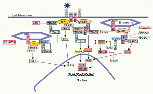
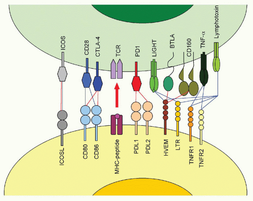
The fiber knob of adenoviruses preferentially binds the coxsackie adenovirus receptor, which is expressed on epithelial cells. In addition, an Arg-Gly-Asp (RGD) motif present within the viral penton can bind α(v)-integrins with lower avidity. The fiber of adenoviruses of subfamily B2, on the other hand, binds CD46, a ubiquitously expressed complement component that also facilitates entry of measles virus and human herpesvirus (HHV)6. The herpes virus mediator (HVEM) is a bimodal switch that can provide both immunostimulatory and immunoinhibitory signals to the immune system. Upon binding to LIGHT (lymphotoxin [LT]-like, exhibits inducible expression and competes with HSV-1 glycoprotein D [gD] for HVEM), HVEM submits stimulatory signals. Upon binding to the B- and T-lymphocyte attenuator (BTLA), it acts as an immunoinhibitor. HVEM also binds HSV-1 gD, thus facilitating entry of this virus. Binding of gD to HVEM takes place on a site that overlaps with the BTLA binding site; therefore, gD can be used to inhibit an immunoinhibitory pathway. HSV-1 may have evolved to block such a pathway, as activation of NF-κB promotes viral replication and assists in transcription of some of the early viral genes.10

The envelope protein of HIV-1 binds cluster of differentiation (CD)4 expressed mainly on T-helper cells. Upon binding, the protein undergoes structural changes that allow for its binding to a coreceptor, which for transmitting virions is CCR5, but following mutations, viruses circulating in an organism can also use CXCR4.11 A mutant allele of CCR5 termed CCR5d32, which results in lack of CCR5 expression on the cell surface and which is found in 10% of Caucasians of European descent, provides resistance to infections with HIV-1.12 Other viral receptors include the nicotinic acetylcholine receptor for rabies virus, heparan sulfate for dengue virus, adeno-associated viruses, and some of the herpes viruses, CD155 for poliovirus, CD81 for hepatitis C virus, CD21 for Epstein-Barr virus (EBV), C-type lectins, such as dendritic cell (DC)-specific intercellular adhesion molecule-3-grabbing nonintegrin (DC-SIGN) and liver/lymph node-specific intercellular adhesion molecule-3-grabbing integrin for Ebola virus, integrin-β3 for Hantan virus or intercellular adhesion molecule (ICAM)-1 for rhinoviruses. Examples for viral receptors including their physiologic functions are listed in Table 39.3.

Upon binding to a receptor, viruses not only need to gain access into the cell, but most of them then have to traverse to the nucleus to initiate their replication. Viruses enter the cell either through endocytosis31 or fusion.32 Clathrin-mediated endocytosis is used by enveloped as well as nonenveloped viruses including adenoviruses, influenza viruses, poxviruses, or rabies virus. In cadherin-mediated endocytosis, the virus-receptor complexes cluster into a cadherin-coated pit on the cell membrane that becomes invaginated, eventually closes, and detaches from the cell membrane. The clathrin-coated vesicles then deliver their cargo to early endosomes from where it travels to late endosomes. Other viruses such as coxsackie B virus, respiratory syncytial virus (RSV), and others enter cells by caveolar endocytosis. Caveolae are invaginations in the plasma membrane that are rich in cholesterol, glycosphingolipids, and claveolin, which are used for uptake of macromolecules into endosomes. In addition, caveolar endocytosis allows for transcytosis of molecules from the basal to the apical side of a cell or vice versa. Human enterovirus has been described to enter cells through a lipid raft dependent pathway, rotavirus infects through a cholesterol- and dynamin-dependent but clathrin-and caveolae-independent pathway, while other viruses enter cells by micropinocytosis or phagocytosis.

Enveloped viruses such as paramyxoviruses, some herpes viruses, or HIV-1 invade cells by direct fusion of the virus envelope with the cell membrane. Fusion is promoted by hydrophobic sequences within a viral surface protein and causes release of the viral genome into the cytoplasm.

Viruses that enter cells through endocytosis end up in endosomes. Mechanisms of escape from endosomes differ for enveloped and nonenveloped viruses. The decrease in pH between early and late endosomes favors conformational changes of viral surface proteins by exposing their hydrophobic residues, which allow for fusion of the viral envelope with the endosomal membrane. This in turn permits escape of the viral core or the genome into the cytoplasm. Nonenveloped viruses disrupt the endosomal membrane either by a pathway called carpet mechanism or by forming pores. In carpet-like disruption of endosomal membranes, viral peptides act like a detergent and thus interrupt the hydrophobic interactions between membrane lipids allowing for the development of micelles and for transient formation
of holes within the membrane.33 Other viruses carry proteins, which form amphipathic α-helices that assemble into a pore within the lipid membrane of the endosome where the hydrophobic parts interact with the lipid bilayer while the hydrophilic parts of the coils form the inner wall of the pore.34 Picornaviruses, parvoviruses, and reoviruses utilize this strategy.


TABLE 39.3 Viral Receptors


Receptor


Virus


Viral Antigen


Physiological Function


References


Herpes virus entry mediator


Herpes simplex virus-1


Glycoprotein D


Receptor for costimualtors/coinhibitors


13,14


Coxsackie adenovirus receptor


Adenovirus C


Fiber


Cell adhesion molecxule


15


Coxsackie B virus


CD46


Adenovirus B2


Fiber


Complement regulatory protein


16


Measles virus


Hemagglutinin


17


CD55


Enteroviruses


Hemagglutinin


Complement regulatory protein


18


CD155


Poliovirus


Virus proteins 1-3


Cell adhesion molecule


19


CD4


Human immunodeficiency virus-1


Glycoprotein 160


T-cell receptor coreceptor


20


Nicotinic acetylcholine receptor


Rabies virus


Glycoprotein


Forms ion channels in neuronal membranes


21


CD81


Hepatitis C virus


Glycoprotein E2


Signal transduction


22


ICAM-1


rhinovirus


Viral proteins 1-4


Cell adhesion molecule


23


CD21


Epstein-Barr virus


Glycoprotein 350/220


Complement component


24


DC-SIGN


Ebola virus


Glycoprotein


C-type lectin, adhesion molecule


25


Dengue virus


Glycoprotein E


26


Hepatitis C virus


Glycoprotein E2


27


P-selectin glycoprotein ligand-1


Enterovirus 71


Selectin receptor; mediates leukocyte rolling


28


SLAM/CD150


Measles virus


Hemagglutinin


Signal transduction


29


Transferrin receptor 1


Lassa fever virus


Glycoprotein


Import of iron


30


CD, cluster of differentiation; DC-SIGN, dendritic cell-specific intercellular adhesion molecule-3-grabbing non-integrin; ICAM, intercellular adhesion molecule; SLAM, signaling lymphocytic activation molecule.


Once within the cell, viruses may be broken down by autophagy,35 a catabolic process involving the degradation of a cell’s own components to convert unneeded pieces into nutrients. During this process, so-called autophagosomes form from membrane structures containing autophagiarelated gene products (Atg), such as the ubiquitin-like Atg8, the Atg4 protease, and the Atg12-Atg5-Atg16 complex. The outer membrane of the autophagosome fuses with a lysosome to allow for degradation of its contents. Formation of autophagosomes is initiated by PI3K and Beclin-1. Most viruses block this pathway by inhibiting PI3K activation, but rhinoviruses and poliovirus sponsor formation of early autophagosomes but block their fusion with lysosomes and then use the structures to egress the cells.36

Once a virus has reached the cytoplasm, it must deliver its genome to the nucleus. Many viruses such as herpesviruses and adenoviruses use microtubules to reach nuclear pores. Very small genomes can diffuse passively though pores into the nucleus, while larger genomes or particles require an energy-dependent process. Some viruses use viral proteins to facilitate nuclear entry. For example, cytomegalovirus (CMV) encodes two proteins, pUL69 and pUL84, that facilitate the transport of its genome to the nucleus. pUL69 binds to UAP56, which facilitates nuclear export of unspliced RNA; pUL84 binds to importin-alpha proteins,37 which can dock to nuclear pores and then be transported through it.

Most viruses initiate their replication in the nucleus as they depend on nuclear enzymes for transcription. Poxviruses and some of the RNA viruses are independent of such enzymes and can replicate their genome in the cytoplasm. HIV-1 replicates in the nucleus after it reverse transcribes its RNA in the cytoplasm.

Replication of different types of virus can be exemplified using the following viruses: adenovirus, a dsDNA virus; adeno-associated virus, an ssDNA virus; reovirus, a dsRNA virus; poliovirus, a positive-sense ssRNA viruses; influenza A virus, a negative-sense ssRNA virus; HIV-1, a positive-sense ssRNA-RT virus; and hepatitis B virus, a dsDNA-RT virus.

Adenovirus transcription is typical for that of some of the larger DNA viruses as it proceeds in stages. Initially, the immediate early gene is transcribed. The resulting gene products alter the host cell to provide a more favorable environment for viral replication and initiate transcription of early viral genes, which have regulatory functions and serve to modify host cell functions or subvert immune responses. Thereafter, the viral genome replicates concomitantly with transcription of the late viral genes that encode
structural proteins. Specifically, the replication cycle of adenovirus starts with expression of E1A, which encodes two polypeptides that bind to cellular proteins, including cellular transcription factors, which in turn changes the cell’s gene expression profile and allows for transcription of the other early viral genes E1B, E2, E3, and E4. E1A promotes apoptosis, while E1B proteins are antiapoptotic. E1B polypeptides turn off host cell protein synthesis and help to stabilize, transport, and selectively translate viral RNA. E2 encodes DNA-binding proteins and a polymerase. E3 gene products are nonessential for virus replication but serve to evade immune responses. E4 encodes seven polypeptides, which collaborate with E1 gene products in promoting viral transcription and modulating host cell functions. E4 gene products are also essential for nuclear export of viral RNA. Adenoviruses also encode one or two virus-associated (VA)-RNA species, which form short hairpin loop structure of approximately 200 bases, are transcribed by polymerase III and stimulate translation of viral genes. VA-RNA can be processed into shorter RNAs and act as micro RNA,38 inhibiting activation of protein kinase R, which inhibits further messenger RNA (mRNA) synthesis through phosphorylation of the translation initiation factor EIF2A. Transcription and translation of early gene products is followed by DNA replication, which is initiated by a terminal protein that is covalently bound to the 5′ ends of the long terminal repeats. Once DNA replication is initiated, the late gene products, which form the viral capsid, are produced from the L1-L4 domains. Viral assembly begins in the cytoplasm and is completed in the nucleus.

Adeno-associated viruses are ssDNA viruses that cause no known disease in humans. They are dependoviruses and require coinfection with another virus, most commonly an adenovirus, to complete their lifecycle. The approximately 4.7 bp genome is flanked by terminal repeats that contain a multipalindromic terminus that forms a loop and thereby promotes priming for DNA replication. The genome contains only two genes; one, the rep gene, encodes four regulatory proteins needed for DNA replication and conversion of the dsDNA intermediate into the final ssDNA. The viral capsid is composed of three virus proteins derived from the cap gene by transcript splicing. Initiation of transcription of the rep gene requires proteins from a helper virus such as gene products from E1, E2, E4, as well as VA-RNA from adenovirus. Final assembly of the adeno-associated virus takes place in the nucleolus.

Reovirus infections are asymptomatic in humans but cause disease in newborn mice. This virus, which contains 10 to 12 segments of dsRNA, replicates in the cytoplasm of infected cells without completely uncoating. RNA is transcribed from the negative strand of the genomic RNA and leaves the capsid to be translated. Secondary transcription occurs later followed by assembly within the cytoplasm.

The viral genome of positive-sense ssRNA viruses, such as poliovirus, a picornavirus, can directly serve as mRNA. Poliovirus RNA lacks the methylated cap structure that is typical for mammalian mRNA but rather has an internal ribosomal entry site. To avoid competition with translation of mammalian mRNA, poliovirus interferes with recognition of the host’s methylated cap, thus inhibiting host cell protein synthesis.39 The poliovirus RNA is translated into a single polypeptide that is cleaved into a replicase, proteases, and structural proteins. The polymerase transcribes the positive-stranded RNA into a minus-sense RNA to serve as template for new positive-stranded RNA. The latter can either be translated, serve as template for minus stranded RNA, or be packaged into new virions. Replication as well as assembly occurs in the cytoplasm.

Influenza viruses are segmented negative-sense ssRNA viruses, which replicate in the nucleus. The RNA-dependent RNA polymerase transcribes positive-stranded RNA segments that are either transported to the cytoplasm for translation or remain in the nucleus to serve as templates for negative-stranded RNA synthesis. Newly produced internal proteins are transported into the nucleus where they, together with RNA segments, form new virus particles. The two viral surface proteins, the HA and the neuraminidase are secreted through the Golgi apparatus to the cell surface where they are then picked up by the envelope once the virus leaves the cell.

HIV-1 is initially reversed transcribed in the cytoplasm by the viral reverse transcriptase into an RNA/negativestranded DNA hybrid. This process is error prone and contributes to the high mutation rate of HIV-1. The RNA is degraded, and a positive-stranded DNA is synthesized allowing for the formation of a dsDNA, which, together with some enzymes, enters the nucleus; there the viral genomes integrates with the help of the viral integrase and serves as a template for synthesis of viral transcripts. Two newly produced viral proteins, Tat and Rev, are essential for efficient protein production: Tat by enhancing transcription and Rev by supporting export of unspliced mRNA from the nucleus, which allows for production of the structural proteins Gag and Env. The full-length viral RNA binds initially to Gag and is then packaged into new virus particles. Env is transported to the cell surface after it is cleaved into two subunits and, with the help of cellular chaperone proteins, folded into a trimer. Assembly of mature virions takes place at the plasma membrane.

Hepatitis B virus (HBV) carries a circular partially dsDNA genome that encodes four structural and two nonstructural proteins through overlapping open reading frames (ORFs). Within the nucleus of an infected cell, the genome is converted into a full dsDNA, which serves as a template for the viral transcripts. The largest mRNA, which is longer than the viral genome, is called the RNA pregenome and is packaged into core particles within the cytoplasm. Within these particles, the pregenomic RNA is reverse transcribed into viral DNA genomes. Upon synthesis, the viral surface protein is transported to the cell membrane and complete assembly of the virion takes place during budding of the virus.

Once replication is completed and full virions have been assembled, viruses need to leave the cells. This again can occur through several pathways. Some viruses, such as HIV-1 or influenza virus, assemble their newly synthesized viral surface proteins on the cell surface and then bud through this part of the cell membrane, picking up not only their own surface proteins but also membrane proteins belonging to
the host cell. Budding eventually destroys the cell membrane and leads to cell death. Other viruses, especially those that are nonenveloped, instruct the infected cell to undergo apoptosis and virus released from dying cells is encapsidated into apoptotic bodies, which are taken up by neighboring cells, thus facilitating infection of new cells. Some viruses are released by exocytosis, a process that resembles reversed pinocytosis in which virus particles are encapsidated into small vesicles that enter the secretory pathways. This form of exit does not kill the cells and is used by so-called nonlytic viruses.


Viral Persistence

Some viruses such as poxviruses or influenza viruses are lytic, which means they inevitably kill the cells they infect. Such viruses cause acute infections in immunocompetent hosts. Other viruses can replicate within a cell without causing its demise, or they can switch between a lytic and a nonlytic infection. These viruses can persist and cause chronic or latent infections, which pose unique challenges to the immune system. In chronic infections, some viruses, such as HIV-1 or hepatitis C virus (HCV), replicate constantly, dodging destruction by the immune system. Other viruses replicate and then persist by turning off synthesis of most of their viral proteins, causing so-called latent infections. Herpesviruses can switch to a latent phase from which they are periodically reactivated to undergo renewed lytic cycles of replication. Adenoviruses persist at low levels in activated T cells, presumably as episomes that remain transcriptionally active.40 Yet other viruses, such as HIV-1 or HPVs, integrate into the host cell genome and thus become an integral part of the cell. In general, DNA viruses with a nuclear replication cycle are able to persist, which may be favored due to the complete lack of DNA degrading enzymes within the nucleus. Rabies virus, a negative-sense ssRNA virus, does not cause chronic infections but kills within days after causing symptoms. Nevertheless, in some individuals years pass between viral transmission and onset of symptoms,41 and it is unknown where and how the virus persists during this long incubation time. Measles virus, another negative-sense ssRNA virus of the paramyxovirus family can cause subacute sclerosing panencephalitis in about 1 out of 100,000 infected individuals within 5 to 15 years after primary infection. Subacute sclerosing panencephalitis is most common in children who are infected early in life, and it has been speculated that the relative immaturity of their immune system allows for the development of a chronic central nervous system infection.42


INNATE ANTIVIRAL IMMUNE RESPONSES

Both innate and adaptive immune responses are essential to wards off pathogens, and individuals with inherited or acquired immunodeficiencies rapidly succumb to virus infections. Even individuals with weakened immunity, such as the very young whose immune system is still immature, the elderly undergoing immunosenescence, or pregnant women whose immune system is transiently suppressed, show markedly increased susceptibility to many viruses.

Immune responses to viruses can roughly be divided into four stages. At first, the immune system has to recognize the threat, then an immediate early response is mounted by cells of the innate immune system, which is followed a few days later by a response from the adaptive immune system. Once the virus is eliminated, the immune response contracts and adaptive immunity enters a stage of immunologic memory. Memory T and B cells, upon reexposure to the same pathogen, mount a response that is more potent and comes up faster than a primary response. In cases where virus persists, the acute phase of the immune response is followed by a chronic immune response.


Early Recognition of a Virus

Viruses like other microbes carry so-called pathogen-associated molecular patterns (PAMPs) that are recognized by pattern recognition receptors (PRRs) expressed on many cell types including cells of the innate and adaptive immune system.43 This recognition system is not as specific as the antigen-recognition receptors of T and B cells, but rather it responds to motifs that are commonly found on pathogens but not within mammals or it identifies molecules present in the wrong compartment within the cell. PRRs can be subdivided into four main types (ie, toll-like receptors [TLRs], retinoic acid inducible gene [RIG]-I-like receptors, nucleotide oligomerization domain [NOD]-like receptors [NLRs], and the interferon-inducible p200 family member absent in melanoma 2).

Ten TLRs (TLR1-10) have been defined in humans and 9 in mice.44 Some TLRs are widely expressed on many different cell types such as TLR1 or 4, while others are expressed mainly on cells of the immune system such as TLR5, antigen-presenting cells (TLR8), or subsets of antigen-presenting cells, such as TLR7 and TLR9, which are primarily expressed in plasmacytoid DCs. Expression patterns of TLRs in humans do not always mirror those in mice. Expression levels of most of the virus-sensing TLRs are upregulated by inflammatory cytokines mainly interferon (IFN)-γ but for TLR3 that is modulated upon cell differentiation. Viruses are sensed by five TLRs. TLR4, which is best known for its response to lipopolysaccharide, also reacts to the fusion protein of RSV45 and to a surface glycoprotein of Ebola virus.46 TLR3 senses double-stranded RNA, TLR9 senses viral and bacterial CpG sequences, and TLR7 (in humans only) and TLR8 (in both humans and mice) reacts to ssRNA. It was initially debated if indeed TLRs directly recognized their PAMP or became instead activated by an intermediate host cell protein. More recent evidence has shown direct binding between TRLs and their ligands. TLR4, which can recognize viral surface proteins, is expressed on the cell membrane where an encounter with such an antigen is most likely. TLR3, 7/8, and 9 are within endosomes where many viruses uncoat, which in turn leads to exposure of their genomes.

Viruses such as herpesviruses,47 West Nile virus,48 or influenza virus49 signal through TLR3; RSV50 and Ebola virus46 can signal through TLR4; influenza virus,51 HIV-1,52
and herpesviruses53 can signal through TLR7 or 8; DNA viruses such as herpesviruses signal through TLR9.54

All TLRs carry an intracellular toll-IL-1 receptor-resistance (TIR) domain, which interacts with TIR domains on intracellular adaptor molecules. TLRs, with the exception of TLR3, signal through myeloid differentiation primary response gene (MyD)88, which in turn interacts with interleukin-1 receptor associated kinases (IRAKs)1, 2, and 4, leading to activation of tumor necrosis factor receptor -associated factor (TRAF)6 and upon additional steps to activation of NF-κB, mitogen-activated protein (MAP) kinases, and Jun-terminal kinases (JNKs). TLR3 signals through TIR-domain-containing adapter-inducing interferon-β (TRIF), which binds to TANK-binding kinase 1 (TBK1), thus activating interferon regulatory factor (IRF)3. In addition, TRIF interacts with receptor-interacting protein 1, which can activate NF-κB. TLR4, in addition to signaling through MyD88, can also bind the TRIF-related adaptor molecule, which recruits TRIF, allowing for signaling to IRF3. Activation of IRF3 results in production of type I IFN, whereas activation of NF-κB induces production of a number of proinflammatory cytokines, such as interleukin (IL)-6, tumor necrosis factor (TNF)-α, and IL-12 (Fig. 39.1).

Deficiencies in TLRs can change an individual’s susceptibility to viral infections. For example, TLR3 deficiency in humans is associated with increased susceptibility to HSV-1 infections,47 whereas mice that lack TLR3 are more resistant to West Nile virus.55 West Nile virus triggers TLR3, and the resulting cytokine response opens the blood-brain barrier, which allows the virus to establish an infection within the central nervous system; in absence of TLR3, the virus remains excluded from the brain.


FIG. 39.1. Toll-like Receptor Signaling Pathways.

TLR signaling and the resulting production of cytokines can be extremely toxic. TLR signaling is therefore tightly regulated.56 Some negative regulators target specific TLRs while others affect common downstream adaptor molecules. Regulators that affect TLRs, which react to viruses, include IRAKM, an IRAK homolog that inhibits IRAK1 and blocks TLR4 and 9, suppressor of cytokine signaling 1, which also suppresses IRAK. Others include phosphoinositide 3-kinase (PI3K), a key regulator of T-cell differentiation, which inhibits JNK and NF-κB functions, toll-interacting protein, which phosphorylates IRAK1, A20 which deubiquitylates TRAF6 and thus affects TLR3, 4, and 9, ST2L which sequesters MyD88, single immunoglobulin (Ig) IL-1R-related molecule, which binds to TRAF6 and IRAK and TRIAD3A, and E3 ubiquitin-protein ligase, which initiates degradation of TLRs (see Fig. 39.1).

Some viruses neither carry PAMPs on their surface for recognition by membrane-bound TLRs nor enter cells through endosomes, and their genomes thus fail to become accessible for recognition by TLRs. Such viruses can be recognized by RIG-I-like receptors, which are located in the cytoplasm.57,58 RIG-I-like receptors are RNA helicases, which respond to ss or dsRNA. Three RIG-I-like receptors
have been identified to data (ie, RIG-I, melanoma differentiation-associated gene 5 [Mda5], and LGP2). RIG-I and Mda5 carry N-terminal caspase activation and recruitment domain (CARD)-like regions, which are involved in downstream signaling and C-terminal RNA helicase domains, which bind RNA and can distinguish between viral and cellular RNA species. LPG2 lacks the CARD domain and is assumed to negatively regulate RNA virus-induced inflammatory responses by blocking binding of RNA to RIG-I. RIG-I recognizes a number of ssRNA viruses such as para- and orthomyxoviruses,59 rotavirus,60 filoviruses,61 and flaviviruses,62 while Mda5 recognizes picornaviruses.63 RIG-I and Mda5 signaling involves binding of their C-terminal CARD domains to the CARD domain on IPS-1, which then through kinases receptor interacting protein 1 or Fasassociated protein with death domain activates NF-κB; they also activate IRF3 and IRF7 through TRAF3/TBK1, which results in production of IFNs and other proinflammatory cytokines (Fig. 39.2).

NLRs are a very large family of PRRs that respond to viral RNA in the cytoplasm.64,65 They contain N-terminal domains for protein-protein interactions such as CARD, pyrin or inhibitor of apoptosis domains, NOD domains for nucleotide binding, and C-terminal leucin-rich repeat domains. NLRs are subdivided according to their N-termini into CARD (CIITA, NOD1, 2, NLRC3-5), pyrin domain (PYD, NLRO1-14), or pyrin or inhibitor of apoptosis domain (NAIP) members. Most NLRs activate cytokine responses, although some are inhibitory and dampen innate immune responses. To give some examples of the specificity of NLRs, NOD-2 senses ssRNA and interacts with paramyxoviruses and myxoviruses.66 NLRC5 interacts with Poly(I:C) and responds to some paramyxoviruses and herpesviruses.67,68NLRX1, an inhibitory NLR, signals upon recognition of Sendai or Sindbis virus and blocks activating signals through RIG-1 like helicases.69 Signaling pathways have not yet been fully characterized for NLRs. NOD-1 and -2, the best known NLRs, bind receptor-interacting serine-threonine kinase 2 resulting in NF-κB and MAP kinase signaling. They also induce autophagy and activate the mitochondrial antiviral signaling protein for induction of type I IFN. Several NLR members containing CARD or PYD domains can assemble into inflammasomes.

Inflammasomes are multiprotein complexes that recruit and activate inflammatory caspases.70 Inflammasomes include PRRs and, as such, are divided into NLRP3 inflammasomes, RIG-I inflammasomes, and absent in melanoma 2 inflammasomes. In these complexes, the PRR activates caspase I through an adaptor, which cleaves the immature forms of IL-1β and IL-18 resulting in biologically active cytokines. NLRP3 inflammasomes were shown to react with influenza virus, rhabdoviruses, and picornaviruses.71,72 Viral RNA can directly activate the caspase activities. The M2 protein of influenza virus, which has ion channel activity, localizes to the trans-Golgi network and reduces the H+ content; the acidity of the Golgi then results in NLRP3 activation.73 Absent in melanoma 2 inflammasomes recognize DNA within the cytoplasm and, as such, play a role in responses to DNA viruses (eg, poxviruses and herpesviruses).74


FIG. 39.2. Signaling through Retinoic Acid-Inducible Gene I Receptors.


The Early Inflammatory Response: Cytokines and Chemokines

Interaction of a PRR with a PAMP initiates a cellular response. Signaling through most of the PRRs results in activation of NF-κB. In resting cells, NF-κB is retained in the cytoplasm through binding to IκB. Upon activation of the IκB kinase (IKK), IκB becomes phosphorylated, then ubiquinated and finally degraded. This in turn releases NF-κB and allows for its entry into the nucleus where it initiates gene expression. NF-κB thus does not require de novo synthesis, which accelerates its activity. A very large number of genes have NF-κB binding sites including genes involved in antigen processing and presentation, lymphocyte effector functions and motility, and cell metabolism. Cytokine genes induced by NF-κB include those for type I IFN, IL-1A and B, IL-2, IL-6, IL-9, IL-11, IL-15, TNF-α, colony stimulating factor (CSF)1 to 3, lymphotoxin B, and the chemokine genes IL-8, CCL2, 5, 11, 15, and CXCL5. In addition, several cytokine-inducing transcription factors, such as IRF1, 2, and 4, are produced in response to NF-κB. IRF3 activated by TLR3, TLR4, or cytosolic PRR signaling is a transcription factor that promotes production of type I IFN and the chemokine RANTES. The MAP kinase activated AP-1 transcription factors also have binding sites specific for regulatory sequences of multiple genes. Most pertinent for early immune responses is probably induction of signal transducers and activators of transcription (Stat)1 and 3, which upon formation of homo- or heterodimers bind to the IFN-gamma activated sequences. Thousands of such motifs can be found in the human genome, and their products affect most cell functions.

Most cells can produce cytokines and chemokines, although, during the initial phase of an immune response, many are synthesized by cells of the innate immune system. The early cytokines have a multitude of functions, which are in part antiviral and in part designed to promote further activation of immune responses. Type I IFNs, which can be produced at capacious amounts by plasmacytoid DCs, bind to the IFN-α receptor. This causes activation of tyrosine kinase (Tyk)2 and Janus kinase (Jak)1, which in turn causes tyrosine phosphorylation and then nuclear translocation of Stat1 and Stat2 proteins. In addition, type I IFN has strong antiviral activity75 and has been licensed for treatment of chronic infections with HBV76 and HCV77 and for treatment of HPV-associated genital warts (Condyloma acuminata).78

IFN-1s downregulate viral promoters. They dampen expression of viral receptors and thus reduce viral entry. They induce expression of the dsRNA-activated protein kinase PKR, which phosphorylates translation initiation factor 2α (eIF2α) causing inhibition of translation of both viral and cellular transcripts. IFN-1s also trigger expression of 2′5′-oligoadenylsynthases that upon binding dsRNA generate AMP-oligomers, which activate RNAseL to cleave both cellular and viral RNAs. IFN-1s induce dsRNA kinase, which inhibits production of viral progeny. IFN-1s lead to synthesis of MxA.79 This protein binds to the nuclear membrane and inhibits trafficking of viral nuclear capsids. In addition, MxA can bind and inhibit the RNA polymerase of influenza virus. APO-Bec3G and F are IFN-induced deoxycytidine deaminases that interfere with the replication of retroviruses.80

In addition to their antiviral activity, IFNs and other early cytokines play a dominant role in initiating both innate and adaptive immunity by causing activation of macrophages, natural killer cells, and DCs. The early cytokines promote proliferation and differentiation of lymphocytes, granulocytes, and antigen-presenting cells, whereas chemokines recruit such cells. IL-6 should be noted, as this cytokine, due to its immunostimulatory effects on B cells, has been implicated in the pathobiology of EBV-associated lymphoproliferative disorders and HHV-8-associated lymphomas in patients with acquired immunodeficiency syndrome.81,82

The apparent redundancy of some of the molecules that contribute to the initial inflammatory response is remarkable. Viruses commonly trigger multiple PRRs, some of which are expressed on or in the same cells, while other are carried by different subsets of cells. For example, influenza viruses thus far are known to signal through TLR3, 7/8, NOD-2, RIG-1, and NLPR3 inflammasomes. Most of the PRRs in the end initiate transcription through NF-κB or through members of the IRF family. Nevertheless, the immune response that is very much guided by the initial inflammatory reaction is unique for each virus, suggesting a finely orchestrated series of events during which the type, strength, and location of PRR signaling elicits for each virus a special mixture of cytokines and chemokines that results in signature immune responses. This is exemplified by recent studies with a vaccine, in which antigen-containing nanoparticles were mixed with ligands for TLR4 and 7.83 This vaccine did not induce a strong effector cell response but drove differentiation of T cells toward memory and B cells toward long-lived plasma or memory cells. Although the slow release of antigens from the nanoparticles may have contributed to this, both TLR ligands were needed to maximize the vaccine’s immunogenicty.


Cells of the Innate Immune System

Cells of the innate immune system, which include DCs, neutrophils, eosinophils, basophils, mast cells, γ/δ T cells, macrophages, and natural killer cells, provide a first layer of defense against virus infections and promote activation of adaptive immunity. Unlike T and B cells, which carry antigen-specific receptors, cells of the innate immune system are, as described previously, activated by PRRs. They act rapidly without undergoing the massive proliferation of T and B cells and then, in general, with the potential exception of natural killer cells,84 fail to establish long-lasting memory.


Granulocytes: Neutrophils, Basophils, Eosinophils, and Mast Cells

Granulocytes play major roles in controlling bacterial and parasitic infections but also influence viral infections. Neutrophils, which are abundant and contribute to more than 50% of circulating leukocytes, release hydrogen peroxide, free oxygen radicals, and hypochlorite, and have been described to limit replication of HSV-1.85 They may also
contribute to the immunopathology, such as of pulmonary RSV infections.86

Basophils release histamine, which increases blood vessel permeability and thus allows for lymphocyte trafficking into inflamed tissues. Basophils are recruited and activated by a number of viruses, such as influenza viruses or RSV.87

Eosinophils, which accumulate in lungs during RSV infection,86 release enzymes, such as ribonuclease, deoxyribonucleases, lipase, as well as plasminogen and peroxidase.

Mast cells release heparin, histamine, and chemokines, and have been reported to respond to Dengue virus infections.88


Gamma/delta T cells

T cells that carry the γ/δ T-cell receptor (TCR) exhibit characteristics of both innate and adaptive immune cells. They are mainly located in the gut and skin but can also be found at low frequencies in blood and lymphatic tissues. Although they are educated in the thymus, their receptor recognizes pathogen patterns or cellular stress molecules independent of major histocompatibility complex (MHC) molecules, and they can expand without the intricate antigen presentation pathways that dictate differentiation of α/β T cells. γ/δ T cells, which evolutionary predate α/β T cells, can secrete cytokines such as IFN-γ, IL-4, or IL-17,89 chemokines such as macrophage inflammatory protein (MIP)-1α; MIP-1β; regulated upon activation, normal T cell expressed and secreted; lymphotoxin (LT)90; and lytic enzymes (ie, perforin and serine esterases for target cell lysis). These T cells play diverse roles in virus infections. To name a few, they promote Th1 responses following infection with coxsackievirus B391; they can kill cells infected with HSV-192; they have been described to lyse cells infected with influenza virus93; and they provide resistance to humans against HIV-1 infections94 and to mice against infections with West Nile virus95 or vaccinia virus.96


Macrophages

Monocytes upon activation by proinflammatory cytokines, specifically IL-6 and macrophage-CSF (M-CSF), differentiate into macrophages. These cells, as their name “large eaters” implies, are phagocytic. Phagocytosis is a specialized form of endocytosis during which the engulfed particles are transported to lysosomes where they are degraded by a toxic and acidic soup composed of oxygen radicals, nitric oxide, proteases, and defensins. Macrophages, in addition, release cytokines, such as IL-1, 6, 10, and 12, type I IFN, and TNF-α, as well as chemokines, such as MIP-2, IL-8, and cytokine-induced neutrophil chemoattractant-1. Some viruses replicate in macrophages (eg, HIV-1, influenza viruses, rhinoviruses, and Ebola viruses).


Dendritic Cells

A crucial role of DCs is their ability to present antigen to naïve T cells. A few years ago, DCs were divided into myeloid DCs, plasmacytoid DCs, and lymphoid DCs, although it is now accepted that lymphoid DCs do not belong to a separate lineage.

Viral antigens are mainly presented by myeloid DCs. Myeloid DCs, which differentiate from Lin-CD115+Flt3+ CD117lo precursors, can further be divided into subsets according to phenotypic markers, functions, and anatomic site of origin.97

Three subsets of myeloid DCs have been identified in human skin. Langerhans cells, which are CD1hi CD14- CD207+, are located in the epidermis. They look and act like DCs, and they are originally derived from bone marrow precursors but unlike other DC subsets, numbers of Langerhans cells are maintained by local proliferation. They only express discreet amounts of TLRs, induce CD8+ T cell responses in vitro and trigger proliferation of naïve CD4+ T cells. Interestingly, CD4+ T cells induced by Langerhans cells do not produce the typical Th1, Th2, or Th17 cytokines but rather produce IL-22,98 which causes skin inflammation as well as wound healing through induction of keratinocyte proliferation.

The dermal layer of skin contains CD14+ CD11bhi DCs, which differentiate from CD34+ precursors and express TLR 1, 2, 4, 5, 6, 8, and 10. They produce a multitude of proinflammatory cytokines including IL-6 and IL-12, which allows them to induce differentiation of naïve B cells into plasma cells. They also prime CD4+ T cells that can promote B-cell differentiation. The other DC subset in the dermis is CD103+CD11blow, which can cross-present antigen and drive proliferation and differentiation of CD4+ T cells into Th1 cells.

Mouse spleen contains two subsets: CD8+CD205+ DCs and CD8+33D1+CD11b+ DCs; whereas the former can cross-present antigen in association with MHC class I and thus drive activation of CD8+ T cells, the latter is more efficient at processing antigen for MHC class II presentation for CD4+ T cell activation.

Lymph nodes also contain these two subsets and in addition migratory DCs such as Langerhans cells, which transport antigen from other locations to lymph nodes. Lymphatic tissues in addition contain follicular DCs.99 These cells are not derived from hematopoietic progenitors but are of mesenchymal origin. They are an integral part of B-cell follicles, where they present antigen to naïve B cells and maintain long-lived plasma cell responses by initially capturing and then slowly release antigen-antibody complexes.

The intestine has three populations of DCs. CD103+ CD1blow DCs can be found in Peyer patches and CD103+ CD11bhi and CD103-CD11bhi DCs in the lamina propria. The latter subset is known to transport antigen from the intestine to draining lymph nodes. Other tissues such as lung, kidney, and spleen contain similar subsets.

It was initially thought the DCs are terminally differentiated, nondividing cells. Recent studies have shown that DCs can divide and that this division is driven by fms-related tyrosine kinase (Flt)-3, which is also a key factor for DCs differentiation from bone marrow precursors.97

Plasmacytoid DCs were originally though to originate from lymphoid progenitors as they share many of the characteristics of lymphocytes: they lack the typical dendrites of other DC subsets, but look like lymphocytes; they express B-cell markers, such as B220 and T cell markers (eg, CD4), as well as transcription factors involved in lymphocyte development, such as terminal deoxynucleotidyltransferase, recombination activating genes 1 and 2, Ig supergene family members, and the pre-T-cell antigen receptor alpha chain. But plasmacytoid DCs also have characteristics of myeloid
DCs and can even differentiate into myeloid DCs.100 It is assumed that plasmacytoid DCs can arise from both lymphoid and myeloid bone marrow precursors, although the latter probably provide the dominant source. Plasmacytoid DCs, which can be distinguished from myeloid DCs by expression of plasmacytoid DC antigen-1 (CD317) in mouse or blood DC antigen-2 (CD303) or -4 (CD304) in human, circulate in blood and are present in lymphatic tissues. Unlike myeloid DCs, they express CD62L and CCR7, which allows them to cross high-endothelial venules to enter T-cell-rich areas in lymphatic tissues. While myeloid DCs take up antigen to present it to cells of the adaptive immune system, plasmacytoid DCs do not phagocytose antigen. Their primary role might be regulatory by producing cytokines most notably type I IFNs, which assist maturation of myeloid DCs.

DCs present in tissues are immature. They take up antigen, but they do not produce cytokines, nor are they able to present antigen efficiently to naïve lymphocytes. Their maturation starts either through interactions of a PRR with a PAMP, through cytokines, ligation of CD40, receptor activator of NF-κB (RANK) or even, as has been observed in vitro, vigorous shaking.101 DC maturation is associated with marked changes in their gene expression profiles102 and their biological functions. Maturing DCs stop phagocytosis and endocytosis. They start secreting chemokines and thus initiate an inflammatory response at the site of infection. They upregulate expression of CCR7 and migrate from tissue to the T-cell-rich zones of draining lymph nodes. They increase synthesis of molecules that are involved in antigen processing and presentation, and translocate MHC class II molecules that are preformed and stored within intracellular vesicles to the cell surface. They also, in a presumably orchestrated but yet ill-defined pattern, start to increase expression of cell surface molecules that regulate immune responses (Fig. 39.3). Such molecules include CD80, CD86, CD40, inducible T-cell costimulator (ICOS) ligand, ligands 1 and 2 for programmed death 1 (PDL1, PDL2), HVEM, and others that are less well defined thus far. Interactions of T and B cells not just with their cognate antigen, but also with these ligands, is crucial for the induction of an adaptive immune response that is of sufficient magnitude to combat the threat without being overwhelming and thus causing undue damage. These interactions are also important to determine the ultimate differentiation fate of responding T and B cells.


FIG. 39.3. Costimulatory (blue lines) and coinhibitory (red lines) T cell (green)-dendritic cell (yellow) interactions.

Signaling through CD40, which is expressed at low levels on immature DCs, facilitates full maturation of DCs; its expression increases upon activation of TLR pathways. CD40 is upregulated on myeloid DCs upon interactions with TLR4 agonists103 and on both plasmacytoid and myeloid DCs upon TLR7 and 9 signaling.104 The CD40 ligand (CD40L) is induced upon CD40 stimulation.105 Ligation of CD40 causes signaling through TRAF6, which results in activation of MAP and Jun kinases and in production of CD86 and cytokines, such as IL-6 and IL-12. CD40 ligation also results in activation of NF-κB and in many aspects seems to complement signaling through TLRs.106

Ligation of CD40L upregulates expression of RANK (also called TNF-related activation-induced cytokine [TRANCE]
receptor), which is a member of the TNF-receptor family. RANK interacts with RANK ligand (also called TRANCE), which is induced on T cells upon their TCR ligation. Signaling through TRANCE induces B-cell lymphoma-extra large expression, which in turn promotes DC survival.107

Signaling between DCs and cells of the adaptive immune system involves a number of B7 family members of costimulatory molecules that are expressed by mature DCs.108 CD80 and CD86 on DCs interact with the activating CD28 and inhibitory cytotoxic T-lymphocyte antigen (CTLA)-4 molecules on T cells. Ligation of CD28 activates Akt/PI3K and thus mammalian target of rapamycin (mTOR) while CTLA-4 inhibits this pathway. Activation of these pathways is needed to adjust the increased bioenergetic needs of differentiating lymphocytes by augmenting glucose uptake and glycolysis through the Krebs cycle and inhibiting forkhead box O, thus allowing for cell cycle entry. Another means by which CD80 and CD86 may influence immune responses is through reverse signaling into B7-expressing DCs.109

The ICOS ligand pathway has some overlapping functions with CD28. Ligation of ICOS on T and B cells upregulates PI3K and thereby influences cellular metabolism.110 ICOS through PI3k/Akt and Rho family members also affects lymphocyte polarization and migration.111 ICOS also plays a role in controlling T-helper-cell development and function.112 ICOS also plays a critical role in the development of both Th17 cells and follicular T-helper cells by inducing the transcription factor c-Maf and the cytokine IL-21.113,114 Patients with a defect in ICOS expression exhibit a profound defect in B-cell maturation and Ig isotype switching.115

PDL1 and PDL2 are coinhibitors expressed on DCs and some other cells that interact with PD1 expressed on T cells, B cells, macrophages, and some types of DCs. PD1 ligation induces cell cycle arrest.116 PDL1 regulates development, maintenance, and function of regulatory T cells (Tregs) through downregulation of Akt and mTOR, and upregulation of phosphatase and tensin homolog.117 PD1 expressed on CD4+ T cells regulates selection and survival of PDL2+ B cells in germinal centers, and affects the magnitude and the quality of the plasma cell response.118


FIG. 39.4. Stimulatory (+) and Inhibitory (−) Signals through Herpes Virus Mediator Interactions.

Another inhibitory pathway involves the BTLA13 expressed by B and T cells. BTLA provides inhibitory signals upon binding to HVEM expressed on DCs. HVEM also interacts with glycoprotein gD of herpes virus and with the immunoinhibitory receptor CD160.13,119 Distinct regions of HVEM interact with two immunoactivators of the TNF family members (ie, LIGHT and lymphotoxin-A) (Fig. 39.4). BTLA, unlike other immunoinhibitory molecules, is expressed on naïve T cells, whereas CD160 is induced upon their activation. Blockade of the HVEM-BTLA/CD160 pathways during T-cell stimulation results in enhanced primary T-cell responses both in young and aged mice.120,121,122 Signaling pathways initiated by BTLA ligation remain poorly understood. It is known thus far that the cytoplasmic tail of BTLA binds growth factor receptor-bound protein (Grb)-2, which in turn recruits p85 of PI3K.123 HVEM is also expressed on Tregs, which through ligation of BTLA suppress effector T cells.124 Another inhibitory molecule on DCs is CD48, which interacts with 2B4 on T and natural killer cells.125 A number of additional inhibitory molecules have been identified on T and B cells and include LAG3, PIR-B, GP49, KLRG1, NKG2A, and NKG2D,125,126,127,128 but their matching ligands remain elusive.

Through the expression of costimulatory and coinhibitory receptors, DCs not only activate but also terminate adaptive immune responses. DCs thus play a dominant role in maintaining tolerance through a number of pathways. Thymic DCs negatively select for T cells with receptors that have high reactivity to self-proteins, and they promote thymic Treg selection. Presentation of antigen by immature DCs induces T-cell anergy and promotes the activity of Treg. Immature as well as mature CD123+ DCs can express indoleamine dioxygenase (IDO),129 which catalyzes the degradation of the L-tryptophan, an amino acid that is
essential for T cells, into N-formylkynurenine. Production of IDO is initiated by CD80/CTLA-4 interactions, TGF-β, or IL-10. IDO inhibits generation of effector T cells but instead promotes activation of Tregs.

DCs influence the homing behavior of T cells. DCs within the intestine activate T cells that preferentially home to gut-associated lymphoid tissues. Such homing is mediated by expression of CCR9 and α4β7 on T cells. Skin or lamina propria-derived DCs, unlike DCs in the spleen or in central lymph nodes, express the integrin chain of CD103, which induces T cells with gut or skin homing preference.130


Natural Killer Cells

Although it was initially assumed that natural killer cells originate from bone-marrow precursors, it is now understood that most of them develop in lymph nodes and tonsils. Natural killer cells express TLR2, 3, 4, and 9, but not 7 or 8, and can be activated by PAMPs. They can also be activated by proinflammatory cytokines and chemokines such as IFNs, IL-12, IL-15, IL-18, IL-2, and CCL5.

Numerous activating and inhibiting receptors, some of which are constitutively expressed, while others are induced, regulate effector functions of natural killer cells. Natural killer cell receptors belong to the family of killer cell Iglike receptors in humans or C-type lectin receptors, such as CD94/NKGD2 and Ly49, in mice.127,128,129 CD244, also known as 2B4, can also serve as a natural killer receptor. Receptors encoded by the same gene families can be activating or inhibitory (Table 39.4).


TABLE 39.4 Natural Killer Cell Receptors


Gene


Protein


Superfamily


Ligand


Function


KIR2DL1


CD158a


Ig superfamily


HLA-A, -Bw, Cw, G


Inhibitory


KIR2DL2,


CD158b1


KIR2DL3,


CD158b2


KIR2DL4,


CD158d


KIR2DL5A,


KIR2DL5B


KIR3DL1,


CD158e1


KIR3DL3


CD158z


LILRB1


LIR1


Ig superfamily


HLA-G


Inhibitory


LILRB1


LIR2


PILRalpha


Ig superfamily


Herpes virus, CD99-like protein


KLRD1/KLRC


CD94/NKG2


C-type lection


HLA-E


Inhibitory


KLRG1


Mafa


C-type lectin


MIC and MHC class I-like proteins


Inhibitory


KIR2DS1,


CD158e2


Ig superfamily


HLA-A, -Bw, Cw, G


Activating


KIR2DS2


CD158j


KIR2DS4


CD158i


KIR3DS1


CD158e2


PILRbeta


Ig superfamily


CD99-like protein


Activating


NCR1


NKp46, CD335


Ig superfamily


Pathogens


Activating


NCR2


NKp44, CD336


NCR3


NKp40, CD337


KLRK1


NKG2D


C-type lectin


MIC and MHC class I-like proteins


Activating


KLRC4


NKG2F


SLAM6


Ly108, NTBA


CD2-like


NTBA


Activating


SLAM7


CD319


molecule


CD319


SLAMF4


CD244


CD48


CD, cluster of differentiation; HLA, human leukocyte antigen; Ig, immunoglobulin; MHC, major histocompatibility complex; MIC, monolayer immune complex; NTBA, NK-T-B antigen; SLAM, signaling lymphocytic activation molecule.


All of the inhibitory natural killer cell receptors carry an immunoreceptor tyrosine-based inhibitory motif in the cytoplasm, which upon phosphorylation recruits the lipid phosphatase SHIP-1 or the tyrosine phosphatases SHP-1 or SHP-2. This in turn results in dephosphorylation of proteins bound to activating natural killer cell receptors. Activating receptors thus largely function through lack of signaling through inhibitory receptors. Activating receptors belong to the same families. The cytoplasmic tails of activating receptors are shorter than those of inhibitory receptors, and they do not carry an immunoreceptor tyrosine-based inhibitory motif. Signaling through combinations of activating receptors is needed to elicit natural killer cell functions. The inhibitory natural killer cell receptors respond to a variety of ligands, including classical MHC class I molecules, nonclassical MHC molecules (Qa-1 in mouse and HLA-E in human), adhesion molecules, cadherins, lectins, CD markers (CD44, 99, and 66), and sugars such as a-2,8-linked disialic acid. Activating receptors respond to some of the same ligands as inhibitory receptors, such as classical and nonclassical MHC determinants. Some also respond to Igs, CD112, CD155, themselves (ie, NTB-A and CD319 encoded by Slam6/7),130 or C-type lectins, such as AICL. They also recognize viral proteins or stress proteins released by cells in response to an infection. Ly49H, an activating receptor encoded by Kira8,
recognizes the m157 gene product of mouse CMV, while NKp46 recognizes influenza virus HA in humans.131 NKG2D recognizes the stress-induced MHC class I-related MICA and MICB proteins released in response to CMV infection.132

Some activating receptors contain immunoreceptor tyrosine-based activation motifs, which through Syk result in activation of the Ras/Raf/Erk and PI3K/Akt pathways leading to cytokine production and degranulation.133 Others, such as NKG2D, signal through DAP10, a PI3K adaptor protein that contains a small Tyr-Ile-Asn-Met (YINM) motif, which binds either PI3K or Grb2.134 Ligation of both is needed for cytolysis of NKG2D targets. Interestingly, IL-15 also promotes NKG2D-dependent lysis by causing Jak3-mediated phosphorylation of DAP10.135 Signaling through CD244, a signaling lymphocyte activation molecule (SLAM) family member, which depending on circumstances can be activating or inhibitory, involves an immunoreceptor tyrosine-based switch motif, which upon recruitment of SLAM-associated protein causes activation through PI3K, while recruitment of EAT2 or ERT may cause inhibition.136

Natural killer cells contribute to early viral control through direct lysis of virus-infected cells and through the production of cytokines such as IFN-γ, IL-10, IL-18, TNF-α, MIP-1α, and MIP-β. Interestingly, activated natural killer cells can not only lyse virus-infected cells, but they can also kill immature DCs.137 It is assumes that this type of natural killer cell-mediated DC editing prevents presentation of viral antigens by not yet fully matured and hence tolerogenic DCs to T cell. Natural killer cells are clearly crucial for the early defense again viral infections and their lack has been shown to result in enhanced susceptibility to infections with a number of different viruses, such as Sendai virus, influenza virus, or CMV, a finding that was confirmed in human patients with natural killer cell deficiency.138,139

Recent publications have shown that natural killer cells upon stimulation with antigens such a murine CMV undergo expansion, persist for months, and mount a recall response upon reexposure with the same antigen, suggesting that they cells can differentiate into memory cells, a function that thus far had been reserved for cells of the adaptive immune system.140,141


Natural Killer T Cells

Natural killer T (NKT) cells are a subset of natural killer cells that share characteristics of T and natural killer cells. NKT cells referred to as type 1 or iNKT cells express an invariant TCR that recognizes glycolipids presented by CD1d.142 Other NKT cells, referred to as type 2 NKT cells express a more variant TCR that does not recognize CD1d-associated ligands. NKT cells express T-cell lineage markers and, for example, in humans, iNKT cells can be CD4+, CD8αβ+, or CD8αα+, but they also express natural killer cell receptors. NKT cells release cytokines, mainly IL-4 and IFN-γ, that affect the differentiation fate of CD4+ T cells, and they can be lytic through secretion of perforin and granzyme or through the Fas/FasL pathway. Upon activation, NKT cells upregulate CD40L and can thus contribute to DC maturation through CD40. In turn, IL-12 secreted by DCs can drive activation of NKT cells. NKT cells are involved in resistance to a number of viruses including HSV-1 and -2, CMV, HIV-1, HBV, HCV, lymphocytic choriomeningitis virus (LCMV), and encephalomyocarditis virus,142 but they may also contribute to virus-associated immunopathology, such as upon infections with RSV.143


ANTIGEN-SPECIFIC IMMUNE RESPONSES

The innate immune system, which can cause extensive symptoms or even death through massive release of cytokines, is our main defense system during the early phase of a viral infection until cells of the adaptive immune system (ie, T and B cells) differentiate into effector cells. This takes time and in a primary infection requires at least 4 to 5 days. Antigen-driven activation of T and B cells depends on signals from the innate immune system and is very much shaped in its flavor by characteristics of the initial inflammatory reaction and the antigen-presenting cells. Although cells of the adaptive immune system can express PRRs, their activation is driven by recognition of antigens with exquisitely specific, clonally expressed receptors, which in the case of B cells have a virtually unlimited repertoire. Prior to an infection, cells with a specific receptor for a given pathogen are only present at very low numbers. Therefore, upon activation, B and T cells first proliferate extensively with an approximate doubling time of 4 to 6 hours to accumulate to numbers suited to eliminate viruses that, if unchecked, can replicate by far more efficiently than lymphocytes.


T Cells

T cells based on surface markers are divided into CD4+ T cells, which have primarily regulatory functions, and CD8+ T cells, which have mainly effector functions. T cells express an antigen-specific receptor that unlike B-cell receptors (BCRs) does not recognize soluble antigen, but rather peptides derived from degraded viral proteins that associate with MHC molecules. This ensures that T cells only respond to cell-associated antigens, which makes them uniquely suited to respond to intracellular parasites such as viruses.


Cluster of Differentiation 4+ T Cells

CD4+ T cells can be divided into Th cells, which promote activation of immune responses, and Treg cells, which have inhibitory functions.

Cluster of Differentation 4+ T-Helper Cells: Subsets. CD4+ Th cells are divided into Th1, Th2, Th17, and follicular Th (Tfh) cells144,145,146 (Fig. 39.5). Th cells originate from a common precursor in the periphery, the naïve CD4 T cell. The inflammatory cytokine milieu during antigen-driven activation dictates its differentiation into any of these subsets. Th1 cells are induced when antigen activation occurs in presence of IL-12 or IL-18. Naive cells differentiate into Th2 cells in a milieu that contains IL-4. Th17 cells were discovered more recently. Naive cells develop into Th17 cells in presence of transforming growth factor (TGF)-β and IL-6. IL-1β and IL-23 enhance/stabilize their differentiation. Once activated, Th subsets block differentiation of uncommitted naïve cells into other subsets. Th1 cells
prevent Th2 differentiation through IL-12, Th2 cells block Th1 development through IL-4, and Th1 derived IFN-γ and Th2 derived IL-4 both inhibit Th17 formation. Tfh cells are only found in B-cell follicles in spleens, lymph nodes, and Peyer patches, where they are instrumental in germinal center formation by secretion of IL-21 and IL-4.

Only gold members can continue reading. Log In or Register to continue

Stay updated, free articles. Join our Telegram channel

Aug 29, 2016 | Posted by in IMMUNOLOGY | Comments Off on Immunity to Viruses

Full access? Get Clinical Tree

Get Clinical Tree app for offline access