The classification applies only to carcinomas. There should be histological confirmation of the disease. See Head and Neck Tumours. See Head and Neck Tumours. The pT and pN categories correspond to the T and N categories.
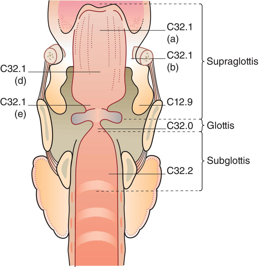
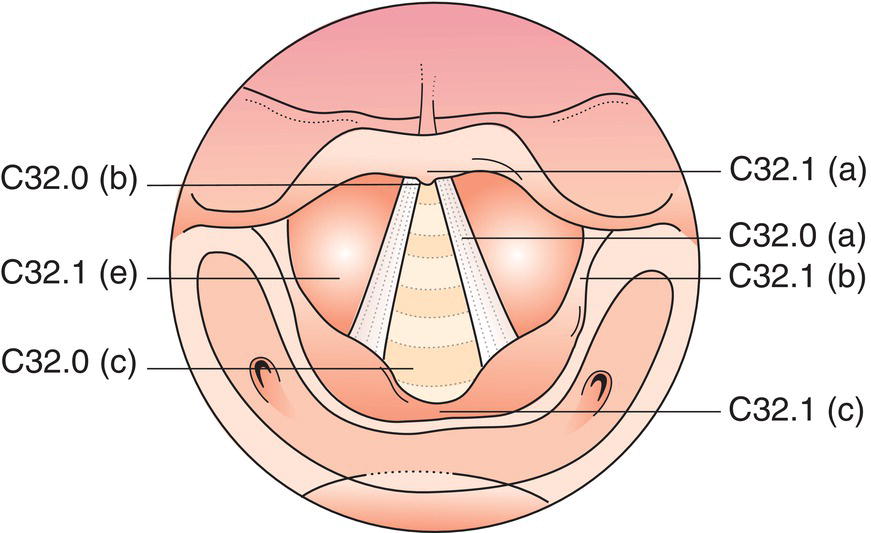
LARYNX (ICD‐O C32.0, 1, 2, C10.1)
Rules for Classification
Anatomical Sites and Subsites
(C10.1), and laryngeal surfaces]
Epilarynx
(including
marginal zone)
Supraglottis
excluding
epilarynx
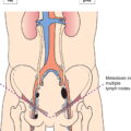
Regional Lymph Nodes
TNM Clinical Classification
T – Primary Tumour
TX
Primary tumour cannot be assessed
T0
No evidence of primary tumour
Tis
Carcinoma in situ
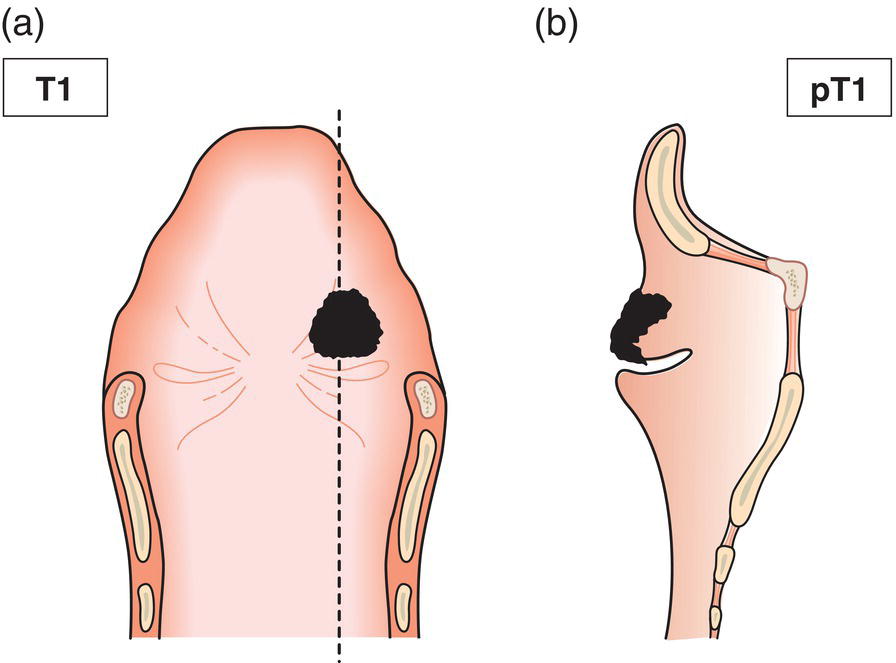
Supraglottis
T1
Tumour limited to one subsite of supraglottis with normal vocal cord mobility (Figs. 80, 81)
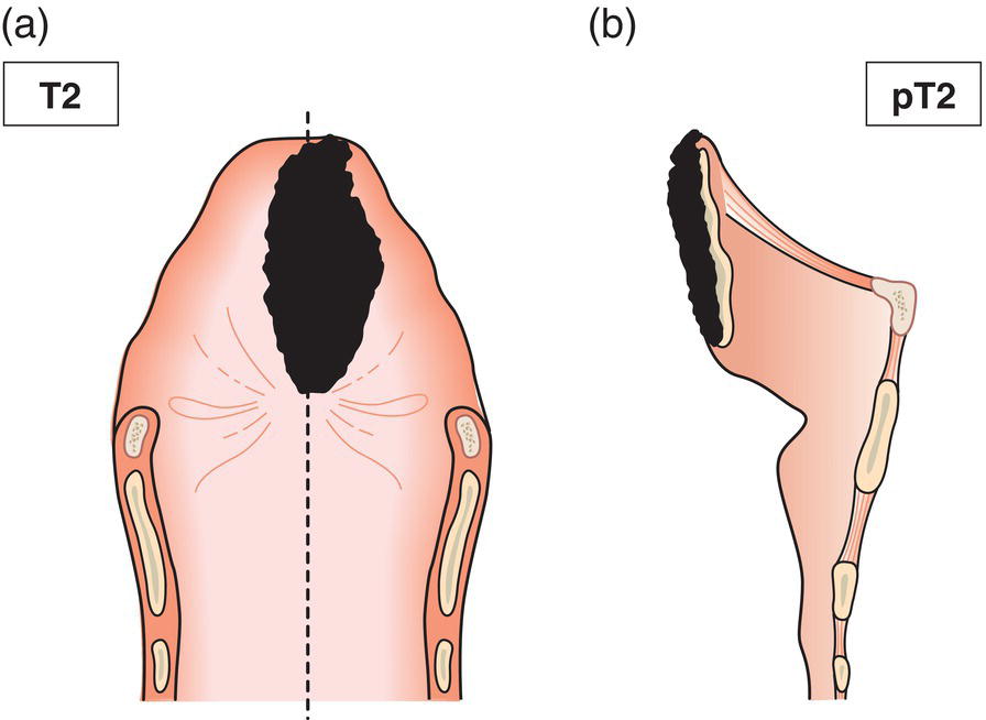
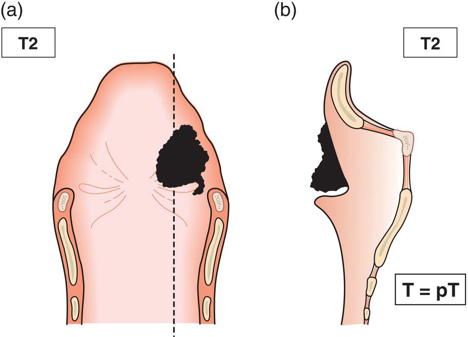
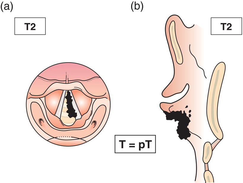
T2
Tumour invades mucosa of more than one adjacent subsite of supraglottis or glottis or region outside the supraglottis (e.g., mucosa of base of tongue, vallecula or medial wall of piriform sinus) without fixation of the larynx (Figs. 82, 83)
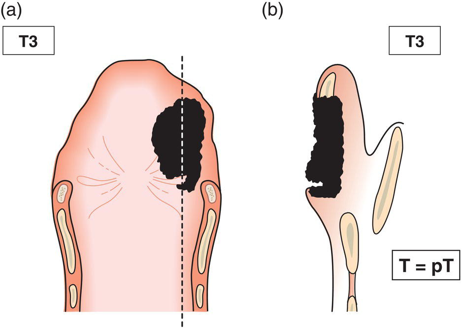
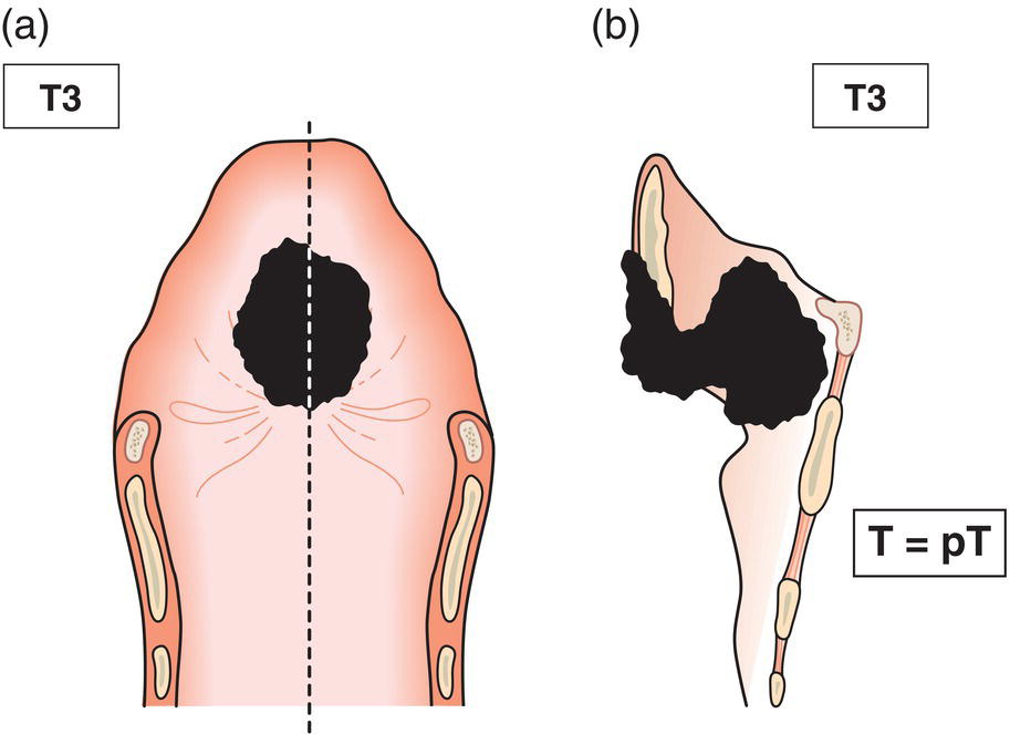
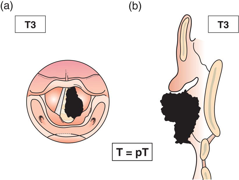
T3
Tumour limited to larynx with vocal cord fixation and/or invades any of the following: postcricoid area, pre‐epiglottic space, paraglottic space and/or inner cortex of thyroid cartilage (Figs. 84, 85)
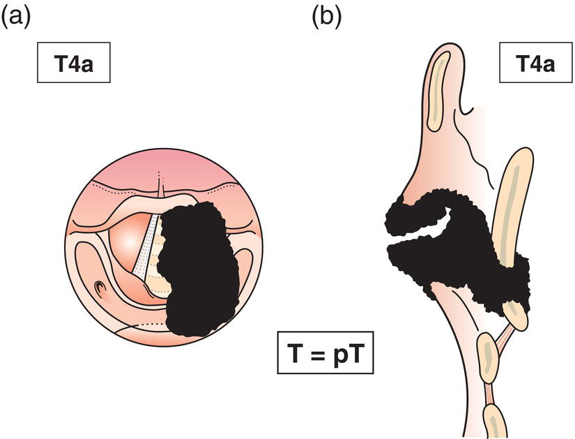
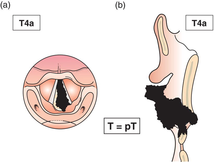
T4a
Tumour invades through the thyroid cartilage and/or invades tissues beyond the larynx, e.g., trachea, soft tissues of neck including deep/extrinsic muscle of tongue (genioglossus, hyoglossus, palatoglossus and styloglossus), strap muscles, thyroid, oesophagus (Fig. 86)
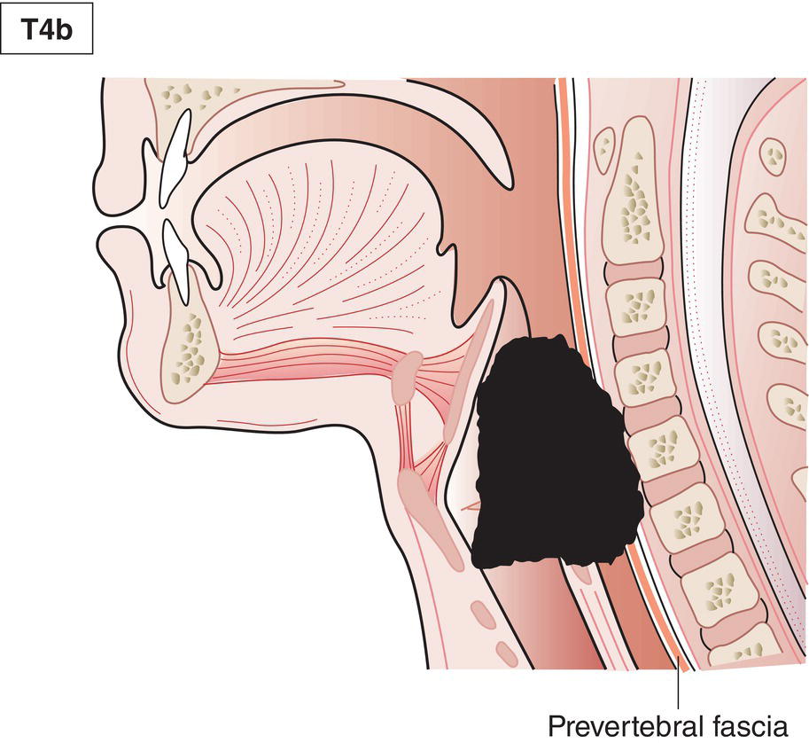
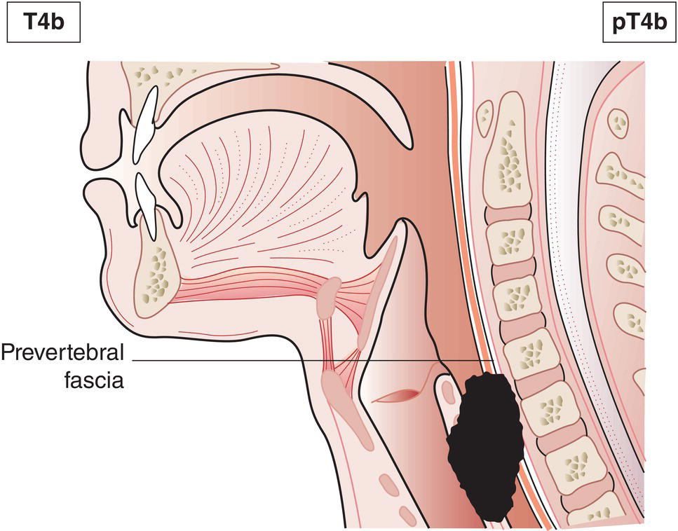
T4b
Tumour invades prevertebral space, mediastinal structures, or encases carotid artery (Fig. 68) 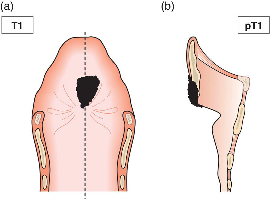
Glottis
T1
Tumour limited to vocal cord(s) (may involve anterior or posterior commissure) with normal mobility (Fig. 87a)
T1a
Tumour limited to one vocal cord (Fig. 87b)
T1b
Tumour involves both vocal cords (Fig. 87c)
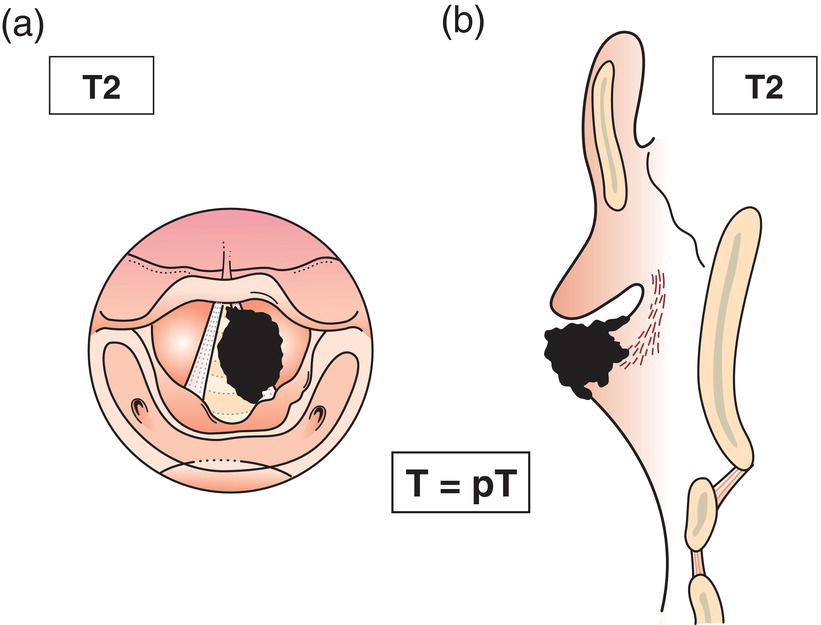
T2
Tumour extends to supraglottis and/or subglottis, and/or with impaired vocal cord mobility (Fig. 88)
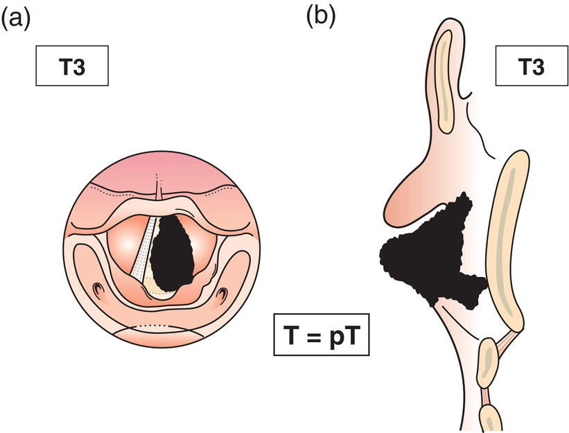
T3
Tumour limited to larynx with vocal cord fixation and/or invades paraglottic space, and/or inner cortex of the thyroid cartilage (Fig. 89)
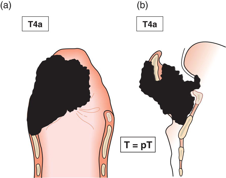
T4a
Tumour invades through the outer cortex of the thyroid cartilage, and/or invades tissues beyond the larynx, e.g., trachea, soft tissues of neck including deep/extrinsic muscle of tongue (genioglossus, hyoglossus, palatoglossus and styloglossus), strap muscles, thyroid, oesophagus (Fig. 90)
T4b
Tumour invades prevertebral space, encases carotid artery, or mediastinal structures (Fig. 91) 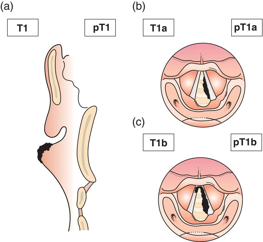
Subglottis
T1
Tumour limited to subglottis (Fig. 92)
T2
Tumour extends to vocal cord(s) with normal or impaired mobility (Fig. 93)
T3
Tumour limited to larynx with vocal cord fixation (Fig. 94)
T4a
Tumour invades cricoid or thyroid cartilage and/or invades tissues beyond the larynx, e.g., trachea, soft tissues of neck including deep/extrinsic muscle of tongue (genioglossus, hyoglossus, palatoglossus and styloglossus), strap muscles, thyroid, oesophagus (Fig. 95)
T4b
Tumour invades prevertebral space, mediastinal structures, or encases carotid artery (Fig. 96) 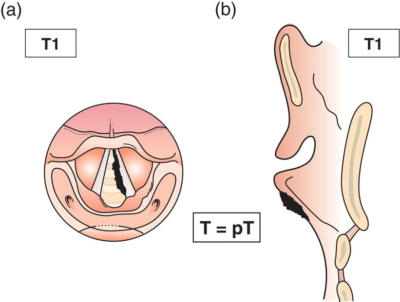
N – Regional Lymph Nodes
pTN Pathological Classification
Summary
Related posts:


Stay updated, free articles. Join our Telegram channel

Full access? Get Clinical Tree


