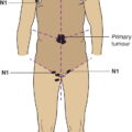
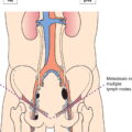
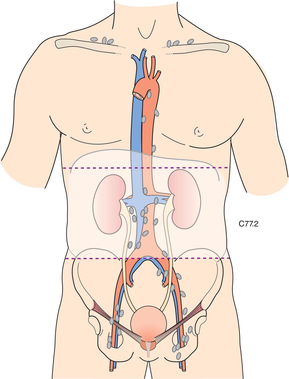
The classification applies only to renal cell carcinoma. There should be histological confirmation of the disease. The regional lymph nodes are the hilar, abdominal para‐aortic, and paracaval nodes. Laterality does not affect the N categories.
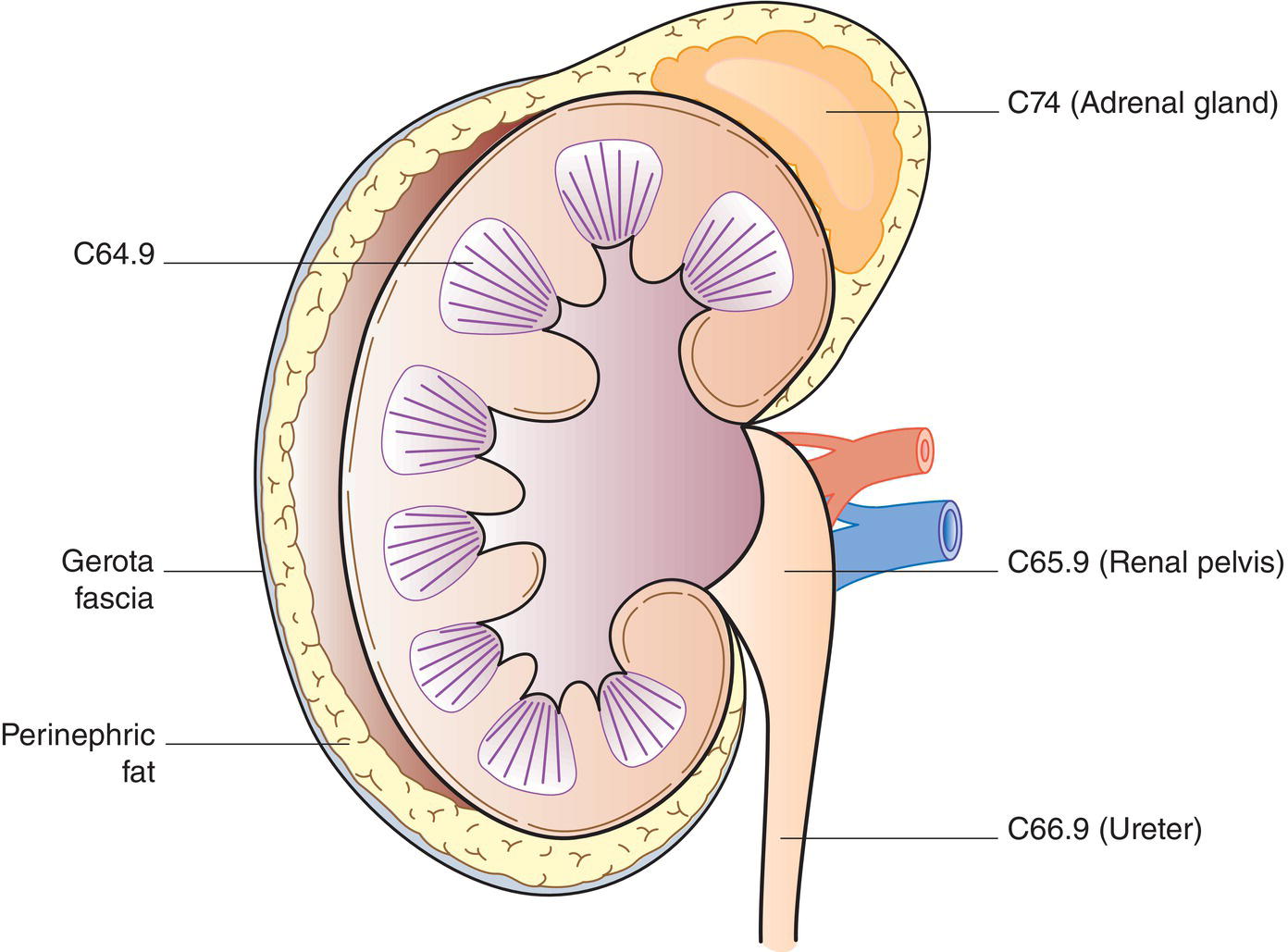
KIDNEY (ICD‐O‐3 C64) (FIG. 506)
Rules for Classification
Regional Lymph Nodes (Fig. 507)
TNM Clinical Classification
T – Primary Tumour
TX
Primary tumour cannot be assessed
T0
No evidence of primary tumour
T1
Tumour 7 cm or less in greatest dimension, limited to the kidney
T1aTumour 4 cm or less (Fig. 508)
T1b Tumour more than 4 cm but not more than 7 cm (Fig. 509)
T2
Tumour more than 7 cm in greatest dimension, limited to the kidney (Fig. 510)
T2a Tumour more than 7 cm but not more than 10 cm
T2b Tumour more than 10 cm, limited to the kidney
T3
Tumour extends into major veins or perinephric tissues but not into the ipsilateral adrenal gland and not beyond Gerota fascia
T3a Tumour extends into the renal vein or its segmental branches, or tumour invades the pelvicalyceal system, or tumour invades perirenal and/or renal sinus fat (peripelvic fat) but not beyond Gerota fascia (Fig. 511) Related posts:
![]()
Stay updated, free articles. Join our Telegram channel

Full access? Get Clinical Tree

 Get Clinical Tree app for offline access