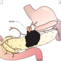
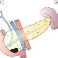
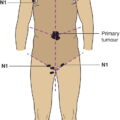
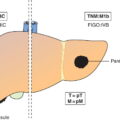
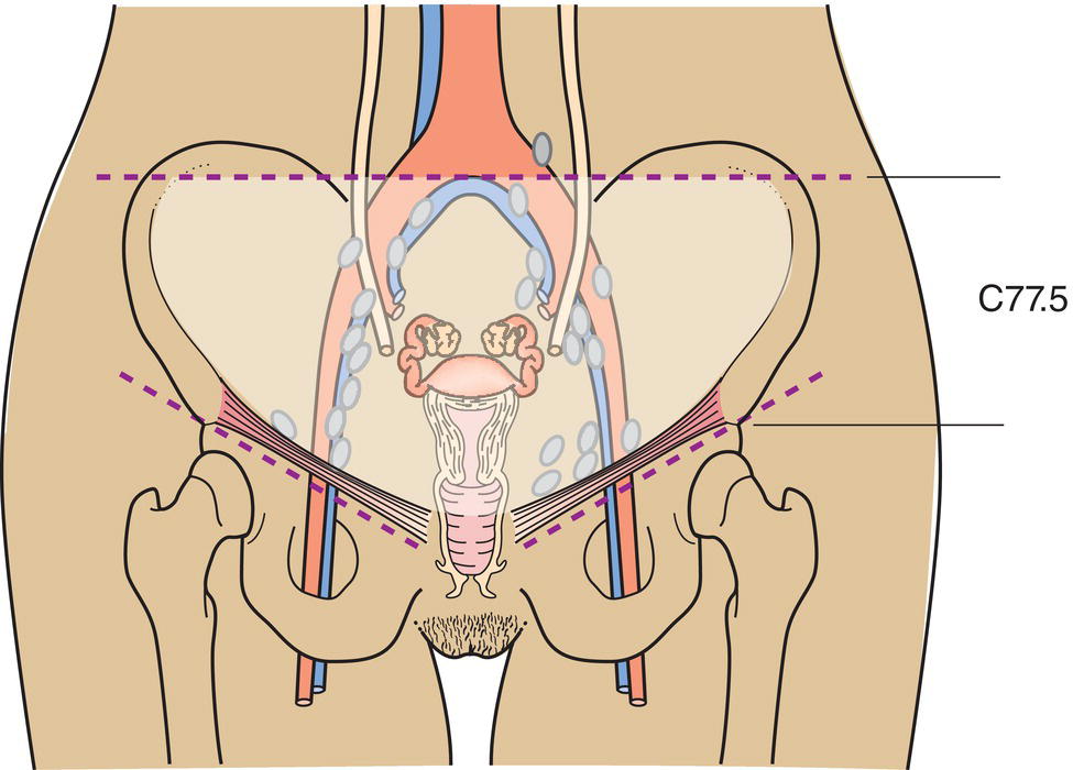
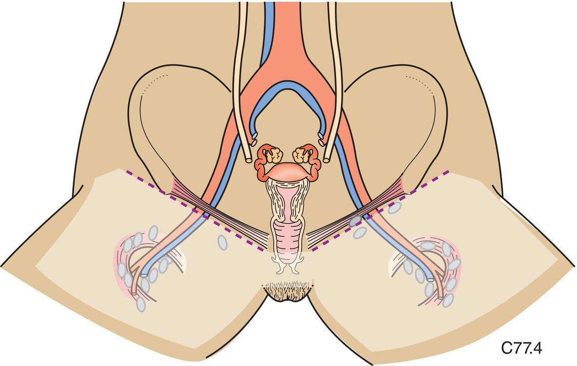
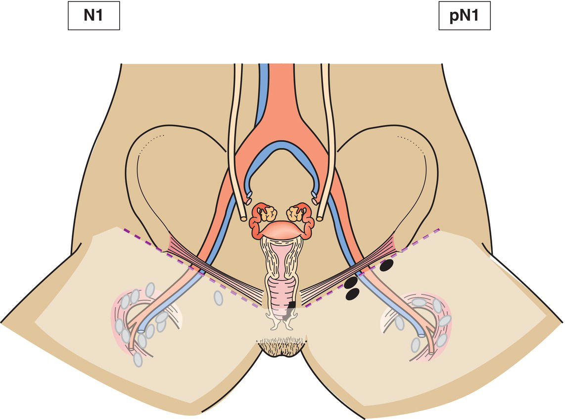
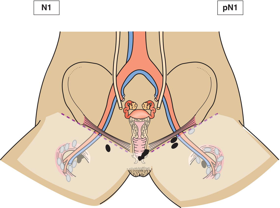
The definitions of the T and M categories correspond to the FIGO stages. Both systems are included for comparison. The classification applies to primary carcinomas only. Tumours present in the vagina as secondary growths from either genital or extragenital sites are excluded. A tumour that has extended to the portio and reached the external os (orifice of uterus) is classified as carcinoma of the cervix. A vaginal carcinoma occurring 5 years after successful treatment (complete response) of a carcinoma of the cervix uteri is considered a primary vaginal carcinoma. A tumour involving the vulva is classified as carcinoma of the vulva. There should be histological confirmation of the disease. The FIGO stages are based on surgical staging. TNM stages are based on clinical and/or pathological classification. Upper two‐thirds of vagina: The pelvic nodes including obdurator, internal iliac (hypogastric), external iliac, and pelvic nodes, NOS. (Fig. 415) Lower third of vagina: The inguinal and femoral nodes. (Fig. 416) The pT and pN categories correspond to the T and N categories. Note
VAGINA (ICD‐O‐3 C52) (Fig. 414)
Rules for Classification
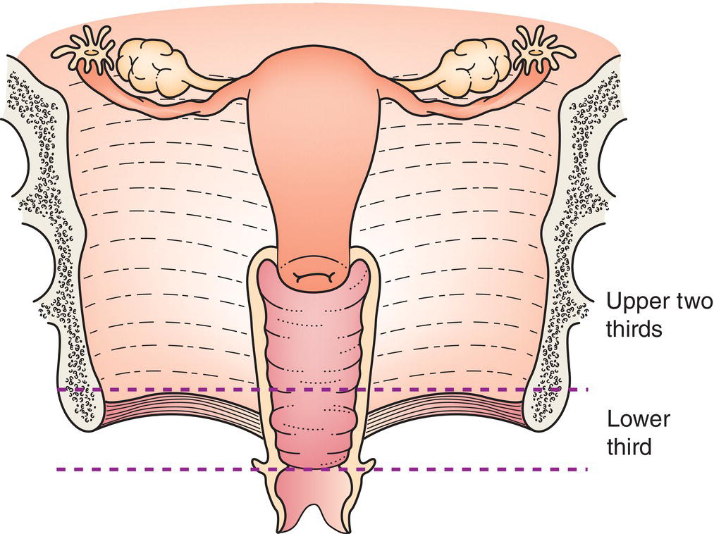
Regional Lymph Nodes
TNM Clinical Classification
T – Primary Tumour
TNM Categories
FIGO Stages
Definition
TX
Primary tumour cannot be assessed
T0
No evidence of primary tumour
Tis
*
Carcinoma in situ (preinvasive carcinoma)
T1
I
Tumour confined to vagina (Fig. 417)
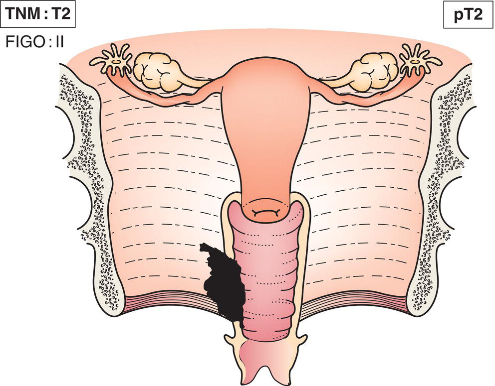
T2
II
Tumour invades paravaginal tissues (paracolpium) (Fig. 418)
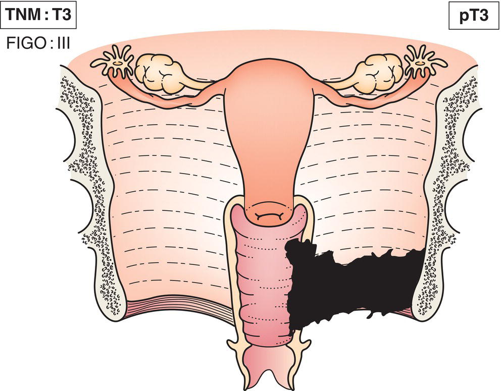
T3
III
Tumour extends to pelvic wall (Fig. 419)
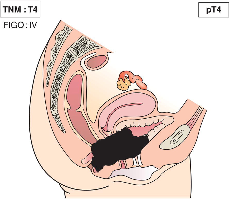
T4
IVA
Tumour invades mucosa of bladder or rectum, or extends beyond the true pelvis** (Fig. 420)
Note
*FIGO no longer includes Stage 0 (Tis).
**The presence of bullous oedema is not sufficient evidence to classify a tumour as T4.
M1
IVB
Distant metastasis 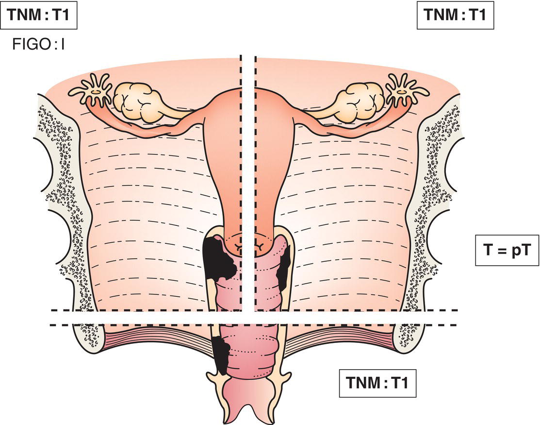
N – Regional Lymph Nodes
NX
Regional lymph nodes cannot be assessed
N0
No regional lymph node metastasis
N1
Regional lymph node metastasis (Figs. 421, 422, 423 ) 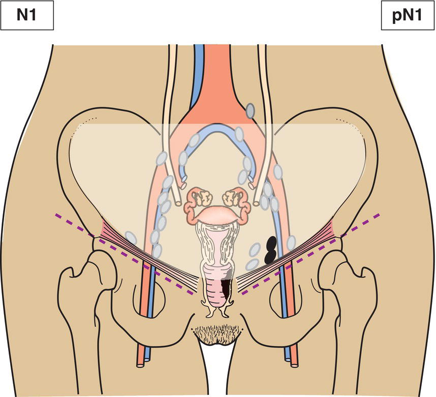
M – Distant Metastasis
M0
No distant metastasis
M1
Distant metastasis
TNM Pathological Classification
pM1
Distant metastasis microscopically confirmed
pM0 and pMX are not valid categories.
pN0
Histological examination of an inguinal lymphadenectomy specimen will ordinarily include 6 or more lymph nodes; a pelvic lymphadenectomy specimen will ordinarily include 10 or more lymph nodes. If the lymph nodes are negative, but the number ordinarily examined is not met, classify as pN0.
Summary
Stay updated, free articles. Join our Telegram channel

Full access? Get Clinical Tree


