div class=”ChapterContextInformation”>
2. Endocrine Hypothalamus
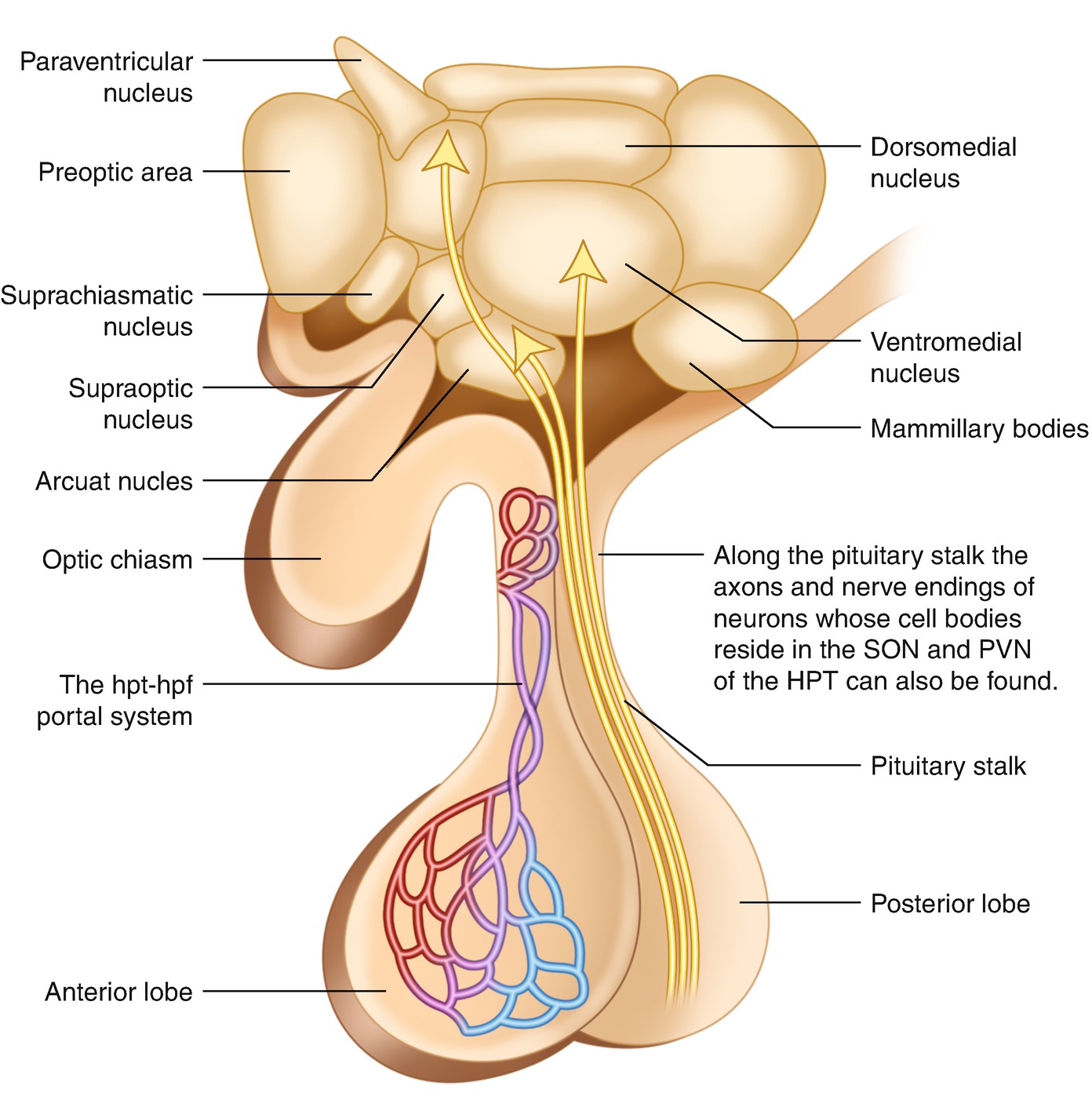
The human hypothalamus, the portal hypophysial vessels and the pituitary gland
The relationship between the adenohypophysis (the anterior pituitary) and the main nuclei of the HPT allows control of the anterior pituitary function by the hypothalamic hypophysiotropic hormones secreted into the portal hypophyseal vessels.
Magnocellular system → Neurohormones
Parvocellular and Arcuate system → Hypophysiotropic hormones
Besides its endocrine role, the hypothalamus is involved in many nonendocrine functions such as regulation of body temperature, thirst, and food intake and is connected with many other parts of the nervous system.
Hence, the hypothalamic hormones can be divided into those secreted by the neurohypophysis directly into the general circulation (neurohormones) and those secreted into hypophysial portal blood vessels (hypophysiotropic hormones).
The former are synthesized in the magnocellular neurons in the SON and PVN and includes vasopressin or antidiuretic hormone (ADH) and oxytocin (both are nonapeptides). The neurosecretory granules with ADH and oxytocin travel down the long axons through the stalk to the posterior hypophysis where the granules are stored and then released into the general circulation. The posterior lobe of the pituitary (neurohypophysis) is made up of the endings of neurons whose cell bodies reside in the supraoptic and paraventricular nuclei of the hypothalamus.
Hypophysiotropic hormones and their main functions
Hormone and structure | Abbreviation | Function |
|---|---|---|
Thyrotropin-releasing hormone (tripeptide hormone) | TRH | Stimulates release of TSH |
Corticotropin-releasing hormone (a 41-amino-acid peptide) | CRH | Stimulates release of ACTH and other products of its precursor molecule, POMC |
Gonadotropin-releasing hormone (a 9-amino-acid peptide) | GnRH, LHRH | Stimulates release of FSH and LH |
Growth hormone-releasing hormone (the major isoform of GHRH is 44 amino acids in length) | GHRH | Stimulates release of GH |
Growth hormone inhibiting hormone somatostatin, tetradecapeptide somatostatin 14 (found mostly in the hypothalamus) and, somatostatin 28 (found in the gut) | SST | Inhibits release of GH |
Prolactin inhibiting hormone (dopamine) | PIH (DA) | Inhibits release of prolactin |
Most of the anterior pituitary hormones are controlled by stimulatory hormones, but GH and especially prolactin (PRL) are also regulated by inhibitory hormones. Some hypophysiotropic hormones are multifunctional. The hormones of the hypothalamus are secreted episodically and not continuously, and in some cases, there is an underlying circadian rhythm.
2.1 Physiologic Puberty
Puberty is best considered as one stage in the continuing process of growth and development that begins during gestation and continues until the end of reproductive life.
After an interval of childhood quiescence—the juvenile pause—the hypothalamic pulse generator increases activity in the peripubertal period, just before the physical changes of puberty commence. This leads to increased secretion of pituitary gonadotropins and, subsequently, gonadal sex steroids that bring about secondary sexual development, the pubertal growth spurt, and fertility.
The normal age at onset of puberty for girls: 8–13 years and for boys: 9–14 years.
The physical changes associated with puberty in females include:
Breast development (thelarche) = the first noted sign of puberty, pubic hair development, adrenarche (the secretion of adrenal androgens), menarche
Development of internal and external genitalia
Pubertal growth spurt, bone mass acquisition
Gynoid body fat disposition

Stay updated, free articles. Join our Telegram channel

Full access? Get Clinical Tree


