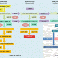
Neoplasia
Vascular
Pituitary adenoma
Pituitary tumor apoplexy
Pituitary carcinoma
Sheehan’s syndrome
Craniopharyngioma
Intrasellar carotid artery aneurysm
Pituicytoma
Fibroma
Subarachnoid hemorrhage
Glioma
Meningioma
Ischemic stroke
Paraganglioma
Genetic
Teratoma
Combined pituitary hormone deficiencies
Chordoma
Angioma
Isolated pituitary hormone deficiencies
Sarcoma
Ependymoma
Infectious
Germinoma
Viral
Cysts
Fungal
Rathke’s cleft and dermoid
Tuberculosis
Ganglioneuroma
Syphilis
Astrocytoma
Bacterial (Others)
Pituitary mestastasis
Primary empty sella functional
Brain damage
Surgery
Drugs
Radiotherapy
Glucocorticoid excess
Radiosurgery
Megestrol acetate
Traumatic brain injury (TBI)
Suppressive thyroxine treatment
Infiltrative/inflammatory disease
Lymphocytic hypophysitis
Dopamine
Granulomatous hypophysitis
Anabolic sex steroids
Xanthomatous hypophysitis
GnRH agonists
Sarcoidosis
Nutritional
Langerhans cell histiocytosis
Obesity
Giant cell granuloma
Malnutrition
Wegener’s granulomatosis
Caloric restriction
Hemochromatosis
Chronic/Acute critical Illness
Idiopathic
Table 8.2
Genetic forms of multiple pituitary hormone deficiencies
HESX1 | OTX2 | LHX3 | LHX4 | SOX3 | SOX2 | PROP1 | POU1F1 | |
GH | + | + | + | + | + | +/− | + | + |
LH/FSH | +/− | +/− | + | +/− | +/− | + | + | − |
PRL | +/− | − | + | − | +/− | − | + | + |
TSH | +/− | +/− | + | +/− | +/− | − | + | +/− |
ACTH | +/− | +/− | +/− | +/− | +/− | − | +/− | − |
ADH | +/− | − | − | − | +/− | − | − | − |
Inheritance | AR/AD | AD | AR | AD | XL | AD | AR | AR/AD |
Pituitary involvement | Normal/Hypoplastic AP; normal/ectopic PP | Normal/hypoplastic AP; altered stalk; Ectopic PP | Hypoplastic, normal or enlarged AP | Normal/hypoplastic AP; normal or ectopic PP | Hypoplastic AP; Ectopic PP | Normal/hypoplastic AP; Hypothalamic hamartoma | Hypoplastic, normal or enlarged AP and normal PP | Normal/hypoplastic AP |
Extra-pituitary phenotype | SOD; normal optic nerves | Anophthalmia or no ocular pathology; Chiari malformation | limited neck rotation or no; SD | Cerebellar anomalies; Chiari malformation | Variable learning difficulties; HCC | Anophthalmia; Microphthalmia; DD; SD; HCC; Aesophageal atresia | No involvement | No involvement |
Diagnosis
Clinical Presentation
The clinical presentation of hypopituitarism is often vague and nonspecific, leading to a further delay in diagnosis. Nonspecific symptoms include a feeling of general poor health, increased lethargy, feeling cool, chronic tiredness, reduced appetite, weight loss, and abdominal pain [2, 3]. Hypopituitarism can sometimes develops acutely, leading to a rapid onset of symptoms (excruciating headache, meningism, and cardiovascular collapse) necessitating admission and intensive care management, as is often seen in patients with tumor apoplexy.
The signs and symptoms of underlying diseases can sometimes follow hypopituitarism [3]. Symptoms attributed to the local effects of tumoral masses in the sellar region with suprasellar extension, such as headaches, rhinorrhea, and visual disturbances (typically bilateral hemianopsia, but can also occur as unilateral) frequently remain unrecognized by patients, mostly men, for a long period of time.
Deficits of anterior pituitary hormones may be secondary to hormone excess caused by functioning pituitary tumors, which produces a complex picture combining hormone excess and deficiencies, such as suppression of gonadotropins in hyperprolactinemia, growth hormone deficiency (GHD) caused by cortisol excess in Cushing’s syndrome [4] or growth hormone (GH) secreting macroadenoma that causes acromegaly and hypogonadism [5]. The presence of central Diabetes Insipidus (DI) usually indicates a non-pituitary lesion affecting the hypothalamus or pituitary stalk. Preoperatively, pituitary adenomas rarely cause DI.
Somatotropin Deficiency
Children
GHD in childhood promotes short stature and delayed bone age with slow growth velocity. Idiopathic GHD is the most common etiology. GH does not appear to have a relevant role in fetal growth. Therefore, in general, children are born with normal length, weight, and general appearance. However, microphallus and cryptorchidism may be present, especially with gonadotropin associated deficiency. Prolonged jaundice, hypoglycemia-associated seizures (when GHD occurs in conjunction with Adrenocorticotropic Hormone (ACTH) deficiency), and midline abnormalities suggest a congenital etiology.
Recognition of GHD is more common from the first 12–18 months after birth, with slow growth as an early sign and a consequent downward shift in the normal growth curve. Children tend to present with adiposity around the trunk. They have immature body and facial traits, a high-pitched voice, prominent forehead, depressed mid-face development, delayed dentition, and small hands and feet.
Adults
The severity of the clinical manifestations of GHD in adults depends on the timing of onset. In general, patients present nonspecific symptoms, such as fatigue, decreased energy, low mood, and altered body composition with increased fat and decreased lean body mass and muscle strength, as well as reduced bone mineral density, compromised metabolism of glucose and lipids, and poor quality of life [6]. Childhood-onset GHD patients have a lower lean body mass, bone mineral content, and better quality of life compared to adult-onset GHD patients.
Gonadotropin Deficiency
The clinical presentation of male hypogonadism depends on the time of onset of androgen deficiency. In men with recent onset hypogonadism, the physical examination is usually normal, while diminished facial and body hair, gynecomastia, and small soft testes are features of longstanding hypogonadism [2]. The principal signs and symptoms of androgen deficiency in men are loss of libido, decreased sexual potency, loss of body hair (axillary and pubic), infertility, and low bone mineral density. The threshold testosterone level below which symptoms of androgen deficiency and adverse health outcomes occur and testosterone administration improves outcomes in the general population is currently not known [7].
Female adolescents have primary amenorrhea and lack of breast development, whereas in adult women, gonadotropin deficiency leads to reduced secretion of estradiol, resulting in infertility and oligo/amenorrhea. Low estrogen is also responsible for genital atrophy and decreased breast volume in chronic hypogonadism. There is a reduction of pubic and axillary hair, especially when concomitant dysfunction of the corticotroph axis is present.
At the prepubertal age, no obvious clinical signs or symptoms are present until the normal age of puberty onset (9–14 years in boys and 8–13 years in girls), when a lack of signs of normal pubertal development are then observed. It should be emphasized that micropenis with or without associated cryptorchidism is an important clinical clue that suggests congenital hypogonadotropic hypogonadism (where there is lack of the normal fetal secretion and postnatal surge of gonadotropins) rather than acquired hypogonadotropic hypogonadism [8].
Thyrotropin Deficiency
The clinical picture of central hypothyroidism is very similar to primary hypothyroidism, but is often milder. Symptoms include cold intolerance, dry skin, decreased appetite with mild weight gain, and fatigue [9]. The presence of goiter usually indicates primary thyroid disease. In children, decreased growth velocity with impairment of neurological development is an important sign.
Corticotropin Deficiency
ACTH deficiency leads to decreased glucocorticoid levels. Mineralocorticoid secretion is preserved, since it is primarily modulated by the renin–angiotensin system. Hyperpigmentation is typical of primary adrenal disease and is absent in central disease. Symptoms of ACTH deficiency are largely nonspecific, including weakness, fatigue, anorexia, weight loss, arthralgia, postural hypotension, and tachycardia [10]. Hyponatremia, hypoglycemia, and eosinophilia may also occur. Ultimately, if left untreated, ACTH deficiency may lead to death due to vascular collapse, since cortisol is needed to maintain vascular tone. Mild ACTH deficiency may remain clinically unnoticed when cortisol production is sufficient for preventing symptoms in the absence of clinical stressors (e.g., infections). Hence, laboratorial evaluation is recommended in all patients at risk of ACTH deficiency.
Antidiuretic Hormone (ADH) Deficiency
ADH deficiency results in polyuria (urine volume >3 L/day in adults) and polydipsia. If the thirst mechanism is not present, as is the case in some patients with hypothalamic lesions, then lack of polydipsia leads to a high risk of life-threatening dehydration and hypernatremia [11].
Diagnostic Testing
The diagnosis of hypopituitarism can often be made through simultaneous measurements of basal anterior pituitary and target gland hormone levels. Each axis should be assessed in patients suspected of having partial or complete loss of pituitary function, because the impairment in these patients is often partial rather than complete.
Low or inappropriately normal serum levels of pituitary hormones in conjunction with low peripheral hormones indicate hypopituitarism. FSH, LH, estradiol (women), testosterone (men), prolactin, TSH, free thyroxine (FT4), 9 am cortisol, and insulin-like growth factor-I (IGF-I) tests form the baseline parameters to assess. In addition, dynamic studies are necessary in most cases for documenting hypopituitarism, particularly for assessing GH secretory reserve and the ACTH-adrenal axis (Table 8.3) [5].
Table 8.3
Hormone testing for pituitary function
Criteria for hormone deficiency | |
Somatotropic axis | |
Baseline | |
IGF-I | Low/low-normal |
GH | No usefulness |
Provocative tests | |
Clonidine test (only for children) | <7–10 μg/L |
Insulin tolerance test | Children: <7–10 μg/L |
Adults: <5.1 μg/L | |
Transition period: <6.1 μg/L | |
GHRH-Arg test (only for adults) | Adults: |
Lean <11.5 μg/L | |
Overweight <8.0 μg/L | |
Obese <4.2 μg/L | |
Transition period: <19.0 μg/L | |
Glucagon test | Children: <7–10 μg/L |
Adults: <2.5–3 μg/L | |
Gonadotropic axis | |
Baseline | |
Male | |
Testosterone | Low |
FSH/LH | Low or inappropriately normal |
Female | |
Estradiol | Low |
FSH/LH | In younger women: low or inappropriately normal |
In postmenopausal women: inappropriately low | |
Provocative test | |
GnRH | Not useful in adults |
Thyrotropic axis | |
Baseline | |
Free T4 | Low, low-normal |
TSH | Low, normal or slightly increased |
Provocative test | |
TRH | Not useful |
Corticotropic axis | |
Baseline | |
Cortisol (morning) | <3 μg/dL (<80 nmol/L) |
>18 μg/dL (>500 nmol/L): hypocortisolism excluded | |
ACTH (morning) | Low or normal |
Provocative tests | |
Insulin tolerance test | Peak Cortisol <18 μg/dL (<500 nmol/L) |
250 μg ACTH test | Peak Cortisol <18 μg/dL (<500 nmol/L) |
Overnight metyrapone test | 11-deoxicortisol <7 μg/dL (<200 nmol/L), low cortisol |
CRH (human or ovine) | ACTH: Peak <2–4× baseline |
Cortisol: Peak <20 μg/dL (555 nmol/L) | |
Antidiuretic hormone | |
Dynamic test | |
Water deprivation | Maximal Urinary Osmolality (MUO) < 300 mOsm/kg/H2O plus >50 % increase in MUO after desmopressin (Complete DI) |
Somatotropin Deficiency
Children
GHD in children is based on auxological data, which is considered the gold standard in such diagnosis [12]. An appropriate differential diagnosis must be performed ruling out other causes of growth failure, such as hypothyroidism, Turner syndrome, and systemic diseases.
Evaluation should be considered when patients present with one of the following conditions: (1) short stature of more than 2.5 standard deviations (SD) below the mean; (2) growth failure, which is defined as height velocity less than 2 SD below the mean for age; (3) a combination of less severe short stature (2–2.5 SD below the mean for age) and growth failure (growth velocity less than 1 SD); (4) clinical picture suggesting hypothalamic-pituitary dysfunction, such as hypoglycemia, microphallus, intracranial tumor, or history of cranial irradiation with decelerating growth; and (5) evidence of deficiency in other hypothalamic–pituitary hormones [13].
The pulsatile nature and short half-life of GH preclude the random measurement of serum GH levels as a useful tool for diagnosing GHD. Thus, IGF-I and IGF-binding Protein 3 (IGFBP-3) are appropriate initial tests for GHD in children providing that conditions such as poor nutrition, hypothyroidism, and chronic systemic diseases are excluded. These hormones reflect an integrated assessment of GH secretion because of negligible diurnal variation [14].
IGF-I and IGFBP-3 measurements should be interpreted in relation to reference ranges that are standardized for sex and age. An important drawback to using serum IGF-I for GHD diagnosis is that its values are low in very young children and overlap in GHD patients and normal children. In this context, IGFBP-3 levels, which are less related to age, are more discriminatory than IGF-I levels at the lower end of the normal range [15].
These tests present less than adequate sensitivity, although specificity is high. Thus, in patients with severe GHD, IGF-I and IGFBP-3 levels are invariably reduced; On the other hand, patients with milder abnormalities of GH secretion demonstrate normal levels of IGF-I and its binding protein in a significant percentage of cases [16].
Despite these limitations, measurement of IGF-I and IGFBP-3 levels associated with provocative testing in an appropriate clinical context is now commonly performed when investigating GHD in childhood.
GH Stimulation Testing in Children
Provocative GH testing has several caveats. They are not physiological, since the secretagogues used do not reflect normal GH secretion; the cutoff level of normal is arbitrary and the tests are age dependent. Furthermore, the tests rely upon GH assays of variable accuracy and are all uncomfortable, cumbersome, and risky for the patient [12, 17]. Therefore, there is currently no gold standard provocative GH test for GHD in children. As a result, subnormal responses to two secretagogues are necessary for diagnosis, with the exception of patients presenting with a central nervous system disorder, multiple pituitary hormone defects, or a known genetic defect. In these cases, one test is sufficient to establish the diagnosis [18].
These stimulation tests are performed after an overnight fasting. After the pharmacologic stimulus, serum samples are collected at intervals designed to capture the peak GH level. A “normal” response is defined by a serum GH concentration of greater than 7–10 mcg/L, although the ideal threshold may vary with the assay used. Of note, all patients should be euthyroid and should not be under supraphysiological doses of glucocorticoids before any testing is performed (Tables 8.3 and 8.4).
Table 8.4
Protocols of dynamic tests for investigation of anterior pituitary (GH and ACTH) and posterior pituitary (ADH) deficiencies
Provocative tests | Dosage | Time of hormone collection | Side effects/drawbacks |
|---|---|---|---|
GH | |||
Clonidine (only for children) | 5 μg/kg, up to 250 μg, PO | GH: 0, 30, 60, 90 min | Drowsiness; false negative results |
Insulin tolerance test | Regular insulin 0.05–0.15 IU/kg, IV | GH: 0, 15, 30, 60, 90, 120 min | Severe hypoglycemia and medical surveillance required |
Glucagon | 0.03 mg/kg (up to 1 mg) IM/SC; if >90 kg, 1.5 mg | GH: 0, 60, 90, 120, 150, 180, 210, 240 min | Late hypoglycemia; very prolonged test; not well validated in adults |
GHRH-ARG (only for adults) | GHRH (1 μg/kg, IV bolus) + Arginine (0.5 g/kg, up to 30 g, IV, over 30 min) | GH: 0, 30, 60, 90, 120 min | Very influenced by adiposity |
ACTH | |||
ACTH1-24 | 250 μg IV/IM | Cortisol: 0, 30 and 60 min | Adrenal atrophy is required |
Insulin tolerance test | Regular insulin 0.05–0.15 IU/Kg, IV | Cortisol: 0, 15, 30, 60, 90, 120 min | See above |
Overnight metyrapone | 30 mg/kg, PO, at midnight (maximum 3 g) | 11-deoxycortisol and cortisol: 8 am | Limited availability; adrenal crisis |
CRH (human or ovine) | 1 μg/kg, up to 100 μg, IV | Cortisol and ACTH: 0, 15, 30, 60, 90, 120 min | Flushing; expensive |
Dynamic test | Procedure | Side effects/Drawbacks | |
ADH | |||
Water deprivation | Nothing allowed by mouth; patient voids; weight is recorded; Serum Na+ and urine Osm are measured at baseline. Weight is checked after each liter of urine is passed. In each voided urine, measure urine Osm and when two consecutive measurements differ <10 % and subject has lost 2 % of BW, plasma sample for Na+, Osm and VP should be drawn. DDAVP 2 μg IV/IM is administered and urine Osm and volume are measured every 30 min in the next 2 h. Dehydration is stopped if patient has lost >3 % of BW or if serum Na+ becomes elevated. | Difficulties in differentiate partial hypothalamic DI from primary polydipsia | |
Clonidine, an α-2 adrenergic receptor agonist, promotes GH release, mainly through GHRH secretion. It is a stronger stimulant for growth hormone release, and therefore false negative results can follow. On the other hand, children presenting with a GH subnormal response to such stimulus rarely secrete normal GH in response to any other stimuli [19]. The test commonly causes hypotension and drowsiness that may last for hours and promote late hypoglycemia.
Insulin-induced hypoglycemia is a potent stimulant of GH release and, therefore, the Insulin Tolerance Test (ITT) is among the most specific tests for GHD. However, safety concerns have prevented the widespread use of this test. The proposed mechanism by which hypoglycemia promotes GH secretion is through the suppression of somatostatin tone and stimulation of α-adrenergic receptors [20]. This test requires constant supervision by a clinician and is contraindicated in children less than 2 years of age.
Administration of glucagon promotes GH secretion through a poorly understood mechanism, with the activation of central noradrenergic pathways as a plausible hypothesis [21]. Glucagon presents mild and transient side effects, such as nausea, vomiting, and sweating, and therefore is a very good choice for infants and young children who are more susceptible to the risks of insulin-induced hypoglycemia.
Adults
In adults, the clinical picture of GHD is subtle and nonspecific, and therefore the diagnosis relies on biochemical testing. Patients with structural hypothalamic and/or pituitary disease, surgery, or irradiation in these areas as well as TBI, SAH, or evidence of other pituitary hormone deficiencies should be evaluated for acquired GHD. Otherwise, the presence of three or more pituitary hormone deficiencies associated with a low IGF-I is highly predictive of GHD, in which case provocative testing is not necessary [22]. In addition, patients should receive adequate replacement of other deficient hormones before GH stimulation testing is performed.
GH Stimulation Testing in Adults
ITT is considered the most validated test currently available and is the diagnostic test of choice for GHD in adults. However, it is contraindicated in patients with seizure disorders or ischemic heart disease and requires monitoring, even in healthy adults. Adequate hypoglycemia (<2.2 mmol/L) is not always achieved, and therefore, larger doses of insulin up to 0.3 U/kg may be necessary in obese patients and those with fasting blood glucose above 5.5 mmol/L [23]. An assay cutoff of 5.1 μg/L is recommended for diagnosis [22].
A GHRH and arginine test (GHRH-Arg test) is a very potent and reproducible test. Arginine potentiates the response to GHRH presumably through the inhibition of hypothalamic somatostatin secretion [24]. This combined test is not affected by gender or age and shows few side effects with no hypoglycemia. On the other hand, the assay cutoff for GHD diagnosis depends on the body mass index (BMI) [25]. In addition, GHRH directly stimulates the pituitary, and patients with GHD of hypothalamic origin, mainly after radiotherapy, could present a falsely normal GH response [26].
Administration of glucagon allows for the assessment of GH and ACTH-cortisol reserves, and has few side effects with minimal contraindications. It is a good choice when other tests are unavailable or contraindicated. In adults, an assay cutoff between 2.5 and 3.0 μg/L is recommended for GHD diagnosis [22].
Transitional Period
In the transition period (i.e., after the cessation of linear growth and completion of puberty), the majority of GHD patients must be retested. Those patients with conditions causing multiple pituitary hormone deficiencies (MPHD) (i.e., three or more pituitary hormone deficits), can continue on GH therapy, but require determination of an adequate dose. Other patients without MPHD but who present with known mutations or irreversible structural hypothalamic-pituitary lesions/damage should be screened for serum IGF-I levels after terminating therapy for at least 1 month. IGF-I levels below −2 SD are sufficient for GH therapy reinstitution. If the IGF-I level is within the normal range, then one provocative testing is mandatory for GH therapy in case of a subnormal response.
In the remaining patients, mostly with idiopathic causes, a serum IGF-I test and one provocative test must be performed, and in case of discordant results, a second provocative test is necessary for the diagnosis of persistent GHD [22, 27].
It is unclear whether different assay cutoffs should be adopted during this transitional period, as opposed to GHD assay cutoffs in adults. Some studies suggest that the assay cutoffs in these cases should be higher than for older adults, with levels of 6.1 μg/L and 19.0 μg/L for the ITT and GHRH-arg, respectively [28, 29].
Gonadotropin Deficiency
In men, low or inappropriately normal levels of gonadotropins combined with low levels of serum testosterone are indicative of secondary hypogonadism. Semen analysis is indicated when considering fertility and may demonstrate a reduced sperm count or possibly azoospermia.
In younger women, oligo/amenorrhoea with low serum estradiol levels and low or inappropriately normal FSH and LH concentrations is consistent with secondary hypogonadism. In postmenopausal women, the absence of the normal rise of FSH and LH levels is sufficient for establishing a diagnosis.
In secondary hypogonadism, serum prolactin should always be measured to exclude hyperprolactinemia, which might occur for several reasons, such as prolactinomas, sellar and parasellar masses causing pituitary stalk compression, and use of drugs with antidopaminergic activity.
In adults, there is no usefulness in performing the gonadotropin-releasing hormone (GnRH) provocative test because it does not provide any additional information [5].
Thyrotropin Deficiency
Evaluation of the thyrotrophic axis is based on the measurement of basal serum TSH and thyroid hormone levels. Central hypothyroidism is diagnosed when serum TSH levels are low or inappropriately normal coupled with low levels of serum free T4. Occasionally, TSH levels may be slightly elevated but usually remain lower than 10 mIU/mL. In these patients, the elevation of serum TSH is associated with decreased bioactivity due to increased sialylation [30]. In patients with concomitant GH and TSH deficiencies, serum-free T4 may be normal (usually at the lower tertile), decreasing only after GH replacement [31, 32]. More recently, it has been proposed that echocardiography can be useful in the evaluation of patients with hypothalamic-pituitary disease and free T4 levels within reference range, as some of these patients present signs of tissue hypothyroidism, a condition that could be named “subclinical central hypothyroidism” [33].
Corticotropin Deficiency
Cortisol secretion follows a circadian cycle, being highest in the early morning and lowest at midnight. Hence, a basal serum cortisol measurement may not reflect disturbances of the hypothalamus-pituitary-adrenal (HPA) axis. In addition, alterations in the levels of cortisol-binding globulin (CBG), which is frequently seen in clinical practice (e.g., higher levels of CBG, and consequently serum total cortisol, during oral estrogen treatment as a contraceptive) may also mask the diagnosis of central hypoadrenalism. Therefore, early morning serum cortisol (between 07:00 and 09:00) may be measured as a first step in the evaluation [10]. Stimulation tests are frequently required for corticotropic assessment. The most commonly used stimuli in clinical practice are insulin-induced hypoglycemia, Metyrapone, synthetic ACTH (ACTH1-24), and CRH (Tables 8.3 and 8.4).
Hypoglycemia is a potent activator of the HPA axis, and the ITT is usually regarded as the “gold standard” for diagnosis (see more details in “GH stimulation testing”).
Stay updated, free articles. Join our Telegram channel

Full access? Get Clinical Tree


