Chapter 11 Heme Biosynthesis and Its Disorders
Sideroblastic Anemia
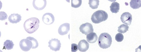
Figure 11-1 NEEDLE-LIKE INCLUSIONS OF PORPHYRIN IN THE CIRCULATING RED CELLS OF A PATIENT WITH CONGENITAL PORPHYRIA AFTER SPLENECTOMY.
(From Merino A, To-Figueras J, Herrero C: Atypical red cell inclusions in congenital erythropoietic porphyria. Br J Haematol 132:124, 2006.)
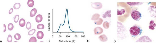
Figure 11-2 A, Peripheral blood smear from a patient with hereditary sideroblastic anemia shows a population of hypochromic and microcytic erythrocytes. B, Erythrocyte volume distribution curve of a patient with hereditary sideroblastic anemia. A dimorphic size distribution is evident. C, Peripheral blood showing Pappenheimer bodies (Prussian blue stain). D, The bone marrow smear stained with Prussian blue shows ring sideroblasts.
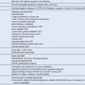
Table 11-1 Classification of Sideroblastic Anemias
| HEREDITARY (NONSYNDROMIC) |
| ACQUIRED |
Idiopathic acquired* (refractory anemia with ring sideroblasts) Associated with previous chemotherapy, irradiation, or in transition myelodysplasia or myeloproliferative diseases |
| DRUGS |
| RARE CAUSES |
| HEREDITARY (SYNDROMIC) |
*Trial of pyridoxine indicated.