Helper T-Cell Differentiation and Plasticity


Helper T-Cell Differentiation and Plasticity


John J. O’Shea


INTRODUCTION

Cluster of differentiation (CD)4 T cells are critical for the elimination of microbial pathogens, but different types of microbes demand distinct responses. To effectively combat these microbes, CD4 T cells differentiate to form subsets of activated cells with specific characteristics. These discrete subsets of CD4 T cells in turn cause the recruitment of different types of inflammatory cells. Each type of inflammatory cell is adapted to eliminate particular types of microorganisms, and this cooperation of T-lymphocytes with other effector cells links adaptive and innate responses for optimal host defense.

In this respect, the action of CD4 T cells role is largely indirect in that they primarily exert their effect by acting on other cells. For this reason, CD4 T cells are referred to as “helper cells,” which can influence macrophages, B cells, CD8 T cells, natural killer (NK) cells, and other effector cells. The “help” provided by CD4 comes largely in the form of cytokines; however, the production of cytokines is a tightly controlled process: distinct subsets of CD4 T cells secrete characteristic repertoires of cytokines. That is, after activation, CD4 T cells divide, differentiate, and adopt restricted patterns of gene expression, which depend upon the nature of the offending microbial pathogen. Consequently, depending upon whether the pathogen is an intracellular microbe, a worm, or an extracellular bacteria or fungus, different types of activated CD4 T cells are generated. Sometimes though, helper cells are not so helpful insofar as CD4 T cells are also important drivers of autoimmunity.

In this chapter, the different fates available to CD4 T cells will be discussed, as will the molecular mechanisms underlying their specification. The degree to which T cells exhibit characteristics of terminal differentiation versus plasticity will also be considered, as will the mechanisms underlying commitment versus flexibility. Finally, the contribution of different helper T-cell subsets to immune-mediated disease will also be discussed.


FROM THE THYMUS TO THE PERIPHERY

Following maturation and selection in thymus, single positive CD4 T cells exit the thymus and migrate through blood to secondary lymphoid tissues including the spleen, lymph nodes, and mucosal-associated lymphoid tissues.1,2 These tissues and organs are composed of zones that are enriched with T- or B-lymphocytes: in the lymph node, the paracortical region contains T cells and dendritic cells (DCs), whereas in the spleen, T cells and DCs reside within the white pulp in the periarteriolar lymphoid sheath.

The trafficking and retention of CD4 T cells in secondary lymphoid tissue is dependent upon selective expression of chemokine receptors and adhesion molecules.3 In lymph nodes and Peyer patches, T cells enter the tissue via specialized blood vessels termed high endothelial venules (HEVs). HEVs express L-selectin and CCL21. Naive T cells, T cells that have yet to encounter foreign antigen, express ligands for L-selectin and CCL21, namely CD62L and CCR7. These ligands promote the entry of naïve T cells into these tissues.4,5,6 Extravasation of naïve lymphocytes through HEVs is dependent upon intercellular adhesion molecules that bind the integrin lymphocyte function-associated antigen-1. Within secondary lymphoid organs, naïve T cells are retained in the T-cell-rich zones by the chemokines CCL21 and CCL19.

If no antigen is encountered, naïve T cells exit the lymph node through the efferent lymphatic vessels and then return to the bloodstream via the thoracic duct. Egress from lymph nodes (and the thymus) requires the seven transmembrane receptor sphingosine 1-phosphate receptor-1.7,8 The concentration of sphingosine 1-phosphate is higher in blood, and this provides a concentration gradient for emerging lymphocytes to follow. The immunomodulatory drug fingolimod,1 a partial agonist of S1P1, acts by blocking egress of T cells from the thymus and lymph nodes.8 Expression of S1P1 is under the control of the transcription factor KLF2, a factor that promotes the naïve state.7,9,10 The transcription factor Foxp1 and miR-125b are two additional factor that preserve T-cell naïveté.11

In the spleen, naïve T cells enter through terminal arterioles, not HEVs; as a result, expression of CD62L is not required. Migration to the splenic white pulp does require integrin and chemokine receptor signaling. Exit from the spleen is thought to occur by migration from white pulp to red pulp with subsequent entry into blood.

Naïve T cells spend relatively short times in the blood before finding a new secondary lymphoid organ to enter. In the absence of encountering cognate antigen, T cells repeat this cycle many times over their lifespan. In this stage, they receive survival signals from low-level, or “tonic,” signaling through the T-cell receptor (TCR) provided by self-peptide/major histocompatibility complex (MHC) complexes and interleukin (IL)-7.


ENCOUNTERING ANTIGEN: NAÏVETÉ LOST

Free antigen can travel by blood to the spleen or by lymph via the afferent lymphatic vessels to lymph nodes. Alternatively, pathogen-associated inflammatory signals activate nonlymphoid resident tissue DCs, causing them to mature. Activated DCs express the receptor CCR7 and migrate to lymph nodes in a CCL21-dependent manner. B cells can also serve as antigen-presenting cells (APCs). Within the lymph nodes, APCs activate naïve T cells bearing the appropriate TCRs in a process that involves direct physical contact involving an array of receptor/ligand interactions including: specific foreign peptide/MHC complex recognized by the TCR; costimulatory molecules CD80, CD86 (ligands for CD28), and adhesion molecules such as CD2; and lymphocyte function-associated antigen-1. In this phase of activation, T cells have prolonged interactions with APCs that last up to several hours.12,13 The ability of CD4 T cells to promote contacts with DCs appears to involve the chemokines CCL3 and CCL4, and ligation of CD40. The adapter molecule SAP (encoded by SH2D1), which regulates signaling by signaling lymphocytic activation molecule-family molecules, promotes stable interactions between T and cognate B cells in nascent germinal centers in the lymph node.14

Within the first 24 hours of encountering antigen, T cells secrete cytokines such as IL-2 and tumor necrosis factor (TNF)&agr;. Transcription factors activated by TCR signals include NF-&kgr;B family members, NFAT proteins, and AP-1, which are critical for cytokine production. At approximately 25 to 30 hours after initial antigen presentation, T cells begin to divide and proliferate. This is followed by the hallmark of lymphocyte activation: clonal expansion of T cells expressing TCRs that recognize cognate peptide.


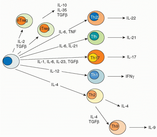
FIG. 29.1. Helper T Cell Fates. In response to the distinct cytokine milieux elicited by different microbial pathogens, naïve T cells become specified to produce selective cytokine repertoires. For instance, intracellular pathogens can induce production of interleukin (IL)-12, which acts via STAT4 to promote differentiation of Thelper (Th)1 cells that selectively produce interferon-&ggr; and not other cytokines. Likewise, helminthes induce IL-4 production, which acts via STAT6 to drive cells toward a Th2 phenotype characterized by IL-4 secretion. Fungi and extracellular bacteria elicit secretion of IL-23 and IL-6, which activate STAT3 and produce CD4 cells that produce IL-17, so-called Th17 cells. Each of these effector cells express transcription factors that enforce commitment to these distinctive phenotypes. These transcription factors can be referred to as “master regulators.” Other cluster of differentiation (CD)4 T cells reside in germinal centers and provide help to B cells. This subset of CD4 T cells is termed follicular helper cells. Another subset of CD4 T cells arises in the thymus and expresses the transcription factor Foxp3. These cells are termed natural regulatory T cells. Foxp3 can also be induced in cells in the periphery and these cells are termed induced Treg cells. Foxp3+ regulatory T cells are critical for constraining immune responses.

Next, T cells must migrate to infected tissues to exert their effect. This occurs by the loss of homing receptors for the secondary lymphoid organs, CD62L and CCR7, and acquisition of new homing molecules such PSGL-1, CD44, CCR5, and CXCR3.15 At this time, T cells transiently downregulate S1P1. Ligation of the TCR induces expression of the integrins, VLA-4 and VLA-5, which bind to fibronectin in extracellular matrices, Activated T cells also upregulate CD44, which binds to hyaluronan. Consequently, T cells that have encountered antigen are retained at the extravascular site where the offending antigen is present.


MANY FATES FOR ACTIVATED CLUSTER OF DIFFERENTATION 4 T CELLS

After initial activation, CD4 T cells can differentiate further such that they begin to selectively produce certain cytokines and not others. A major driver of the selective differentiation of CD4 T cells to populations of cells with distinctive cytokine production is the cytokine milieu generated by DCs and other innate cells. The resultant T cells tend to preserve their phenotype and with time maintain their distinctive phenotypes in a cell-autonomous manner. In this respect, the acquisition of the distinct fates can be thought of as a process of lineage commitment, although the extent to which activated T cells are terminally differentiated or retain a flexible repertoire is the topic of intense ongoing research.

Classically, T cells were viewed as having two major fates: Thelper1 (Th1) cells, which selectively produce interferon (IFN)-&ggr;, and Th2 cells, which produce IL-416,17,18,19,20 (Fig. 29.1 and Table 29.1). In addition to a stereotypic pattern of cytokine production, these lineages express a “master regulator”

transcription factor responsible for the distinctive pattern of gene expression.21,22 Recently, though, newer fates for helper T cells have been recognized, and they are designated based on expression of their eponymous, signature cytokines: Th17, Th22, and Th9 cells.23 These newly appreciated subsets can also express characteristic, lineage-defining transcription factors; however, as will be discussed, the expression of these master regulators is now recognized to be more promiscuous than previously appreciated. In addition to Th1, Th2, Th17, Th9, and Th22 cells subsets are T cells that reside in proximity to B cells in germinal centers, so-called follicular helper T (Tfh) cells. However, the distinction between Tfh cells and cytokine-secreting effector subsets is also a topic of intense investigation. Finally, there are various types of regulatory T cells. Each subset will be briefly discussed with respect to their characteristics features, inductive factors, key transcription factors, and negative regulators.


TABLE 29.1 Th Subsets


Subset


Inducing Cytokines


Sensors


Master Regulator Transcription Factor


Other Transcription Factors


Signature Cytokine


Other products


Inhibitors


Function


Host Defense Against:


Th1


IL-12, IFN-&ggr;


Also IL-18, IFN&agr;/&bgr;, IL-2, IL-27


STAT4, STAT1


T-bet


Hlx


Runx3


Ets family


Eomes


IFN-&ggr;


TNF


IL-12R&bgr;1


IL-12R&bgr;2, IL-18R, CXCR3, CCR5


IL-4


Activation of macrophages, enhanced antigen presentation, B-cell class switching to IgG1, IgG3(human), IgG2&agr;, IgG3 (mouse)


Intracellular microbial pathogens (eg, Mycobacteria, Listeria monocytogenes, Toxoplasmosis gondii, viruses


Th2


IL-4


Also IL-2, IL-25, IL-33, TSLP


STAT6, STAT5


GATA3


Maf


STAT3, Notch, IRF-4, Gfi-1


IL-4


IL-5, IL-13, IL-24, IL-31, CCR3, CCR4, CCR8, ECM1


IFN-&ggr; Mina


Activation and mobilization of mast cells, basophils, eosinophils,


Alternative macrophage activation, barrier function, mucus production,


B-cell class switching (IgE)


Helminths


Th17


IL-6, IL-23, TGF&bgr;, IL-1, TLR2


STAT3


Ror&ggr;t


Ahr, Batf, IKB&zgr;, IRF-4, Runx1, HIF1&agr;,


IL-17a, IL-17f


IL-21, IL-22, CCR6, CCL20


IL-2, IFN-&ggr;, IL-4, Foxp3, T-bet, Ets-1, Tcf-1


Induction of neutrophilia and recruitment of neutrophils, germinal center formation


Extracellular bacteria: Staphylococcus aureus, Klebsiella pneumoniae


Candida


Th22


IL-6, TNF


STAT3


?


Ahr, Rorgt, Notch


IL-22


CCR10, CCR6, CCR4


TGF&bgr;


Induction of defensins


Klebsiella pneumoniae


Th9


IL-4, TGF&bgr;


IL-2


IL-25


Pu.1, IRF-4


Mucus production


Tfh


IL-6, IL-21


STAT3


Also STAT4


Bcl6


PD-1, CXCR5, BTLA, PSGL-1, ICOS


IL-2


Blimp1


B-cell maturation, plasma cell differentiation, Ig class switching


Treg


IL-2, TGF&bgr;, retinoic acids


STAT5


Foxp3


Runx/CBF, Nrfa2, Foxo1, Foxo3, Eos, Helios


IL-10, TGF&bgr;


IL-35


IL-6


Roryt gamma, HIF1a


Maintenance of peripheral tolerance


Tr1


IL-27, IFN&agr;/&bgr;, ICOS


?


Ahr


IFN, interferon; Ig, immunoglobulin; IL, interleukin; TNF, tumor necrosis factor.


As indicated, activated CD4 T cells help eliminate pathogens by recruiting different types of effector cells (macrophages, neutrophils, eosinophils, NK cells, B cells, and CD8 T cells). However, activated, differentiated CD4 T cells are themselves referred to as effector cells. While this terminology may be confusing, it refers to the ability of activated CD4 T cells to rapidly produce maximal levels of their signature cytokines. The limitation of the terminology is that it does not refer to an exact, precisely defined step in maturation, but is rather used largely to distinguish them from naïve and “memory” cells. Unlike CD8 T cells, CD4 T cells are much more heterogeneous; not surprisingly, CD4 memory T cells are also more heterogeneous and our understanding of the transition of CD4 effector cells to memory cells is poorly understood. Memory CD4 cells can be broadly classified as effector memory (Tem) and central memory (Tcm).24,25,26 Tcm express CCR7 and recirculate through lymphoid organs. Tem do not express CCR7 and home to nonlymphoid tissues. Tem rapidly produce cytokines following secondary stimulation, but have limited proliferative capacity. Tcm, by contrast, produce IL-2 and are able to proliferate.


Th1 Cells

Th1 cells are defined by the selective secretion of IFN-&ggr;. IFN-&ggr; activates macrophages to kill phagocytosed microbes. It enhances antigen presentation, by upregulating MHC and costimulatory molecules, amplifying T-cell-dependent immune responses. IFN-&ggr; also further promotes the differentiation of CD4+ T cells to the TH1 subset and activates NK and CD8 T cells, thereby further enhancing production of IFN-&ggr; and inhibiting TH2 and TH17 cell differentiation. IFN-&ggr; promotes B-cell class switching to immunoglobulin (Ig)G1 and IgG3 (IgG2a or IgG3 in mice) and inhibits switching to IL-4-dependent isotypes (eg, IgE). In this manner, Th1 cells effectively protect the host against intracellular pathogens including: Mycobacteria, Toxoplasma, Listeria, and viruses.

In addition to expressing their signature cytokine IFN-&ggr;, TH1 cells also express high levels of IL-12 receptor &bgr;1 and &bgr;2 subunits, IL-18 receptor, E-selectin, P-selectin, CXCR3, and CCR5.

TH1 differentiation is stimulated by intracellular bacteria, viruses, and adjuvants that elicit production of IL-12, IL-18, and type I IFN. IL-12 and to some extent type I IFNs activate the transcription factor STAT4, which has thousands of targets in developing Th1 cells, including the genes encoding IFN-&ggr;, and IL-12 and IL-18 receptors.27 IFN-&ggr;, acting via STAT1, reinforces Th1 differentiation. STAT4 and STAT1 induce expression of T-bet (encoded by the Tbx21 gene), which has been termed the master regulator of Th1 cells.28,29,30,31 Conversely, though, the transcriptional repressor Twist1 is also target of STAT4. Twist1 limits the expression of cytokines including IFN-&ggr;, IL-2, and TNF.32 Similarly, the protease Furin, which processes the anti-inflammatory cytokine transforming growth factor (TGF)-&bgr;, is also regulated by STAT4.27,33 Thus, STAT4 simultaneously promotes and constrains T-cell activation and thereby limits Th1-mediated immunopathology.

T-bet regulates thousands of genes in Th1 cells.34,35 It binds to the Ifng gene and promotes its expression.36,37,38 T-bet also directly binds and inhibits expression of the Socs1, Socs3, and Tcf7 (which encodes TCF-1) genes. Interestingly, it can also associate with the repressor Bcl6 and inhibit IFN-&ggr; expression. T-bet also inhibits expression of Ror&ggr;t, the master regulator of Th17 cells, by blocking the action of Runx1.39 T-bet inhibits Il4 gene expression in a Runx3-dependent manner.36 T-bet is phosphorylated by Itk, and phosphorylated T-bet binds and inhibits the action of GATA3, the master regulator of Th2 cells.40 T-bet is also important for T-cell trafficking and promotes the differentiation of T cells to an effector phenotype.41 As will be discussed, T-bet antagonizes the Tfh characteristics.42,43

It is important to bear in mind that Th1 differentiation can occur in the absence of either T-bet or STAT4, indicating that there are alternative means of regulating IFN-&ggr;. In CD8 cells, a related T-box family member, Eomes, is the major regulator of IFN-&ggr; production. In addition, other transcription factors are preferentially expressed in Th1 cells including Hlx, Runx3, and Ets family members; they also promote IFN-&ggr; production and repress IL-4 transcription. Another factor that influences Th1 specification is the “strength” of TCR signaling, with strong signaling favoring TCR signaling. The mechanisms underlying this are relatively poorly characterized. A downstream signaling molecule engaged following TCR ligation and cytokine receptor occupancy is mammalian target of rapamycin (mTor). Deficiency of mTor inhibits effector cell differentiation including generation of Th1 cells.44 Its regulators include Tor complex (Torc)1 and Torc2. Inhibition of Torc1 signaling inhibits Th1 and Th17 differentiation.45,46,47


Th2 Cells

The signature cytokines for Th2 cells are IL-4, IL-13, and IL-5, which are important for barrier defense at mucosal and epithelial surfaces. IL-4 stimulates the development of TH2 cells and functions as an autocrine growth factor for differentiated TH2 cells. It also stimulates B-cell immunoglobulin class switching to IgG4 in humans (IgG1 in mice) and IgE in both species. Conversely, IL-4 inhibits switching to the IgG2a
and IgG3 isotypes in mice, both of which are stimulated by IFN-&ggr;. IL-4, together with IL-13, promotes the formation of “alternatively activated” macrophages, suppresses IFN-&ggr;-mediated classical macrophage activation and thus inhibits defense against intracellular microbes. IL-4, IL-13, and IL-5 mobilize eosinophils, basophils, and mast cells, and IL-13 increases mucus secretion from airway and gut epithelial cells. These actions contribute to elimination of microbes at epithelial surfaces. In this manner, Th2 cells are important for host defense against helminths and other parasites, but are also the major drivers of allergy and asthma.

TH2 cells also produce IL-24 and IL-31, and express the chemokine receptors CCR3, CCR4, and CCR8.48,49 Th2 cells also expression extracellular matrix protein-1, which regulates cell migration and expression of S1P1.50

For Th2 cells, a major driver of differentiation is IL-4 itself. IL-4 can be produced by an array of innate cells including: NK T cells, mast cells, and basophils.51,52,53,54,55 More recently recognized are innate cells designated natural helper cells that reside in fat associated lymphoid clusters.56,57,58 IL-25, an IL-17-related cytokine, IL-33, an IL-1-related cytokine, and thymic stromal lymphopoietin all promote Th2 responses, principally by driving IL-4 production in innate cells.51,57,59,60,61,62

IL-4 stimulates TH2 development by activating STAT6, which together with TCR-dependent signals, induces expression of the Th2 master regulator, GATA-3. Like STAT4, STAT6 also regulates thousands of other genes in Th2 cells including pivotal transcription factors, cytokines and cytokine receptors, and chemokines and chemokine receptors.27,63,64,65 IL-2 also contributes to Th2 differentiation by activating STAT5, which positively regulates expression of both IL-4 and the IL-4 receptor.66,67,68,69 STAT5’s targets in Th2 cells include both the Il4 and the Il4r gene.70 Additionally, STAT3 has been reported to be a contributor to Th2 differentiation.71

Gata-3 is a critical factor for lineage commitment of Th2 cells, although it also plays essential roles in thymic differentiation. Gata-3 binds to many key Th2 genes including the Il4, Il5, Il13, Gata3, and other genes. 72 GATA3 also actively represses RUNX3, which in turn leads to reduced IFN-&ggr; production.73 GATA3 also induces the transcription factor c-Maf, which can aid in Th2 differentiation; however, c-Maf deficiency does not abrogate production of Th2 cytokines.74 GATA3 also has important roles in thymic development, regulating key factors such as Th-POK, Notch, and TCR subunits.72

The transcription factors Notch, IRF-4, and growth factor independent (Gfi)-1 also aid in Th2 differentiation. Notch has been reported to promote Gata3 expression and to do so in a Stat6-independent manner.75 IRF-4 is a contributor to Th2 differentiation, but it is also important for other subsets.76 Gfi is induced by IL-4 and regulates Th2-cell proliferation.77,78,79 Whereas “strong” TCR signals favor Th1 differentiation, weak TCR signaling favors Th2 differentiation; again, the signaling intermediates responsible remain unclear. In addition, deletion of Torc2 inhibits Th2 differentiation.45

Finally, Mina, a member of the jumonji C (JmjC) protein family, binds and represses the Il4 promoter. Conversely, reducing levels of Mina in T cells leads to Il4 de-repression.80 Eomesodermin (Eomes), a key regulator of IFN-&ggr;production in CD8 T cells, is important for limiting IL-5 production in Th2 memory cells.48 Eomes acts by limiting GATA3 binding to the Il5 promoter.


Th17 Cells

The appreciation that the Th1/Th2 paradigm failed to explain a good deal about immunity and autoimmunity,81,82,83 and the recognition of the existence of a subset of T cells that preferentially produces IL-17 led to a small renaissance in CD4 T-cell biology. This newly appreciated subset, termed Th17 cells, was noted to produce IL-17A, IL-17F, IL-21, and IL-22.84,85,86,87,88,89,90,91,92

IL-17 enhances neutrophil generation by increasing granulocyte macrophage colony stimulating factor production and promotes neutrophil recruitment by production of chemokines.93,94,95 Because neutrophils are a major defense mechanism against extracellular bacteria and fungi, TH17 cells play an especially important role in defense in such infections including Staphylococcus aureus, Klebsiella pneumoniae, and Candida albicans.49,96 The IL-23/IL-17 axis also contributes to host defense against Mycobacteria tuberculosis.97 Additionally, IL-17 stimulates the production of antimicrobial substances, including defensins. IL-17 also contributes to germinal center formation, as well as lymphoid structures induced during inflammation.98,99 As will be discussed, Th17 cells are also very important in the pathogenesis of a variety of autoimmune and immunemediated diseases.

Th17 cells typically express the chemokine receptor CCR6, along with its ligand, CCL20.100 Cytokines that promote Th17 differentiation include IL-6, IL-23, IL-1, and TGF-&bgr;.91,101,102,103,104,105,106,107,108,109,110,111,112 IL-6 and IL-23 activate Stat3, which directly regulates many genes that contribute to the phenotype of Th17 cells including Il17a/f, Il23r, and Il21.113 Stat3 also regulates Rorc, which encodes Ror&ggr;t, the master regulator for Th17 cells and other transcription factors expressed by Th17 cells.114,115 In the mouse, absence of Stat3 in T cells blocks Th17 differentiation.116,117 Th17 cells are abundant in the gut and are regulated by intestinal flora, but the precise mechanisms responsible for driving IL-17 production have not been elucidated.114

Ror&ggr; is an orphan retinoid receptor that is ubiquitously expressed.118 One splice variant, Ror&ggr;t, is selectively expressed in hematopoietic cells. In addition to serving as “master regulator” of a helper-cell subset, like GATA3, Ror&ggr;t has important functions in thymic development, in the development of lymphoid tissue inducer cells, and in development of IL-22-producing NK cells.119,120,121,122 In addition to Ror&ggr;t, Ror&agr; is also a contributor to Th17 differentiation.123

Other transcription factors expressed by Th17 include the aryl hydrocarbon receptor, Batf, I kappaB zeta, IRF-4, and Runx1; absence of any of these impairs IL-17 expression.39,124,125,126,127,128,129 The key metabolic sensor hypoxia-inducible factor (HIF)-1130 is also a positive regulator of Th17 differentiation. HIF-1 associates with Ror&ggr;t and recruits the acetyl transferase p300 to the Il17 gene.130,131 TLR2
signaling also promotes Th17 differentiation.132 As indicated previously, mTor signaling via mTorc1 also promotes Th17 differentiation.45

IL-2 acting via STAT5 is a potent negative regulator of IL-17.133 Regulatory T (Treg) cells can promote Th17 differentiation, and they appear to do so by limiting IL-2.134,135 IL-2 activates STAT5, which binds to the Il17a/flocus and displaces STAT3.136 IL-4 and IFN-&ggr; also inhibit Th17 differentiation.84

Foxp3 negatively regulates Th17 differentiation and does so by directly binding Ror&ggr;t and inhibiting its function.137 The transcription factors Gfi-1, Ets-1, T-bet, and Tcf-1 also inhibit Th17 cell differentiation.39,138,139,140,141


Th22 Cells

IL-22 is an IL-10-related cytokine located in proximity to the IFNG gene, but regulated in a distinctive manner.142 It acts on epithelial barrier cells, having both pro- and anti-inflammatory effects.143,144,145 It is important for host defense against Klebsiella pneumonia.49 Although IL-22 is produced by Th17 cells, some cells produce IL-22 and not IL-17.146,147,148,149 This is especially prominent in skin-homing cells that express the chemokine receptors CCR10, CCR6, and CCR4.150,151 IL-22 is also produced by mucosal innate immune cells including lymphoid tissue inducer and so-called NK-22 cells.59,119,152,153,154,155,156

Production of IL-6 and TNF by plasmacytoid DCs promotes IL-22 production without inducing IL-17 and IFN-&ggr;. Unlike other Th subsets, no clear master regulator that directs this phenotype has been identified, but the aryl hydrocarbon receptor157 and Ror&ggr;t are important for IL-22 production.125,158 Notch drives IL-22 production by enhancing Ahr expression.159 Conversely, Tgf&bgr; inhibits IL-22; it does so by inducing Maf, which directly represses IL-22.160


Th9 Cells

Adding TGF-&bgr; to cultures of T cells incubated with IL-4 generates a population of cells that produce IL-9 but not IL-4. Such cells have been referred to as Th9 cells.161,162,163,164 IL-2 promotes IL-9 production.165 In addition, Th9 cells express the receptor for IL-25 (IL-17RB) and IL-25 promotes IL-9 production.166 Innate cells are major sources of IL-9.167

IL-9 acts on mucosal cells to enhance mucus production.168,169 IL-9 has also been reported to have variable effects on Th17 differentiation and Th17-dependent pathology.170,171 The transcription factors PU.1 and IRF-4 are required for development of IL-9-producing T cells and allergic inflammation.164,172,173


Follicular Helper T Cells

For the subsets of CD4 T cells discussed, activation and instruction by cytokines is associated with the acquisition of particular chemokine receptors that promote exit from lymph nodes and recruitment to sites of inflammation. However, some antigen-experienced CD4 T cells, called follicular helper T cells (Tfh), stay in the lymph nodes and provide “help” to B cells.174,175,176 The signature cytokine for Tfh cells is IL-21. However, this cytokine is not exclusively produced by Tfh cells; it is also produced by other subsets including Th1 and TH

Only gold members can continue reading. Log In or Register to continue

Stay updated, free articles. Join our Telegram channel

May 30, 2016 | Posted by in IMMUNOLOGY | Comments Off on Helper T-Cell Differentiation and Plasticity

Full access? Get Clinical Tree
Get Clinical Tree app for offline access