Healthcare-Associated Infections in Transplant Recipients


Healthcare-Associated Infections in Transplant Recipients


Mini Kamboj

Kent Sepkowitz


Clinical transplantation has become a life-saving treatment option for many chronic organ diseases and advanced hematologic malignancies. Progress in surgical techniques and transplant modalities, novel immunosuppressive agents, improved graft survival, and posttransplant care all have led to an improvement in survival among solid organ transplant (SOT) and hematopoietic stem cell transplant (HSCT) recipients.

However, despite these advances, healthcare-associated infections (HAIs) and opportunistic infections remain a major cause of illness and death. In general, transplant predisposes patients to two types of infection for different reasons. Opportunistic infections exploit the immune defects imposed by transplant. In contrast, HAIs occur because of prolonged hospitalizations for the procedure (e.g., many allogeneic HSCT have lengths of stay >30 days) and abnormal anatomy after surgical placement of a new organ. In addition, the two factors interact, with the immunosuppression further increasing the risk for HAIs. This chapter will focus on the immunologic and anatomic risks for HAIs in both SOT and HSCT recipients.


HISTORICAL PERSPECTIVE AND CURRENT TRENDS


SOLID ORGAN TRANSPLANT

Although successful SOTs were performed early on, including kidney (1954) and liver (1967), it was not until the approval of Cyclosporine (“Sandimmune”) in 1983 that SOT was adopted across the United States as a reliable treatment for end-stage organ disease. The 1-year graft survival improved dramatically with the pharmacologic immunosuppression provided by cyclosporine; however, the long-term survival remained dismal (1,2,3,4). Tacrolimus (FK-506), introduced in the early 1990s, proved superior in potency and safety and has become the standard immunosuppressive agent used for SOT recipients (5,6,7,8,9,10).

The number of individuals undergoing SOT in the United States for liver and kidney has been slowly rising over the last decade. However, the number of people on the waiting lists has increased faster than the number of transplants performed. The maximum increase in the waiting list of individuals for kidney transplant has grown 68.5% in the last decade, followed by liver transplant with an increase of 28.4% (Figure 45.1A, B).

The most recent trends show 5-year graft survival of 70% to 82.5% for kidney transplants, 67.5% for liver, and 73.1% for heart transplants. Pancreatic and intestinal transplants are performed less often and have a 5-year graft survival of 48.3% and 50.6%, respectively (11) (Figure 45.1A, B).


HEMATOPOIETIC STEM CELL TRANSPLANT

The most widely used forms of hematopoietic stem cell therapy are autologous and allogeneic transplants.


  • Autologous transplant involves first harvesting peripheral blood stem cells from the patient before treatment with high-dose chemotherapy and/or radiotherapy, then infusion of the cells after treatment to reconstitute the marrow. The most common hematologic malignancies treated with autotransplant are multiple myeloma and lymphoma.


  • Allogeneic transplant is performed for hematologic malignancies when the marrow itself is diseased—stem cells are obtained from peripheral blood, marrow, or cord blood from a related or unrelated donor with matching HLA type. Manipulation of the stem cell product may be done to reduce the risk of graft vs. host disease (GVHD), such as CD34 selection or T-cell depletion. The most common indications for allogeneic transplants include acute leukemia, myelodysplastic syndrome, aplastic anemia, or congenital immunodeficiency disorders.

The first successful allogeneic HSCT was performed by Nobel Laureate E. Donnall Thomas in 1969 about a decade after having performed a syngeneic HSCT (where the stem cell source is identical twin). In the late 70s, HSCT was expanded to include autotransplant for treatment of chemo-and radiosensitive high-risk solid tumors and lymphomas. The National Marrow Donor Program (NMDP) was established in 1986 to increase the pool of marrow donors and to facilitate identification of an appropriate match. The last two decades have seen marked expansion of HSCT techniques and practices. The most notable changes include:


  • Newer indications, including autoimmune disorders, such as lupus and rheumatoid arthritis, and sickle cell anemia.


  • An expanded armamentarium of immunosuppressive agents including novel immunotherapy agents (e.g., rituximab and ofatumumab—chimeric and human monoclonal antibody against CD20; alemtuzumab—monoclonal antibody against CD52; infliximab—monoclonal antibody against TNF-α; daclizumab—monoclonal antibody to interleukin-2 (IL-2) receptor; and so on).


  • More accurate HLA typing.


  • Discovery of the role of natural killer (NK) cell typing (12).


  • Use of peripheral blood and umbilical cord blood as stem cell sources. The HLA matching criteria for cord blood transplant (CBT) are less stringent (13,14), making CBT
    a critical treatment option for patients without suitable donors.


  • Appreciation of the benefits of graft-vs.-leukemia. This has led to reduced intensity conditioning (RIC) regimens, sometimes referred to as “nonmyeloablative” or “mini” transplant. RIC can be given to an older population as well as to outpatients (15,16,17,18,19). RIC regimens are now used in 40% of allogeneic HSCTs performed.


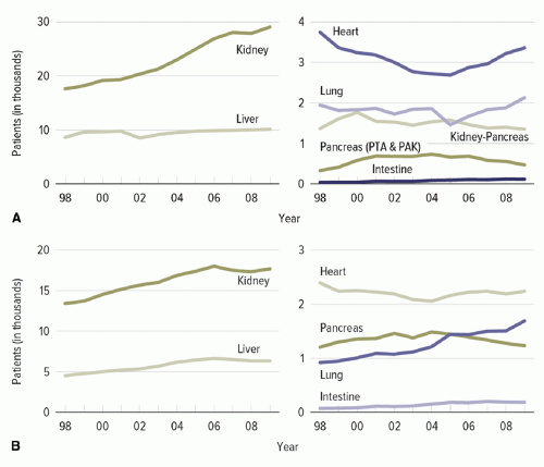
Figure 45.1. (A) Patients added to the waiting list during the year. (B) Transplants performed during the year. The trends in solid organ transplant in the last decade (1998 to 2008) by organ type. With permission from Organ Procurement and Transplantation Network (OPTN) and Scientific Registry of Transplant Recipients (SRTR). OPTN / SRTR 2010 Annual Data Report. Rockville, MD: Department of Health and Human Services, Health Resources and Services Administration, Healthcare Systems Bureau, Division of Transplantation; 2011. http://www.srtr.org/annual_reports/2010/. Accessed October 8, 2012.

In 2009, 7,012 allogeneic transplants (including 1,815 pediatric transplants) and 9,778 autotransplants were performed in the United States. The numbers of allogeneic transplants from unrelated donors have almost doubled in the last 10 years (20).

One-year survival among adults (<50 years) after myeloablative transplant from matched sibling donor is close to 70% and from unrelated donor 60%. Infections are among the major causes of nonrelapse mortality in this population (Figure 45.2A, B) (20).


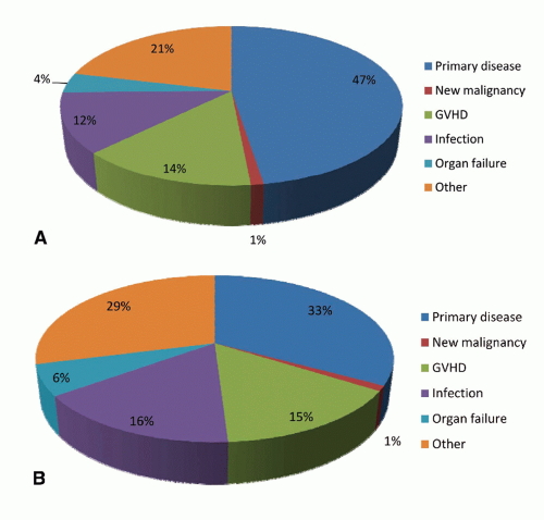
Figure 45.2. The causes of death after allogeneic HSCT performed in the years 2008 (A) and 2009 (B). Adapted from Pasquini MC, Wang Z. Current use and outcome of hematopoietic stem cell transplantation: CIBMTR summary slides, 2011. http://www.cibmtr.org, with permission.


PART I: Risk of Infection Including Healthcare-Acquired Infection in the Transplant Recipient

The risk of infection in the transplant patient is largely determined by the interaction among three factors:


  • The presence of anatomic abnormalities related to surgery or disruption of the mucocutaneous barrier by indwelling devices.


  • Environmental factors, including exposure to organisms in the community or the hospital.


  • The patient’s net state of immunosuppression.


ANATOMIC/TECHNICAL ABNORMALITIES IN THE PATHOGENESIS OF INFECTION

The anatomic/technical factors can be divided into two categories:


  • Those related to surgery itself, such as biliary abnormalities in liver transplant and ureterovesical reflux after renal transplant. Additional examples include complications at the time of the transplant that result in the creation of devitalized tissue, fluid collections, and/or ongoing urine or bile leaks, and posttransplant complications, such as organ ischemia. Unless such abnormalities are promptly eliminated, secondary infection is inevitable (21).


  • Perioperative breaches of mucocutaneous surfaces by vascular access devices, endotracheal tubes, drainage devices, or urinary catheters are associated with secondary infection. Removal of these devices is indicated as early as possible. The incidence of device-related infection is related to the nature of the transplant (small bowel = liver > lung = pancreas > heart > kidney), the complexity of the surgery, and the duration of time that “devices” compromise the integrity of the skin. In addition, hematomas are iron-rich and therefore may promote the growth of Listeria, the Zygomycetes, and other microbes (22,23,24).


ENVIRONMENTAL FACTORS

Although community-acquired infections, such as influenza, are important for transplant patients, the more important exposures are those that occur in the hospital where three important patterns—domiciliary, nondomiciliary, and person-to-person spread—can be demonstrated. The term domiciliary is used to describe acquisition of organisms from the potable water, healthcare workers’ hands, high-touch surfaces in the patient’s immediate environment, and contaminated air, which can occur on the transplant ward or in the patient’s room. Outbreaks in which infection is acquired in a domiciliary mode usually are characterized by clustering of cases in time and space and are therefore relatively easy to recognize. Domiciliary outbreaks of Pseudomonas aeruginosa (and other gram-negative organisms) (25,26,27), vancomycinresistant enterococcus (VRE) (28,29), Legionella (30,31,32), Aspergillus (33,34,35), or mucormycosis (36) infection are well-documented.

Nondomiciliary infection represents nonward-related common-source outbreak, which is acquired when the patient travels within the healthcare facility for an essential procedure and is exposed to excessive levels of potential opportunistic pathogens present in the air. These infections usually are associated with construction and/or areas of moisture or vegetation that favor the growth of such molds as Aspergillus spp., Fusarium spp., or Scedosporium spp. (37,38). Thus, invasive infection due to molds, particularly Aspergillus spp., has been well-documented in transplant patients who have been sent to radiology or endoscopy suites, in holding areas outside cardiac catheterization laboratories, the operating rooms, and a hospital area that is undergoing renovation (39,40,41). Owing to lack of clustering of cases in space, nondomiciliary outbreaks can take longer to detect. The best clue to the presence of an environmental hazard is the occurrence of infection due to one of these opportunistic organisms when the net state of immunosuppression is not, under typical circumstances, great enough to allow such an infection to occur unless an environmental hazard is present (22,42). An example would be the development of invasive pulmonary aspergillosis in a renal transplant patient in the absence of antirejection therapy.

Person-to-person spread of pathogens constitutes the third most common pattern of spread. Transmission may occur via the airborne/droplet, bloodborne, or fecal-oral route. Infections commonly spread in hospitals through the airborne/droplet route include community respiratory virus, varicellazoster virus (VZV), and Mycobacterium tuberculosis. These pathogens may cause rapid spread with occurrence of secondary cases within days for respiratory viruses and after months for M. tuberculosis exposure (43,44,45). Such outbreaks usually occur when there is breach in infection control practices or a delay in recognizing source-case and implementation of appropriate infection control precautions.

Transplant patients with respiratory viruses have a higher risk of complications including lower airway disease, secondary infections, and long-term decline in pulmonary function. Experience with the severe acute respiratory syndrome (SARS), 2009 H1N1 influenza pandemic, and other viruses confirm that the impact of such infections is greater in transplant recipients than that seen in the general population (46,47).

The majority of infections that occur early after transplant (within the first month) are attributed to anatomic or epidemiologic factors, whereas infections related to immunosuppression typically occur later. Constant surveillance of infections among transplant patients is essential, especially for pathogens based on local epidemiology as well as outbreaks in the community.


THE NET STATE OF IMMUNOSUPPRESSION

The net state of immunosuppression is a complex function determined by the interaction of a number of factors.


SOLID ORGAN TRANSPLANT

Numerous factors affect the net degree of immunosuppression. First, underlying disease processes, such as diabetes or systemic lupus erythematosus, may themselves compromise the inflammatory response. Second, Immunosuppressive medications are given routinely to control several conditions that may
come to transplant such as chronic hepatitis, biliary cirrhosis, or inflammatory lung disease. The intensity and duration of such immunosuppressive therapy administered has a direct impact on the net state of immune compromise. Third, active infection with one or more immunomodulating viruses (e.g., cytomegalovirus [CMV], Epstein—Barr virus [EBV], human herpesvirus-6, hepatitis viruses B [HBV] or C [HCV], or HIV) may add to the net state of suppression. Finally, conditions common to the immunocompromised host such as damage to the mucocutaneous surfaces of the body, neutropenia, or metabolic abnormalities such as protein—calorie malnutrition, uremia, or hyperglycemia all may additionally compromise immune function.

These numerous potential contributors to the net state of immunosuppression may be dwarfed by the potent immunosuppression given to prevent graft rejection. The standard of care for SOT recipients is a multidrug regimen consisting of a calcineurin inhibitor (cyclosporine or tacrolimus), prednisone, and either azathioprine or mycophenolate. In addition, antithymocyte globulin (ATG; a polyclonal anti—T cell drug) or OKT3 (a monoclonal anti—T cell drug) can be used as induction therapy, especially in kidney and pancreas transplantation (Table 45.1).

The calcineurin inhibitors are the cornerstones of modern antirejection therapy. They exert their effects through a complex signaling pathway that results in the inhibition of the transcriptional activation of genes required for T-cell activation, proliferation, and function. This results in the inhibition of a large number of proinflammatory cytokines, with the most important of these effects being the blockade of essential functions of IL-2. The infectious disease consequences of these drugs are direct results of these mechanisms: a dose-related inhibition of microbial-specific T-cell cytotoxic activity, thus promoting numerous infections, including those caused by the herpes group viruses, various fungi, mycobacteria, and the many intracellular infections. The key toxicities of the calcineurin inhibitors are injury to the kidneys and hypertension (48,49).


TABLE 45.1 Induction and Maintenance Immunosuppressive Agents Used in Solid Organ Transplant


Organ


Induction Agents


Initial Immunosuppression


Regimen at 1 Year


Comments


Kidney


T-cell depleting antibody (58%)


IL2-RA (21.2%)


IL2-RA and T-cell depleting antibody (3.6%)


Tacrolimus and mycophenolate (81%)


Cyclosporine (5.7%)


mTOR inhibitors (3%)


Tacrolimus and mycophenolate (72.1%)


Cyclosporine and mycophenolate (5.3%)


mTOR inhibitors (6.5%)


Rising use of T-cell depleting antibody for induction


Liver


IL2-RA (14.3%)


T-cell depleting antibody (10.3%)


Tacrolimus and mycophenolate (85.8%)


mTOR inhibitors (2.5%)


Tacrolimus and mycophenolate (39%)


Tacrolimus (34%)


Relatively low use of T-cell depleting regimen


Lung


IL2-RA (40%)


T-cell depleting regimen (∽18%)


Tacrolimus and mycophenolate (52%)


Tacrolimus and azathioprine (23%)


Tacrolimus (8%)


Tacrolimus and mycophenolate (46%)


Tacrolimus and azathioprine (16%)


Tacrolimus (15%)


Intestine


T-cell depleting agents (51.7%)


IL2-RA (3.3%)


IL2-RA and T-cell depleting antibody (1.7%)


Tacrolimus (58.9%)


Tacrolimus and mycophenolate (32.8%)


Tacrolimus (74.6%)


Tacrolimus and mycophenolate (13%)


Heart IL2-RA (26.9%)


T-cell depleting regimen (22.6%)


Tacrolimus and mycophenolate (70%)


Cyclosporine and mycophenolate (15%)


Tacrolimus and mycophenolate (55.6%)


Cyclosporine and mycophenolate (16.2%)


Pancreas


T-cell depleting regimen (71.4%)


IL2-RA (10%)


Tacrolimus and mycophenolate (>80%)


Tacrolimus and mycophenolate (65%)


Higher rejection rates and more intense immunosuppressive regimens


HEMATOPOIETIC STEM CELL TRANSPLANT

The factors similar to those outlined for SOT affect the risk of infection for HSCT. The nature and intensity of the immunosuppressive regimen used before transplant as treatment of the underlying cancer is a major determinant of infections occurring early on. Examples include the use of agents such as purine analogs, alemtuzumab, pentostatin, and rituximab, which are associated with immunosuppressive effects lasting from months to years.

However, additional factors unique to the HSCT population add considerably to the net state of immunosuppression. Additional immunosuppression will occur at the time of conditioning when the patient receives total body irradiation or ATG. Furthermore, the stem cell source (e.g., cord blood grafts can take longer to engraft, increasing the infection risk in the early posttransplant period), graft manipulation such as T-cell depletion (reduces passively transferred T-cell immunity, especially against viruses, and impairs T-cell reconstitution after transplant), and donor matching (determines the risk of GVHD) influence the risk for infection. After transplant, the
occurrence of GVHD and its treatment with pharmacologic immunosuppression, as well as the immunomodulating infections listed above determine the pace of immune reconstitution and susceptibility to infection.

The course of allogeneic HSCT is shown in Figure 45.3 and consists of conditioning therapy (myeloablative or nonmyeloablative) followed by infusion of stem cells, which are mostly derived from peripheral or cord blood. The graft may be manipulated for CD34 selection or T-cell depletion. Engraftment usually occurs between 2 and 3 weeks, depending upon the transplant type and source of stem cells. The time to engraftment or marrow reconstitution is typically longer for CBT as cord blood contains fewer nucleated cells despite the use of double cord units (DUCBT). Furthermore, these cells necessarily are immunologically naive, further delaying immune recovery. In some studies, the prolonged period of neutropenia and lymphopenia after CBT has been associated with a higher risk of infectious complications in the first 100 days after CBT without any substantial impact on nonrelapse mortality (50,51,52,53,54,55).

T-cell depletion reduces the risk of GVHD by eliminating donor T cells, but increases the risk of PTLD (posttransplant lymphoproliferative disorder) and infection, especially invasive fungal infections and severe viral diseases (56,57,58). Most techniques used for T-cell depletion are ex vivo by physical separation or immunologic manipulation of the graft with the use of monoclonal antibodies (anti-CD2, anti-CD3, anti-CD5, anti-CD52, anti-CD25, and anti-CD8). In vivo T-cell depletion is attained with the use of antilymphocyte antibodies (ATG or alemtuzumab) in conditioning regimens.

T-cell depletion is a strategy used to reduce the incidence of GVHD after transplant from HLA nonidentical donors. Prophylaxis for GVHD is routinely used in all other forms of allogeneic transplant. Although graft vs. leukemia (GVL) effect would diminish with the routine use of prophylaxis, the use of posttransplant donor lymphocyte infusions (DLI) allows achievement of the desired immunologic state despite anti-GVHD prophylaxis. Cyclosporine and methotrexate (with or without corticosteroids) or tacrolimus with methotrexate is the most commonly used regimen for prevention of acute GVHD (59,60). Alternate prophylactic agents, in the event of significant toxicity (especially nephrotoxicity with cyclosporine and tacrolimus), include sirolimus and mycophenolate. In the absence of GVHD, prophylaxis is generally continued for 6 months for identical donors and longer in mismatched transplants.


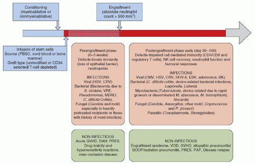
Figure 45.3. The time course of allogeneic stem cell transplant and associated infectious and non-infectious complications.


Corticosteroids (methylprednisolone) are the mainstay for the management of acute GVHD, and more intense immunosuppressive regimens (e.g., cyclosporine, tacrolimus, infliximab, daclizumab, alemtuzumab) are reserved for steroid refractory patients. The mechanism of action of drugs used in the prevention and management of acute GVHD, common adverse effects, and infectious complications are discussed below.


EFFECTS OF PHARMACOLOGIC IMMUNOSUPPRESSION ON THE OCCURRENCE OF INFECTION

The major determinants of the net state of immunosuppression are the dose, duration, and temporal sequence in which the immunosuppressive regimen is administered. A brief description of some of the key agents and novel immunomodulating drugs used in SOT and HSCT that are particularly associated with an increased risk of HAI and other serious infections is provided here. Because of these agents, the range of HAIs in this population is distinct from those found on a general ward, so approaches to surveillance should be tailored accordingly. In addition, they may predispose to inflammatory disorders, such as interstitial pneumonitis that may appear to be an HAI, such as ventilator-associated pneumonia (VAP), thereby complicating surveillance.


Purine Analogs

Purine analogs are structurally similar to purine metabolites and inhibit DNA synthesis and repair. The most widely used agents in this class include fludarabine, pentostatin (ADA inhibitor; ADA plays a role in T-cell and B-cell differentiation), and cladribine (ADA-resistant nucleoside analog) (61,62). These agents are mostly used in the treatment of lymphoid malignancies. In addition, fludarabine commonly is used in high doses along with busulfan in preparative conditioning regimen for myeloablative HSCT and in lower doses, in combination with busulfan/melphalan with or without low-dose radiation, for nonmyeloablative preparative regimens. Purine analogs induce profound neutropenia, CD4 lymphopenia (lasting ≥1 year for fludarabine and 2 to 4 years after cladribine and pentostatin) (63), as well as B-cell and monocyte dysfunction. As a result, they increase host susceptibility to a wide range of pathogens including bacterial (encapsulated bacteria, Listeria spp., Legionella spp., M. tuberculosis, nontuberculous mycobacteria, Nocardia spp.), viral (CMV, herpes simplex virus [HSV], VZV), Pneumocystis jiroveci pneumonia (PCP), and Cryptococcus spp. Because infection has been reported months after the use of these agents, prophylaxis for PCP is routinely recommended (64,65,66,67).


Alemtuzumab

Alemtuzumab (Campath) is a humanized monoclonal antibody targeting CD52 (glycoprotein expressed on B-and T lymphocytes, monocytes, and NK cells). Profound depletion of all lymphocyte subsets is observed after alemtuzumab administration and CD4 counts may remain suppressed for up to 2 years (median, 9 months) (68,69,70). It has been used in induction regimen for SOT to prevent acute rejection as well for the treatment of steroid refractory acute rejection (71,72,73,74,75,76,77,78,79,80,81). In HSCT, alemtuzumab has been used in conditioning regimen in allogeneic transplants, especially RIC, to promote engraftment and reduce the risk of GVHD (82,83,84,85,86). Alemtuzumab renders the host susceptible to a wide range of pathogens owing to the associated immune defects (87,88,89,90,91,92,93,94,95,96,97,98,99,100,101,102).

In one small study of 27 patients receiving treatment with alemtuzumab for lymphoproliferative diseases, Martin et al. (98) reported opportunistic infections (OIs) in 56% of patients (median time to OI after the alemtuzumab dose was 165 days) and non-OIs occurred in 22/27 (82%) patients. Non-OIs included community respiratory virus infections in 22 patients, device-related infections, bacteremia, or other serious bacterial infections including those due to multidrug-resistant organisms (MDROs) in eight patients each. These findings highlight the impact of immunosuppressive regimen on occurrence and outcome of common HAIs and heighten the need for surveillance and prevention in this population.


Antithymocyte Globulin

ATG is a polyclonal immunoglobulin—horse-or rabbit-derived antibodies against human T cells. ATG is used in conditioning regimens for HSCT to prevent GVHD and as an induction agent in SOT for the prevention of acute graft rejection (103). The spectrum of infection seen after ATG is similar to alemtuzumab. The use of ATG increases the risk of EBV-PTLD, with the highest risk in conditioning regimens that use ex vivo T-cell depletion along with ATG (71%) (104,105,106,107). An important complication associated with ATG infusion is the cytokine release “storm” manifesting as high fevers, rigors, and chills, and is often managed by the administration of corticosteroids.


Anti-Interleukin-2Rα Antibodies

The newer agents daclizumab and basiliximab (IL-2Rα receptor antibodies) do not cause cytokine storm and are used in combination with ATG for the prophylaxis of allograft rejection after SOT (mostly renal) and treatment of steroid refractory GVHD (108,109). These drugs act by competitive inhibition of IL-2 binding sites on activated T cells and suppression of IL-2-mediated T-cell responses. A number of infections have been associated with the use of daclizumab, including EBV-PTLD, CMV, influenza-like illness (IFI), toxoplasmosis, mycobacterial infections, or severe respiratory viral infection.

However, most of these infections have occurred in the context of heavy prior immunosuppression for the treatment of refractory GVHD (110,111,112,113). There is insufficient experience with basiliximab, although some studies in renal transplants suggest an overall higher risk of infections (except IFI) with this agent in comparison to alemtuzumab (102).


Rituximab

Rituximab is a chimeric murine-human monoclonal antibody against CD20 antigen that is expressed on B cells. Primarily used in autologous HSCT in the treatment of B-cell lymphomas, rituximab has a major therapeutic role in the management of PTLD and emerging role in renal transplant in the prevention of graft rejection after ABO incompatible match, B cell-mediated graft rejection, and prevention of recurrent glomerular disease in the allograft. The effects of rituximab on B-cell populations may last up to 9 months. During the early use of this agent for the treatment of non-Hodgkins lymphoma (NHL), no definite increased risk of infections was observed except when used for HIV-associated lymphoma, particularly with low CD4 counts
(114,115). Rituximab leads to a decline in neutralizing antibodies that maintain virologic control in patients with chronic HBV infection and those with isolated core HBV antibody positivity. Reactivation is known to occur as long as 1 year after the last dose of rituximab, and screening for HBV infection with serologic testing for hepatitis B surface antigen and hepatitis B core antibody and antiviral prophylaxis are routinely recommended (116,117,118,119,120,121). Rituximab use has also been associated with persistent and relapsing infection with Babesia microti (122). The treatment of babesiosis in this setting can be particularly challenging, requiring long-term antibiotics and risk of emergence of resistance. Other infections with significant association with rituximab administration include enterovirus 71 meningoencephalitis (123), CMV (124,125,126), and Polyomavirus infections (JC and BK) (127,128,129,130,131).


Corticosteroids

The effects of corticosteroids can be divided into two categories: an immunosuppressive effect and an anti-inflammatory one. The key immunosuppressive effect of corticosteroids is the inhibition of T-cell activation and proliferation (thus blocking clonal expansion in response to antigenic stimulation). This is accomplished through the suppression of IL-2 and other pro-inflammatory cytokines. The end result is a striking inhibition of cell-mediated immunity (CMI). The infections promoted by this impairment include herpes group viruses, hepatitis viruses, fungi, mycobacteria, and bacteria that persist intracellularly (e.g., Listeria spp. or Salmonella spp.) (132).

The anti-inflammatory effects of corticosteroids include the following: inhibition of proinflammatory cytokines; inhibition of the ability of polymorphonuclear leukocytes to accumulate at sites of infection and inflammation; inhibition of the proinflammatory arachidonic acid metabolites (e.g., prostaglandins, thromboxane, leukotrienes, and platelet-activating factor); and inhibition of mediators of vasodilatation, including the inducible form of nitric oxide synthase, thus decreasing macrophage nitric oxide production, endothelial permeability, and microvascular leak.

In addition, corticosteroid-induced dermal atrophy may predispose a patient to more catheter-related infections.


Calcineurin Inhibitors

These drugs (cyclosporine and tacrolimus) exert their effects through a complex signaling pathway that results in the inhibition of the transcriptional activation of genes required for T-cell activation, proliferation, and function. This results in the inhibition of a large number of proinflammatory cytokines, with the most important of these effects being the blockade of essential functions of IL-2. The infectious disease consequences of these drugs are direct results of these mechanisms: a dose-related inhibition of microbial-specific T-cell cytotoxic activity, thus promoting the herpes group viruses, fungal, mycobacterial, and other intracellular infections. The key toxicities of the calcineurin inhibitors are injury to the kidneys and hypertension (22,42,49). Posterior reversible encephalopathy syndrome (PRES) is a serious neurologic complication associated with cyclosporine use in transplant recipients (133,134,135,136).


Sirolimus (Rapamycin)

Rapamycin’s targets include RAFT1/FRAP proteins in mammalian cells, which are associated with cell cycle phase G1. Rapamycin is less potent than the other drugs in terms of inhibition of cytokine synthesis, but has potentially useful activity in inhibiting immunoglobulin synthesis and growth factor synthesis, and potentially useful effects in protecting against chronic allograft injury. At present, the primary use of rapamycin is in combination with cyclosporine, thus permitting lower doses of cyclosporine with less renal toxicity (45,46,47). The major difficulties in the use of rapamycin include pneumonitis (clinically indistinguishable from PCP), aphthous ulcers, thrombotic microangiopathy, and significant drug-drug interactions (49,137).


HEMATOPOIETIC STEM CELL TRANSPLANT

During the first month after HSCT, immunosuppression from the preparative or induction regimen is not consequential and the risk of OIs is therefore low, particularly in the first 2 weeks after transplant and in those without intense pretreatment or preexisting infections. Healthcare-associated bacterial infections, such as Streptococcus spp., Enterococcus spp., Candida spp., or Clostridium difficile, predominate during this period of neutropenia and impaired mucosal defenses.


SOLID ORGAN TRANSPLANT

Postsurgical infections or donor-derived infections due to bacteria or fungi (mostly Candida spp.) are the dominant cause of infections in SOT during this period. Anatomic defects and associated functional disruption (e.g., loss of mucociliary action in lung), organ ischemia, impaired lymphatic drainage, and changes in microbiota contribute to the pathogenesis of infection.

The predilection by site and organism is determined by the organ transplanted: cystitis and pyelonephritis (renal) (138,139,140); abdominal abscesses resulting from infected hematomas, anastomotic leaks, enterobiliary reflux (liver)
(141,142,143,144); postprocedure pneumonia (lung) (145,146); mediastinitis, sternal infections, or pneumonia (heart). Indwelling devices, including urinary or intravascular catheters, further increase the risk of infection for all SOT recipients.

HAIs due to a specific organism may be more common by organ type—Candida spp. infections are common after liver and renal transplants. Enterococcus spp., particularly invasive VRE infections, is seen after liver transplant. Aspergillus spp. has the highest incidence among lung transplant recipients occurring in roughly 15% (147,148,149,150).


SPECIFIC PATHOGENS: VIRAL INFECTIONS


RESPIRATORY VIRUS INFECTIONS (RVI)

Community respiratory viruses (CRV), acquired either before initiating transplant or as an HAI, are among the most important causes of morbidity and mortality in patients undergoing transplant. It is unclear if acquisition of infection is higher in immunosuppressed individuals, but the risk of immediate and long-term complications is much higher in this population. Severe pulmonary and systemic complications, including progression to lower airway leading to acute respiratory distress syndrome (ARDS), bacterial superinfection (especially with Streptococcus pneumoniae, Streptococcus pyogenes, or Staphylococcus aureus), and fungal superinfection with invasive aspergillosis occur in transplant recipients. Infection can be rapidly fatal during the preengraftment phase after allogeneic HSCT, in lung-heart/lung and pediatric transplant recipients (46,151,152,153,154,155,156,157,158,159,160,161,162,163,164).

Delayed complications of CRV include bronchiolitis obliterans syndrome (BOS) and chronic rejection in lung transplant recipients as well as long-term airflow decline in lung in HSCT (165,166,167). Transplant recipients may also persistently shed virus, creating unique infection control challenges. The biologic implications of persistent shedding on the host are not completely understood, however, emergence of antiviral resistance and sustained transmission of resistant virus are a potential nosocomial threat (168,169,170).

Widespread application of polymerase chain reaction (PCR)-based diagnosis has identified noncultivable respiratory viruses as a cause of previously undiagnosed respiratory illness. PCR testing improves the diagnostic yield of CRV by 2-to 3-fold compared to previous methods such as direct fluorescent antibody (DFA) testing or viral culture in transplant recipients with respiratory illness, mostly by greatly improving the sensitivity for detection of paramyxoviruses, human metapneumovirus, rhinovirus, and coronaviruses and to some extent influenza and respiratory syncytial virus (RSV) (171,172). The improved sensitivity of detection with PCR-based methods is sustained throughout the course of respiratory illness and beyond, creating a challenge in terms of determining the duration of isolation for patients with resolution of respiratory symptoms, but persistently positive PCR-based tests. The biologic viability and transmission potential of viruses during this phase of illness is not known and hence varied infection control strategies are used across transplant centers.

The old infection control paradigm of enhanced precautions and targeted active surveillance during “flu season” has been challenged by the same molecular diagnostic methods. These tests have revealed substantial year-round community spread of a shifting group of respiratory pathogens, and thus the incessant need for heightened surveillance even outside of traditional “flu season” in high-risk populations. The same technology inevitably will identify even newer emerging viruses (e.g., bocavirus, mimivirus, rhinovirus C, and so on) in causing respiratory illness.

The role of established respiratory viral pathogens in causing clinical disease among transplant recipients, management, and strategies to avoid and control nosocomial transmission is discussed below.


INFLUENZA A AND B


Epidemiology

Influenza viruses remain a risk to individuals undergoing transplant. Influenza viruses are negative-sense, single-stranded RNA viruses with a segmented genome. Antigenic drift and major genetic reassortment among influenza viruses makes influenza one of the most unpredictable and challenging pathogens. Influenza viruses are of two types (A and B) that cause disease in humans. In the Northern Hemisphere, influenza infections typically occur in winter through early spring, peaking in January or February (80% of all seasons since 1976), although both early and late peaks occur. For example, the 2009 H1N1 pandemic influenza A virus first spread in the United States in spring-summer of 2009 (173,174,175,176,177,178).

During an influenza season, two influenza A strains (mostly H3 and H1) cocirculate along with one or two influenza B viruses; one of the circulating viruses usually dominates. The Centers for Disease Control and Prevention’s (CDC) influenza surveillance system generates weekly reports on circulating strains, antigenic characterization, and antiviral susceptibility of select isolates (179). Testing of isolates for antiviral resistance from immunocompromised hosts, such as transplant recipients, is given priority by local health departments due to the higher likelihood of encountering resistant strains.


Mode of Transmission

Influenza virus is spread from person to person by large-particle droplet transmission (sneezing or coughing) and occasionally by contact (fomites and healthcare workers’ hands). Airborne transmission of influenza may occur, however, the superiority of N95 respirators compared to surgical masks for protection against influenza has not been established (180).


Influenza in Transplant Recipients

Influenza causes complications in persons undergoing transplant including increased mortality. The highest risk periods include early after transplant and in patients with lymphopenia (absolute lymphocyte count <200 mm3). Secondary bacterial and fungal infection, ARDS, and allograft rejection after influenza infection may also occur (153,164,181,182). Healthcare-associated outbreaks are common on transplant wards and can have particularly devastating outcomes (43,183,184,185).

Pandemic influenza H1N1/2009 caused substantial morbidity and mortality in transplant recipients with mortality similar to previously established risk groups, including elderly patients or those with other high-risk comorbid conditions. This complication rate was similar to that seen with standard seasonal influenza (186,187,188,189,190).


Among HSCT recipients, the incidence of pandemic influenza H1N1/2009 cannot be accurately measured from various studies, but was estimated to be around 5%. The high rate may be related to the frequent testing of HSCT recipients as opposed to normal hosts for whom sniffles or a day of fever might be ignored. Approximately 50% of infected patients required hospitalization, 32% to 35% had evidence of lower airway disease, 11% to 15% required intensive care unit (ICU) stay/mechanical ventilation, and the overall mortality was 6% to 7% (46,188). Reports of rapid emergence of H275Y mutation among transplant recipients has been described after oseltamivir and peramivir use for the treatment of pandemic influenza H1N1/2009, sometimes as early as within first 2 weeks of treatment (191,192,193). The H275Y neuraminidase gene mutation confers resistance to neuraminidase inhibitors, oseltamivir, and peramivir but not zanamivir (194,195).

The treatment of influenza should be started early and may prevent progression to pneumonia, though this has not been clearly demonstrated (196). M2 inhibitors (rimantadine and amantadine) and neuraminidase inhibitors (oseltamivir, zanamivir, and peramivir) are the currently available agents. The choice of agent should be based on the susceptibility profile of circulating strains; M2 inhibitors should be avoided if this information is not available or when treating influenza B (M2 inhibitors have no activity against influenza B). Despite lack of data on any clear benefit, it is common to extend the duration of treatment in transplant recipients or use combination treatment with an M2 inhibitor and a neuraminidase inhibitor in severe cases. The threshold for antiviral resistance testing should be low and should be done early if suspected.

The optimal management approach for long-term shedders of influenza has not been determined. No clear benefit has been demonstrated with peramivir that was released for emergency use authorization (EUA) during the 2009 pandemic and the safety and tolerability of this agent in transplant recipients is not determined (197). The efficacy and safety of oseltamivir when used for prophylaxis in transplant recipients is established (198,199).


Prevention of Healthcare-Associated Transmission and Outbreak Control

Influenza vaccination of healthcare workers and household contacts of the transplant recipient remains the single most important preventive strategy against influenza infection in transplant recipients. Trivalent influenza vaccine (TIV) is the preferred vaccine for household contacts and healthcare workers caring for transplant patients (200,201,202,203,204). There is theoretical concern regarding the transmission of vaccine virus from the cold-adapted live attenuated influenza vaccine (LAIV), however, no episodes of secondary transmission have occurred among transplant recipients (205). Reversion to wild-type strain or loss of cold adaptation has also not been demonstrated. LAIV should generally be avoided in contacts of transplant recipients who require protective isolation, but probably is safe for contacts of all other transplant recipients (206).

In transplant recipients, vaccination with TIV is safe, but antibody and cell-mediated immune response is suboptimal within the first 6 months after transplant and in persons on significant immunosuppression thereafter, especially corticosteroids such as for the management of chronic GVHD (207). In SOT, TIV vaccine is safe and may be administered between 3 and 6 months after transplant (201,202,203,204,207). High-dose influenza vaccine, which contains four times the hemagglutinin antigen compared to standard TIV, has been found in some studies to be more immunogenic against influenza A strains in immunocompetent elderly individuals with comorbid conditions (nononcologic). No studies on the immunogenicity, clinical efficacy, or safety of high-dose vaccine currently exist among transplant recipients (207,208,209,210). Although licensed for use, the CDC and the Association for Professionals in Infection Control and Epidemiology, Inc. (ACIP) currently do not prefer high-dose vaccine over standard dose due to lack of data on clinical effectiveness (208).

The management of influenza outbreaks on transplant wards requires a multifaceted approach (44).


  • Symptom screening—All admissions to the floor should be screened for respiratory symptoms and daily symptom assessment performed for all admitted patients as well as for all healthcare workers who work on the floor, including those, such as food and nutrition and radiology workers, who spend only limited time on the floor. Patients with respiratory symptoms should not be admitted to transplant ward and symptomatic healthcare workers furloughed until no symptoms and negative repeat test for influenza. Visitors should be limited and appropriately screened for symptoms daily.


  • If sustained transmission occurs 48 hours after implementing these measures, all nonessential admissions should be postponed. In addition, the number of healthcare workers entering the patients’ room must be limited and floating staff discontinued. A designated triage area should be created at the entrance to the ward for screening of visitors and nursing leadership designate staff for daily healthcare worker screening.


  • Isolation precautions—Infected persons should be placed under droplet precautions (i.e., single room, mask, gowns, and gloves) and susceptible patients should be placed under reverse or protective precautions (i.e., single room, mask, and gloves).


  • Antiviral treatment should be started on all infected persons irrespective of the duration of symptoms in an attempt to reduce the period and intensity of viral shedding.


  • Prophylaxis should be offered to all susceptible persons regardless of their vaccination status. M2 inhibitors should be avoided unless the virus is known to be susceptible. Asymptomatic persons receiving prophylaxis do not have any visitation or work restrictions.


  • Vaccination should be offered and made readily accessible for all susceptible individuals.


  • Patient and staff education should be emphasized (particularly hand hygiene, cough etiquette, symptom screening, and vaccination).


  • Viral testing for all symptomatic persons should be done promptly.


Influenza Infection in Donors

Transplantation of solid organs procured from donors simultaneously infected with 2009 H1N1 has generally been shown to be safe (211). However, during the 2009 pandemic, the International Society of Heart and Lung Transplantation advised against using potential lung donors who had been diagnosed as having influenza (clinical or virologic diagnosis). If appropriate antiviral therapy was administered, they could be considered as potential heart donors (212). The organ procurement and
transplantation Ad Hoc Disease Transmission Advisory Committee set interim guidelines advising against recovering lungs and intestine from donors known to be infected with novel H1N1 virus and lungs from donors with seasonal influenza (213).

Influenza transmission by stem cell transplant has never been conclusively demonstrated and no specific donor restrictions are suggested. Influenza virus has rarely been recovered from blood by conventional or amplification-based assays, suggesting low transfusion-associated risk (214).


RESPIRATORY SYNCYTIAL VIRUS


Epidemiology

RSV is among the most common CRV infection encountered in transplant recipients (164). Most children are infected at <1 year of age and reinfections occur throughout life. RSV can be particularly severe at extremes of ages, in persons with co-morbid conditions and in transplant recipients. In the Northern Hemisphere, RSV infections typically occur in fall and early winter (October to December), although late peaks can occur.


Mode of Transmission

RSV is transmitted by fomites and subsequent mucosal inoculation and by large-particle droplets. Contact and droplet precautions prevent healthcare-associated transmission. Prolonged shedding lasting for months can occur in transplant recipients; there is no consensus regarding whether persons who chronically shed RSV should be isolated. However, we recommend extending isolation precautions for the entire duration of shedding. In the absence of molecular-based testing, DFA for RSV has high sensitivity (95% compared to culture) and can be relied upon to make infection control decisions.


RSV in Transplant Recipients

The risk factors for severe infection among transplant recipients include myeloablative allogeneic HSCT, mismatched donor, advanced age, infection during the peritransplant or preengraftment period, and lymphopenia (156,157,164,215). Among SOT, lung transplant and age <1 year are risk factors for severe disease.

For allogeneic HSCT, upper respiratory infection (URI) progression to lower airway disease occurs commonly after infection in the first month after transplant (∽75%) (216), and the mortality with lower airway involvement is high (19% to 100%) (155,159,164,216,217,218). Lung transplant recipients have a high rate of serious disease, but this is not observed in other SOTs (219,220,221). Other complications of RSV infection include allograft rejection and BOS in lung transplant recipients and, for long-term survivors, prolonged and often permanent respiratory dysfunction (166,222,223,224).


Prevention of Healthcare-Associated Transmission and Outbreak Control

The principles for the prevention of healthcare-associated transmission and outbreak control are similar for all respiratory viruses, including influenza, and are outlined above (see “Influenza” section) (225,226). Screening of asymptomatic persons before transplant is not recommended during RSV season. However, for persons with RSV-URI, HSCT should be delayed (227). Limited data from a small, randomized controlled trial and uncontrolled studies suggest benefit with early treatment of RSV-URI with ribavirin; this can delay progression to lower airway disease (228,229,230,231,232,233,234,235). Oral and inhaled ribavirin has been used safely among transplant recipients (236,237). Owing to concerns regarding teratogenicity, inhaled ribavirin should be used with caution to prevent exposure to the drug among pregnant healthcare workers or those planning to become pregnant (females) as well as men who plan to father a child. Optimally functioning scavenging devices should be used and if contact cannot be avoided, airborne precautions should be instituted (44). Palivizumab has not been demonstrated to prevent progression of RSV infection or death (238).

Multiple strains of RSV can cocirculate during the same season; therefore, molecular typing may assist in outbreak investigations (239,240). No antiviral prophylaxis or vaccine exists for the prevention of RSV infection. Palivizumab has been administered during RSV outbreaks among asymptomatic HSCT recipients for the prevention of infection in susceptible patients, but the efficacy is unknown. The approach may be considered for the most vulnerable patients if sustained transmission has already occurred (241). RSV immunoglobulin (RespiGam) is no longer available for use.


OTHER PARAMYXOVIRUSES—PARAINFLUENZA

Parainfluenza viruses (PIV) cause infection year-round. There are four serotypes (PIV 1-4), and multiple parainfluenza types can cocirculate at the same time. In our experience, infections due to PIV3 are most common and peak during summer months. PIV4 has been isolated mostly during the fall and winter (Figure 45.4). PIV are transmitted by large-particle droplets
and fomites. DFA and viral culture have poor to moderate sensitivity (50%) for the detection of PIV, especially PIV4. PCR greatly improves the detection of these viruses from respiratory samples (44,172,223).


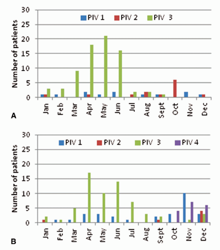
Figure 45.4. The seasonal distribution of parainfluenza viruses by subtype in the years 2010 (A) and 2011 (B).

Similar to other paramyxoviruses, PIV are common and can cause severe disease among allogeneic HSCT and lung transplant recipients and can trigger allograft rejection and BOS in lung recipients, resulting in long-term reduction in pulmonary function (242,243,244,245,246,247). Asymptomatic shedding of PIV is known to occur (223,230). Viral testing of all patients and healthcare workers, regardless of symptoms, is essential to control parainfluenza outbreaks.

No antivirals have proven efficacy for the treatment or prophylaxis of parainfluenza and no vaccine is available.


OTHER PARAMYXOVIRUSES—HUMAN METAPNEUMOVIRUS

Human metapneumovirus (HMPV) is a paramyxovirus that was first described in 2006 as a cause of severe lower airway disease among transplant recipients (248). A number of reports since have emerged among HSCT and lung transplant recipients, and the disease spectrum appears similar to other paramyxoviruses (249,250,251,252,253). HMPV has two major genetic lineages (A and B) and typically circulates around late winter-spring (February to May) (249,254). DFA has poor sensitivity (50%) compared to PCR and cultivation is difficult. Diagnosis typically is made using a molecular test. The mode of transmission of HMPV is not clearly known, but droplet precautions seem to be an effective strategy for preventing healthcare-associated transmission (249). No effective prophylactic pharmacologic strategies or vaccine exist (255,256).


ADENOVIRUS

Adenovirus is a DNA virus with 53 different serotypes (species; A-G). The clinical presentation as well as virulence of various serotypes may differ: enteric adenovirus serotypes (serotypes 40 and 41) typically cause gastrointestinal symptoms and oculopathogenic types present with conjunctivitis (257,258,259). Immunity to one serotype does not confer protection from another, meaning that a single patient can develop several adenovirus infections in a year.

Infection among transplant hosts may be primary, but mostly is the reactivation of virus that is latent in lymphoreticular tissue, especially in children who may shed the virus for a long duration after infection regardless of the state of immunosuppression (260,261). No seasonal variation in disease has been noticed among transplant recipients, further supporting reactivation of latent infection as the main etiology of disease. The incidence of detectable adenovirus viremia after allogeneic HSCT occurs in 20% to 26% of children and 9% in adults (higher in T-cell depleted grafts; 19% to 20% in adults), and 5.8% and 10% in adult and pediatric liver transplants, respectively (96,261,262,263,264,265,266).

Invasive adenovirus infection is associated with high mortality in stem cell transplant recipients (260). Nasopharyngeal (NP) shedding of adenovirus (detected in 13% of all HSCT candidates regardless of symptoms) before transplant has been shown to be a strong risk factor for adenovirus viremia. In a single study, a strong association was shown in a cohort of pediatric transplant recipients between NP shedding of adenovirus and subsequent viremia. Therefore, some centers have a “screen and postpone” approach for patients with NP swabs/aspirates that show adenovirus (267).

Adenovirus causes pneumonitis, hepatitis, colitis, and hemorrhagic cystitis in HSCT recipients. Among allogeneic recipients, mismatched donor, T-cell depletion, use of ATG/Campath, and GVHD increase the risk for invasive disease (96). Cidofovir is the most commonly used treatment for adenovirus in this population and most effective when used preemptively with the detection of low or early viremia (263,268,269). The efficacy in treating invasive infection has not been demonstrated.

In SOT, adenovirus disease mostly manifests as interstitial nephritis or hemorrhagic cystitis in renal transplant; hepatitis in liver transplant; or, rarely, as pneumonitis in lung recipients. The overall incidence of disseminated end-organ adenoviral disease is low despite the detection of viremia in 6% to 8% of asymptomatic SOT recipients (270,271,272,273,274,275).

Adenovirus is highly contagious and may be transmitted via large-particle droplets, contact with body secretions, fecal-oral spread, direct mucosal inoculation (conjunctiva), and donor organ (276). Outbreaks due to adenovirus are known to occur on transplant wards (277). For outbreak investigations, rapid serotyping may be enabled by PCR-based amplification and sequencing of conserved region bracketing the highly variable region (HVR) of hexon gene. Serotyping can enhance infection control efforts by rapidly categorizing strains isolated from different patients to determine whether an increase in episodes is related to endogenous reactivation of latent virus (different strains in all patients) or from healthcare-associated acquisition (same strain seen in all patients) (257).

The optimal isolation approach for adenovirus-infected persons is not known; we recommend that it be determined by site of isolation of virus-contact precautions for stool and urine, and droplet and contact precautions for respiratory, ocular, or disseminated infection (278).


HUMAN RHINOVIRUS AND HUMAN CORONAVIRUS

Rhinovirus and coronaviruses are the common cold viruses. Rhinovirus is a picornavirus, and currently three genotypes are recognized to cause human infection (A, B, and C) with no cross-protection. Rhinovirus C is the most recently discovered genogroup of rhinovirus; it causes severe airway disease in infants and children (279,280,281,282,283

Only gold members can continue reading. Log In or Register to continue

Stay updated, free articles. Join our Telegram channel

Jun 16, 2016 | Posted by in INFECTIOUS DISEASE | Comments Off on Healthcare-Associated Infections in Transplant Recipients

Full access? Get Clinical Tree

Get Clinical Tree app for offline access