Granulocytes and Mast Cells


Granulocytes and Mast Cells


Patrizia Scapini

Nicola Tamassia

Carlo Pucillo

Marco A. Cassatella


INTRODUCTION

Polymorphonuclear leukocytes or granulocytes are hematopoietically derived blood cells that typically act at the frontline of innate defense in host response to foreign microorganisms. Granulocytes contain heterogeneous cytoplasmic granules, which are storage pools for cell-specific intracellular enzymes, preformed receptors, cationic proteins, and other cell-specific molecules. According to their granular staining properties, polymorphonuclear leukocytes are classified into three different populations: neutrophils, eosinophils (Eos), and basophils (Bas). Neutrophil granules stain preferentially with neutral dyes, eosinophilic granules stain with acidic colorants such as eosin, and Ba granules stain with basic dyes.

Mast cells (MCs), similarly to polymorphonuclear leukocytes, represent another crucial effector cell type of the innate immune system that also stores elevated amounts of preformed inflammatory mediators within their cytoplasmic granules. However, while polymorphonuclear leukocytes are mainly peripheral blood-circulating cells, MCs are tissue resident cells distributed throughout the vascularized tissues or serosal cavity. Granulocytes and MCs differ in their functions and roles during the inflammatory process, MCs, Bas, and Eos being, for instance, essential components of allergic inflammation. Interestingly, recent data have revealed that granulocytes and MCs may also play key roles in orchestrating the transition from innate to adaptive immunity. These latter observations have caught the attention of immunologists who are currently reevaluating the importance of granulocytes and MCs, and very intensively working to clarify their multifaceted aspects in immunity.


NEUTROPHILS

Neutrophils are well known to function as the first line of defense against invading pathogens, principally bacteria and fungi, but also viruses.1 These cells, together with monocytes, macrophages, and dendritic cells (DCs), feature the characteristic properties of the “professional” phagocytes and utilize several effector mechanisms to destroy pathogens, including the generation of massive amounts of reactive oxygen species (ROS) in combination with the discharge of many potent antimicrobial enzymes or factors.1 Because of their powerful microbicidal equipment, neutrophils are often depicted as harmful cells that can cause damage to the surrounding tissues during acute inflammation (as observed in those inflammatory diseases dominated by neutrophils).1 Nonetheless, extensive research performed in the last 20 years has recognized neutrophils as highly versatile and sophisticated cells displaying a significant synthetic capacity as well as an important role in linking the innate and adaptive arms of the immune response.2,3


Neutrophil Generalities

Neutrophils are the most abundant (40% to 70%) circulating leukocyte type in human blood, normally present at 2.5 to 7.5 × 10⁹ cells/L.4 Morphologically, these cells can be identified by the peculiar shape of their nucleus, which is polymorphous and usually consists of three to five sausage-shaped masses of chromatin connected by fine threads (Fig. 20.1). Mature neutrophils are terminally differentiated, nondividing cells that develop and mature in the bone marrow from pluripotent CD (cluster of differentiation)34-positive (CD34+) stem cells, under the regulatory effects of several colony-stimulating factors (CSFs), including granulocyte-macrophage CSF (GM-CSF), granulocyte CSF (G-CSF), interleukin (IL)-3, and IL-6.4 Neutrophils can be cytofluorimetrically identified by their characteristic morphology (high side scatter) and expression pattern of plasma membrane proteins such as CD66b, CD11b, CD15, and CD16, in conjunction with the lack of expression of CD2 and CD19. In humans, circulating polymorphonuclear leukocytes are 10 to 20 µm in diameter, display a half-life previously thought to correspond to 7 to 12 hours, but more recently reevaluated and extended to up to 90 hours,5 and exist in a dynamic equilibrium with a “marginated” pool that is sequestered within the microvasculature of many organs.1,4 In the resting uninfected host, the peripheral neutrophil population is maintained within a constant number by several mechanisms. One of them consists in programming neutrophils to spontaneously undergo apoptosis1,3 to be, in turn, cleared by tissue macrophages located in the bone marrow, spleen, and liver.6 In this latter context, a feedback loop involving an IL-23/IL-17/G-CSF axis, crucial for the regulation of neutrophil production, has been recently identified in mice.7 According to this model, the uptake of apoptotic neutrophils by macrophages and DCs would determine a downregulation of their IL-23 production. Consequently, the Th17 subset of proinflammatory T-lymphocytes would be poorly sustained and thus much less IL-17A would be generated. As IL-17A positively regulates fibroblast- and endothelial cell-derived G-CSF, which is essential for controlling both granulopoiesis and neutrophil survival, the final outcome of this circuit—triggered by the massive neutrophil
apoptosis at peripheral sites—would consist in a decrease in the levels of neutrophils released from the bone marrow.7 On the other hand, the number of circulating neutrophils can dramatically increase (even up to 10-fold) under acute inflammatory conditions (eg, during a bacterial infection), from accelerated neutrophil production and release from the bone marrow.4 Moreover, even the lifespan of neutrophils is significantly extended under inflammatory conditions, as various host- and pathogen-derived mediators such as G-CSF, GM-CSF, interferon (IFN)-γ, tumor necrosis factor (TNF)-α, lipopolysaccharide, and nucleic acids inhibit neutrophil apoptosis and hence prolong their survival.8


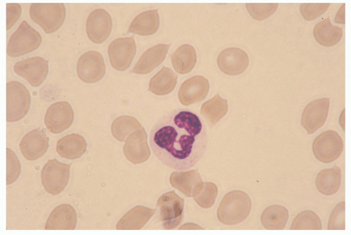
FIG. 20.1. A Polymorphonuclear Neutrophil Circulating in Peripheral Blood. From Anderson’s Atlas of Hematology; Anderson, Shauna C., PhD. Copyright 2003, Wolters Kluwer Health/Lippincott Williams & Wilkins.

During an acute inflammatory response, neutrophils are rapidly recruited to the site of injury by a coordinated sequence of events that begin with the elaboration of various mediators able to specifically promote their migration from the intravascular compartment. Mediators derive from numerous sources (tissue macrophages, endothelial cells, activated plasma components) and include vasoactive amines, proinflammatory lipids, small polypeptides, chemotactic factors, and cytokines such as TNFα, IL-1β, or IL-17A (the latter being one of the most abundant products of Th17 cells).1,3 Chemotactic factors, in particular, are generated in temporally distinct waves and include C5a, leukotriene-B4 (LTB4), formyl-Met-Leu-Phe (fMLF), as well as neutrophil specific chemokines, such as CXCL8/IL-8, CXCL1/GROα, CXCL5/epithelial cell neutrophil-activating protein-78, etc.1,3 Once recruited at an inflammatory site, neutrophils function as mobile arsenals that recognize, phagocytose, and ultimately destroy their targets. If the acute inflammatory response correctly subsides, then neutrophils may actively participate in its resolution (see following discussion). If not, an uncontrolled and continuous release of the proinflammatory cargo (ROS and proteases) by neutrophils recruited at the site of infection/injury may eventually lead to destruction of bystander tissue, and thus to exacerbation of the ongoing inflammation, ultimately provoking the onset of chronic inflammatory/autoimmune diseases.9


Neutrophil Microbicidal Mechanisms

To destroy and eliminate invading pathogens, neutrophils essentially utilize two fundamental mechanisms10: an oxygen-dependent process that is mediated by the generation of ROS, which include O2 (superoxide anion), hydrogen peroxide, singlet oxygen, and other products derived from the metabolism of hydrogen peroxide; and an oxygen-independent process consisting in the release into the phagocytic vacuole of lytic enzymes; and antimicrobial polypeptides stored in their intracellular granules.10 The oxygen-dependent process, also referred to as the “respiratory burst,” is defined as an increase of a mitochondrial independent oxidative metabolism that leads to the generation of O2, which occurs through the activation of the phagocytic NADPH oxidase, an enzymatic system that is unique to phagocytes (neutrophils, monocytes, macrophages, DCs, and also Eos). Nicotinamide adenine dinucleotide phosphate (NADPH) oxidase is a multiprotein complex formed by a flavocytochrome-b558 (a heterodimer of gp91phox and p22phox chains, where phox stands for phagocyte oxidase), three cytoplasmic components (namely p40phox, p47phox, p67phox) and either Rac1 or Rac2 from the Rho family of low-molecular-weight GTPases (Fig. 20.2). Upon cell stimulation, the cytosolic components of the complex become phosphorylated and assemble together with the cytochrome and Rac1/2 on the plasma membrane, thus forming the active enzyme that produces superoxide anion radicals, by catalyzing the transfer of electrons from NADPH to molecular oxygen (see Fig. 20.2). O2 is converted by superoxide dismutase into hydrogen peroxide, which, in the presence of myeloperoxidase and halogens, is then metabolized into hypochlorous acid. The latter represents one of the neutrophil’s
major weapons against microbes, as it also synergizes with granule proteins to kill pathogens in the neutrophil phagolysosome. O2 can also reacts with other cellular radicals, such as nitric oxide, to form different species of cytotoxic oxidant, such as peroxynitrite. The critical role of NADPH oxidase and its products in host defense is best illustrated by the plight of patients with chronic granulomatous disease, in which mutations in any of the NADPH oxidase complex subunits (gp91phox, p22phox, p40phox, p47phox, and p67phox) leads to a severe immunodeficiency characterized by defective killing of phagocytosed pathogens for the lack of ROS generation.10 These infections typically involve microorganisms for which oxidant-mediated killing is particularly critical for effective host defense, such as Staphylococcus aureus, Aspergillus spp., Nocardia, and a variety of gramnegative enteric bacilli.


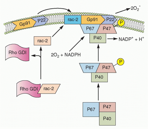
FIG. 20.2. Nicotinamide Adenine Dinucleotide Phosphate (NADPH) Oxidase Assembly. In the resting neutrophil, the cytochrome b subunits gp91-phox and p22-phox are tightly bound in the membrane. p47-phox, p67-phox, and rac-s complex are in the cytosol. Upon activation, Rho GDP-dissociation inhibitor (GDI) releases rac-2, and p47-phox becomes phosphorylated. This causes translocation of rac-2, p47-phox, and p67-phox to the membrane and complex formation with the cytochrome components, thereby completing the assembly of the active oxidase. (After Burg ND, Pillinger MH. Clin Immunol. 2001;1:7-17.)

The release of potent proteolytic enzymes contained in their granules represents the other crucial mechanisms utilized by neutrophils to eliminate pathogens following phagocytosis.10 Neutrophil granules are subdivided into peroxidase-positive granules (based on the presence of myeloperoxidase, their marker), also called primary or azurophil granules (owing to their affinity for the basic dye azure A), and peroxidase-negative granules that include the specific (secondary) granules, the gelatinase (tertiary) granules, and the secretory vesicles11 (Fig. 20.3). The different types of granules appear at progressive stage of neutrophil development,11 with the primary granules, as suggested by the name, being the first ones to appear during hematopoiesis at the promyelocyte stage. As highlighted in Figure 20.3, granules are released in a hierarchical order and under separate control by mature neutrophils, depending on the type of stimulus.11

Azurophil or primary granules undergo limited exocytosis in response to stimulation and are packaged with acidic hydrolases and antimicrobial proteins that contribute primarily to the killing and degradation of engulfed microorganisms into the phagolysosome.11,12 These granules contain myeloperoxidase, an enzyme that catalyzes the formation of hypochlorous acid, hydrolases, lysozyme, matrix metalloproteinases, and three structurally related serprocidins (serine proteases with microbicidal activity): proteinase-3, cathepsin G, and elastase. The latter proteins can degrade a variety of extracellular matrix components, including elastin, fibronectin, laminin, type IV collagen, and vitronectin. Azurophil granules also contain antimicrobial molecules such as bactericidal/permeability-increasing protein (which is important for killing gram-negative bacteria) and α-defensins, a family of small cysteine-rich antibiotic peptides with broad antimicrobial activity against bacteria, fungi, and certain enveloped viruses.11,12

Specific (or secondary) granules, which are formed at the myelocytic stage, are smaller and less dense than the azurophil ones, and contain unique constituents, such as collagenase, haptoglobin, vitamin B12-binding protein, as well an extensive array of membrane-associated proteins including cytochromes, signaling molecules, and receptors.11,12 Secondary granules also contain an arsenal of antimicrobial substances, such as lactoferrin, neutrophil gelatinase-associated lipocalin, lysozyme, hCAP18 (cathelicidin human cationic antimicrobial protein of 18 kDa, the proform of LL-37), and pentraxin-3. The inhibitory effect of antimicrobial proteins include depriving ions essential for microbial survival, degrading structural components of microorganisms (eg, peptidoglycan), and disrupting the integrity of target cell membrane by punching pores in the membrane or by perturbing membrane integrity. Neutrophil-specific granules also contain an important family of soluble proteinases, known as matrix metalloproteinases (MMPs), which include neutrophil collagenase-2 (MMP-8), gelatinase-B (MMP-9), and leukolysin (MMP-25). These proteinases are generally stored as inactive proenzymes and undergo proteolytic activation following granule fusion and interaction with azurophilic granule contents. Neutrophil MMPs disrupt major structural components of bacteria and/or extracellular membranes, and are therefore crucial not only for bacterial killing, but also for neutrophil extravasation and migration.11,12

Tertiary granules are produced at the metamyelocyte stage of differentiation and are smaller, lighter, and more easily exocytosed than the other granule classes.11,12 These granules are indeed important primarily as a reservoir of matrixdegrading enzymes and membrane receptors needed during polymorphonuclear leukocyte extravasation and diapedesis. The primary constituent of tertiary granules is gelatinase, a latent metalloenzyme with the capacity for tissue destruction. Finally, the secretory vesicles are smaller than the other granules, are generated by endocytosis during the late stage
of nuclear neutrophil segmentation in the bone marrow, and are the most readily mobilizable.11,12 These vesicles are preferentially directed to the plasma membrane, as reflected in the density of vesicle-associated membrane protein (VAMP), a fusogenic protein associated with the granule membrane. Secretory vesicles do not contain toxic substances, but mainly plasma proteins like albumin and receptors (including β2-integrins, the complement receptor [CR]1, receptors for formylated bacterial peptides [fMLF-R], CD14, the Fc portion of γ-immunoglobulins (Igs) [FcγRIII/CD16], and the metalloprotease leukolysin). Heparin-binding protein (also known as CAP37 or azurocidin), whose release is essential for the polymorphonuclear leukocyte-induced increase in vascular permeability at the initial stage of extravasation, is also stored in the secretory vesicles.


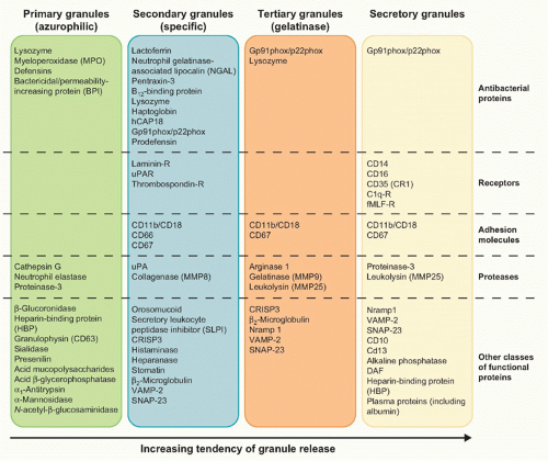
FIG. 20.3. Main Constituents of Neutrophil Granules.

An additional nonphagocytic microbicidial mechanism used by neutrophils to capture and destroy microbes in the extracellular space consists in the ability of neutrophils to form so-called neutrophil extracellular traps (NETs).13 The latter structures consist of nuclear chromatin decorated with antimicrobial peptides and enzymes (eg, bactericidal/permeability-increasing protein, elastase, pentraxin3, cathepsin G, and many others) that lacks membranes and cytosolic markers.13 NETtosis, a novel type of neutrophil death mechanism that occurs under settings of extreme neutrophil stimulation (different from necrosis, apoptosis, and also independent from caspase activation), underlies the generation of NETs.14 Accordingly, the nuclear envelope, granules, and cell membranes gradually dissolve during NETtosis, allowing the nuclear contents to mix and condense in the cytoplasm before being released into the extracellular space.14 NETs, in turn, bind to various grampositive and gram-negative bacteria (such as Staphylococcus aureus, Salmonella typhimurium, Streptococcus pneumoniae,
and group A streptococci), as well as to pathogenic fungi (such as Candida albicans). Similarly to what happens in the phagolysosome, the high local concentration of antimicrobial peptides and enzymes is responsible for the killing of the pathogens trapped by NETs.13 The observation that neutrophils from patients with chronic granulomatous disease do not form NETs has suggested, on the one hand, that ROS-mediated signaling/cascades are involved in NET generation, and, on the other hand, that the lack of NETs might contribute to the pathogenesis of chronic granulomatous disease.3,14 Whatever the case, it is noteworthy to remark that both the oxygen-dependent and -independent effector mechanisms in host defense toward pathogens are also utilized by neutrophils for their cytotoxic and tumoricidal activities.


Neutrophil Receptors

Under inflammatory conditions, neutrophils sense a wide range of extracellular ligands that, through the interaction with specific receptors, subsequently trigger a number of effector functions, including adhesion, migration, phagocytosis, survival, cell activation, gene expression modulation, target cell killing, and mediator production and release.1,3,15 For instance, agonist-stimulated neutrophils may trigger not only degranulation, but also the release of arachidonic acid and/or other eicosanoids (eg, prostaglandin [PG]E2), via the activation and/or the upregulation of PLA2 and COX-2, respectively. Upon appropriate stimulation, neutrophils can also generate LTA4 through the action of 5-lipoxygenase, as well as LTB4 by converting LTA4 via the action of LTA4 hydrolase.15 A nonexhaustive list of neutrophil receptors includes 1) receptors for proinflammatory mediators (eg, the anaphylotoxin complement component C5a, LTB4, platelet-activating factor [PAF], substance P, and fMLF); 2) receptors for cytokines, such as IFNγ, IL-1, IL-4, IL-6, IL-10, IL-13, IL-15, IL-18, TNFα, G-CSF, GM-CSF, and many others; 3) receptors for chemokines, including CXCR1 and CXCR2; 4) receptors/adhesion molecules for the endothelium; 5) receptors for tissue matrix proteins; and 6) opsonin receptors, such as FcγRs and those for the major cleavage fragments of the complement system (see following discussion). Neutrophils also express a variety of pattern recognition receptors (PRRs), including all toll-like receptors (TLRs), with the exception of TLR3, cytoplasmic ribonucleic acid helicases involved in viral ribonucleic acid recognition such as MDA5 and RIGI, and deoxyribonucleic acid binding cytoplasmic proteins (IFI16 and LRRFIP1).3


The Role of Neutrophils in Acute Inflammation

In order to carry out their prototypical defensive role in acute inflammation, bloodstream neutrophils must extravasate. To do so, they attach to activated endothelium, transmigrate through postcapillary venules (diapedesis), and then migrate toward a corresponding chemotactic gradient to the injury site where they recognize their target, engulf, and finally destroy it.


Neutrophil Extravasation

In general, leukocyte recruitment during inflammation, also called extravasation, is a multistep and highly complex phenomenon characterized by a number of predetermined steps occurring in the vessel lumen, known as leukocyte capture (or tethering), rolling, activation, and firm adhesion (arrest)16 (Fig. 20.4). These latter steps are not discrete phases of inflammation; rather, they simply represent a sequence of events from the perspective of each leukocyte. Moreover, each of these steps appears to be necessary for the effective leukocyte recruitment; blocking any of them can severely reduce leukocyte accumulation in the tissue.17 Adhesion of blood leukocytes to the endothelium during inflammation requires specific leukocyte-endothelial interactions involving different families of adhesion molecules. The latter include members of the selectin family and their cognate carbohydrate and glycoprotein ligands, which mediate the initial leukocyte deceleration along the vessel wall (a process called “rolling”), as well as members of the integrin family and their cognate Ig superfamily ligands, which mediate the subsequent high-affinity adhesion and arrest of leukocytes to the venules16,17 (Table 20.1).

Under noninflammatory conditions, neutrophils (as well as other leukocytes) travel primarily through the center of the blood vessel lumen, where the flow is fastest. In response to proinflammatory signals, however, both the neutrophils and the blood vessels undergo a series of changes. As a consequence of vascular dilatation, blood flow first increases and then slows, thus facilitating the interactions between leukocytes and the endothelial cells. The process of capture/tethering and rolling then ensues, in which L-selectins on neutrophils, and P- or E-selectins on endothelia, interact with sialyl-Lewisx moieties or PSGL-1 on their respective cell partners. Although these interactions are reversible and transient, they prepare neutrophils for a tighter binding, integrin-mediated step.16,17 The most important neutrophil integrins are four, each one composed of an identical β-subunit β2, known as CD18) noncovalently linked to different α-subunits: CD11a/CD18 (αLβ2, lymphocyte function antigen), CD11b/CD18 (αMβ2, CR3), CD11c/CD18 (αXβ2, CR4), and CD11d/CD18.18 When neutrophils encounter their specific chemoattractants (eg, CXCL8), displayed bound to glycosaminoglycans on the vessel wall, neutrophil integrins are converted from an inactive to an active conformation16,17 (see Fig. 20.4). Activated integrins can interact with their counterreceptors on the surface of the TNFα and/or IL-1β-activated endothelium (eg, intercellular adhesion molecule-1 [ICAM-1] and intercellular adhesion molecule-2), leading to the strong adhesion and arrest of the cells. Neutrophils then flatten and transmigrate between and through the endothelial cells of postcapillary venules into the surrounding tissue (see Fig. 20.4), also secreting a broad range of MMPs that degrade the basement membrane. Transmigration involves homophilic interaction of CD31/PECAM-1 and JAM-A on neutrophils and endothelial cells, where CD31/PECAM-1 and JAM-A act sequentially to mediate neutrophil migration through the venular walls.17,18 Once in the interstitial compartment, neutrophils migrate along the chemotactic gradient toward the site of
injury or infection and, once arrived, begin to react with the etiopathogenic agent.


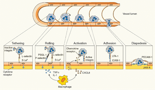
FIG. 20.4. Leukocyte Transmigration. Following an inflammatory stimulus, tissue-resident macrophages and other cells release inflammatory mediators such as tumor necrosis factor α and interleukin-1, which induce the rapid expression of preformed P-selectin (and transcription-dependent E-selectin expression) on the endothelium. The interaction between selectins and their glycoprotein ligands initiates leukocyte tethering and rolling. Activation by chemokines—and other leukocyte activators (eg, leukotriene-B4 or platelet-activating factor)—presented on endothelial cells causes leukocyte integrin activation, thus resulting in transition from cell rolling to cell firm adhesion, in view of the strength of integrin-mediated binding with endothelial immunoglobulin superfamily members. Leukocytes can then transmigrate through the endothelial monolayer and chemotactically move toward the inflammatory stimulus. Examples of adhesion molecules involved in each step are depicted.

The fundamental importance of adhesive glycoproteins in vivo is testified by those individuals affected leukocyte adhesion deficiency (LAD) disease, who display an abnormally high susceptibility to bacterial infections.19 Several types of LAD disease have been identified. In LAD-I, mutations of the β2 integrin typically eliminate the expression of all four integrin complexes. Because of the multiple defects in adhesion-related functions, patients with LAD-I develop recurrent bacterial and fungal infections, mostly with Staphylococcus aureus or gram-negative enteric microbes. Neutrophilia with paucity of neutrophils at inflamed or infected sites is characteristic of LAD-I, while typical clinical features include frequent skin and periodontal infections, delayed separation of the umbilical cord and omphalitis, and deep tissue abscesses. LAD-II is caused by mutations in the membrane transporter for fucose and thus is associated with loss of expression of fucosylated glycans on the cell surface. Fucosylated proteins such as sialyl-Lewis X (CD15s) are ligands for endothelial selectins and are important for the rolling phases of leukocyte extravasation. Patients with LAD-II also have leukocytosis and form pus poorly, although infections tend to be less severe in patients with LAD-II than in patients with LAD-I. Finally, LAD-III has been recently described. LAD-III is an autosomal recessive disorder caused by mutations in the human kindlin-3 gene, which codes for a protein essential for integrin activation. Consequently, LAD-III is characterized by impaired adhesion of leukocytes to the endothelium of inflamed tissues and by severe bleeding. Curiously, a number of LAD-III patients additionally suffer from osteopetrosis.


Neutrophil-Mediated Phagocytosis

Neutrophils play a critical role in host protection as they eliminate microorganisms through phagocytosis (the cellular process of engulfing particles larger than 0.5 µm). While many cells in our body are capable of phagocytosis, neutrophils do it to an extent sufficient to be considered “professional phagocytes” (eg, a single neutrophil can engulf up to 10 to 12 particles [eg, bacteria]).20 Phagocytosis is triggered either through receptor (eg, mannose receptor or Dectin-1) recognition of certain polysaccharides present on the surface of some yeast cells and/or upon the binding of opsonized microorganisms through, for instance, FcγRs and CRs.20 Neutrophils constitutively express the low-affinity
FcγRs (FcγRIIA/CD32A and FcγRIIIA/CD16A), and, when exposed to IFNγ or G-CSF, the high-affinity FcγR (FcγRI/CD64) as well.3 CRs expressed by neutrophils are CR1 (also known as CD35), which binds to complement components C1q, C4b, C3b, and mannan-binding lectin; CR3, which binds to iC3b, intercellular adhesion molecule-1, and some microbes; and CR4, which binds to iC3b. By expressing these latter receptors, neutrophils are able to recognize and bind, in a cooperative manner, IgG-opsonized particles and/or complement-opsonized microbes, and then activate their phagocytosis. During the phagocytic process, the foreign particle is internalized, initially through membrane recruitment to the site of particle contact, and then via membrane extensions outward to surround the particle and form a new vesicle called a cytoplasmic phagosome.20 (Fig. 20.5). The phagosome then undergoes fusion with neutrophil granules to form a phagolysosome, a protected space in which proteolytic enzymes and other bactericidal components are discharged and pathogen degradation occurs. At the same time, NADPH oxidase assembles on the phagosomal membrane after phagocytosis and starts to generate ROS into the phagolysosome to kill bacteria by oxidizing microbial proteins and lipids. The activity of NADPH oxidase also leads to the acidification of the phagosome, which enhances the effectiveness of pH-sensitive antimicrobial compounds. Thus, neutrophil mechanisms of pathogen destruction within the phagosome are multiple and involve granule fusion, toxic oxygen radical production, activation of latent proteolytic enzymes, and the activity of antibacterial proteins (see Fig. 20.5). Remarkably, an activation of gene transcription and a selected generation of cytokines also occur during phagocytosis, a feature that neutrophils utilize for boosting a more effective innate immune response. For instance, recruited neutrophils that phagocytose
a pathogen also respond by producing chemokines, in particular CXCL8, to amplify their own recruitment, but also CCL3, CCL4, and CCL19 that serve to recruit monocytes and DCs.21


TABLE 20.1 Main Adhesion Molecules Involved in Leukocyte-Endothelial Cell Interaction


Adhesion Molecule


Distribution


Ligands and Counterreceptors


Function


E-selectin (CD62E)


Endothelial cells


PSGL-1, ESL-1, CD44a, CD43a


Rolling


P-selectin (CD62P)


Endothelial cells, platelets


PSGL-1, PNAd


Rolling


L-selectin (CD62L)


All leukocytes except effector and memory effector T cells


PNAd, MAdCAM-1, PSGL-1, E-selectin, P-selectin


Rolling


Selectin ligands


PSGL-1


All leukocytes


All selectins (essential for P-selectin)


Rolling


sLex


Myeloid cells, some memory T cells, HEVs


All selectins


Rolling


PNAd


HEV, some sites of chronic inflammation


L-selectin, P-selectin


Rolling


Integrins


αMβ2(MAC-1; CD11b/CD18)


Granulocytes, monocytes, some activated T cells


ICAM-1, fibrinogen, C3b, JAM-C


Adhesion, transmigration


αLβ2(LFA-1; CD11a/CD18)


All leukocytes


ICAM-1, ICAM-2, JAM-A


Adhesion, transmigration


αDβ2(CD11d/CD18)


Monocytes, macrophages, eosinophils


ICAM-1, VCAM-1


Adhesion


αxβ2(p150,95; CD11c/CD18)


DCs


Fibrinogen, C3b


Adhesion


α4β1(VLA-4)


Most leukocytes


VCAM-1, fibrinogen, JAM-B


Rolling, adhesion


α4β7(LPAM-1)


Lymphocytes, NKCs, mast cells, monocytes


MAdCAM-1, fibronectin, VCAM-1


Rolling, adhesion


Immunoglobulin superfamily


ICAM-1 (CD54)


Most types of cells


LFA-1 Mac-1, fibrinogen


Adhesion, transmigration


ICAM-1 (CD102)


Endothelial cells, platelets


LFA-1 Mac-1


Adhesion, transmigration


VCAM-1 (CD106)


Endothelial cells


VLA-4, α4β7αDβ2


Rolling, adhesion


MAdCAM-1


HEVs in PP and MLN


α4β7, L-selectin


Rolling


PECAM-1


Endothelial cells, platelets, leukocytes


PECAM-1


Transmigration


JAM-A


Endothelial cells, platelets, most leukocytes


JAM-A


Transmigration


JAM-B


Endothelial cells, HEVs


JAM-B, JAM-C


Transmigration


JAM-C


Endothelial cells, HEVs, platelets, monocytes, DC, some T cells


JAM-C, JAM-B


Transmigration


CD, cluster of differentiation; DC, dentritic cell; ESL, HEV, high endothelial venule; ICAM, intercellular adhesion molecule; JAM, LFA, MAdCAM, mucosal addressin cell adhesion molecule; NKC, PECAM, PNAd, PSGL, sLex, VCAM, VLA.


a CLA decorated.


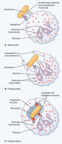
FIG. 20.5. Phagocytosis. The figure shows ingestion, digestion, and destruction of foreign particulate matter (a bacterium, in this example) by a neutrophil. A: Cell membrane receptors bind to antibody and complement molecules previously attached to the bacterial surface. B: The cell membrane creeps around the bacterium and envelopes it. C: The bacterium is trapped in a special space, the phagocytic vacuole, into which lysosomes discharge proteases, which together with oxidants kill it. Then, digestive enzymes dissolve it. (Thomas H. McConnell, The Nature Of Disease Pathology for the Health Professions, Philadelphia: Lippincott Williams & Wilkins, 2007.)


Role in the Resolution Phase of Inflammation

Locally activated neutrophils not only amplify the inflammatory process, but, surprisingly, actively participate in its resolution phase.22 To do so, during the late, final phases of a resolving, acute inflammatory reaction neutrophils, for instance, switch their eicosanoid biosynthesis potential from LTB4 to lipoxins, with profound modifications of their effector functions. In fact, lipoxin A4 (LXA4) and LXB4 stop neutrophil chemotaxis, adhesion, and transmigration through endothelium (by decreasing P-selectin expression), inhibit Eo recruitment, stimulate vasodilation (by inducing synthesis of PGI2 and PGE2), inhibit LTC4– and LTD4– stimulated vasoconstriction, inhibit LTB4 inflammatory effects, and inhibit the function of natural killer (NK) cells.22 Neutrophils also contribute to the biosynthesis of resolvins (such as resolvin E1, resolvin E2, resolvin D1, and resolvin D2) and protectin D1, which all inhibit neutrophil transendothelial migration and tissue infiltration, as well as stimulate resolution and reduce the magnitude of the inflammatory response in vivo.22 Furthermore, neutrophils might also serve as major producers of anti-inflammatory cytokines such as transforming growth factor (TGF)β and IL-1 receptor antagonist, the latter being an endogenous inhibitor of IL-1β signaling and mediated effects.21 Finally, neutrophils must be cleared from the inflammatory site as inflammation resolves. Indeed, neutrophils undergo apoptosis and are engulfed by tissue macrophages, which then reprogram into the M2 phenotype and start to generate antiinflammatory cytokines such as TGFβ and IL-10.6,23


Novel Neutrophil Effector Functions

The functions described previously for neutrophils in host defense are fundamental for combating infectious diseases. However, more recent discoveries on neutrophils as source of a variety of cytokines21 have revealed that these cells are not only key components of the inflammatory response, but also crucial effectors of innate and adaptive immune regulatory networks.23


Neutrophil-Derived Cytokines and Chemokines

Numerous in vitro and in vivo studies, focusing on novel aspects of the neutrophil biology and function, have recently shed new light on the potential role that neutrophils can exert in the modulation of innate and adaptive immune responses.23 It is now unequivocal that neutrophils are not, as for a long time thought, terminally differentiated cells “devoid of transcriptional and protein synthesis activity.”23a In fact, besides the several preformed or rapidly generated inflammatory mediators described previously, neutrophils display the capacity to de novo synthesize and release also several chemokines and cytokines with immunoregulatory properties.21,23 It is, however, important to mention that, at least in vitro, neutrophils usually produce, on a per-cell
basis, fewer molecules of a given cytokine than mononuclear leukocytes.21 However, considering that neutrophils clearly predominate over other cell types under inflammatory conditions in vivo, it becomes obvious that the contribution of neutrophil-derived cytokines can be of foremost importance. To date, a wide range of stimuli able to induce characteristic signatures of chemokine and cytokine synthesis by neutrophils have been identified. Among these, cytokines themselves, chemotactic factors (fMLF, LTB4, PAF, C5a, and CXCL8), phagocytic particles, microorganisms (such as fungi, viruses, and bacteria), and PRR ligands can all induce the synthesis and release of chemokines and cytokines by neutrophils.21 Considering that neutrophils usually represent the first cell type infiltrating at the site of infections, a stimulus-specific response of neutrophils in terms of cytokine production might direct the evolution of certain types of inflammatory and immune reactions to support the transition from innate to adaptive immunity.

Table 20.2 lists all the cytokines that, to date, have been shown to be released by neutrophils in vitro, either constitutively or following appropriate stimulation, or in vivo. Numerous in vivo observations, in fact, not only have confirmed and reproduced the in vitro findings, but often have clarified their biological meaning and implications. As outlined in Table 20.2, neutrophils can produce proinflammatory, anti-inflammatory, immunoregulatory, angiogenic, and fibrogenic cytokines, chemokines, and ligands belonging to the TNF superfamily. The role of these molecules in mediating various neutrophil-dependent immunoregulatory functions is partially described in the following chapter. For instance, chemokines are particularly represented among the cytokines produced by neutrophils and include those primarily chemotactic for neutrophils themselves, monocytes, DCs, NK cells, and T helper (Th)1 and Th17 cells. It follows that a role for neutrophils in orchestrating the sequential recruitment to, and activation of, distinct leukocyte types in the inflamed tissue is plausible, as already demonstrated to occur in several experimental models.21,23


Neutrophils in Immunoregulation

There is now wide experimental evidence that neutrophils have the capacity to modulate the migration, maturation, and function of several leukocyte types including DCs, T cells, and B cells.23 Regarding DCs, it is noteworthy to mention that neutrophils have been shown to produce biologically active CCL20 and CCL19, two structurally related CC-chemokines that have been suggested to play a fundamental role in trafficking of, respectively, immature and mature DCs to mucosal surfaces and lymphoid organs. Likewise, neutrophils release several antimicrobial compounds, such as lactoferrin, LL-37, and cathepsin G, that have been found to act as chemoattractants for immature DCs. In addition, neutrophils can proteolytically activate prochemerin to generate chemerin, one of the few chemokines that attracts both immature DCs and plasmacytoid DCs.3 Neutrophils can also modulate DC maturation and function either through the release of several mediators or through direct physical interaction between Mac-1 (CD11b/CD18) and DC-specific intercellular adhesion molecule.24 Neutrophils also act as transport vehicle for pathogens and, in turn, deliver antigens to DCs, thus playing an important role in the activation of T-cell immune responses controlled by DCs.24


TABLE 20.2 Cytokines Expressed in Resting or Activated Neutrophils


C-X-C Chemokines


GROα/CXCL1


CINC-2α/GROχ/ MIP-2α/CXCL2


CINC-2β/GROγ/MIP-2β/CXCL3


PF4/CXCL4


ENA-78/CXCL5


GCP-2/CXCL6


IL-8/CXCL8


MIG/CXCL9 IP-10/CXCL10 I-TAC/CXCL11


C-C Chemokines


MCP-1/CCL2MIP-1α, CCL3


MIP-1β, CCL4


TARC/CCL17


PARC/CCL18


MIP-3α, CCL19


MIP-3β, CCL20


MDC/CCL22


Proinflammatory


Cytokines


TNFβ


IL-1α, IL-1β


IL-6(?),IL-7, IL-9


IL-16(?), IL-17A/F(?)


IL-18


MIF


Anti-inflammatory


Cytokines


IL-1ra


IL-4(?), IL-10(?)


TGFβ1, TGβ2


Immunoregulatory


Cytokines


IFNα, IFNβ, IFNγ(?)


IL-12


IL-23(?)


Other Cytokines


Oncostatin M


GDF (?)


NGF, BDNF, NT4


PBEF/visfatin/NAMPT


amphiregulin


TNF Superfamily


Members


FasL


CD30L


TRAIL


LIGHTa


Lymphotoxin-β


APRIL, BAFF/BLyS


RANKL


Colony


Stimulating


Factors


G-CSF


M-CSF(?)


GM-CSF(?)


IL-3(?)


SCFa(?)


Angiogenic


and Fibrogenic


Factors


VEGF


BV8/Prokineticin-2


HB-EGF


FGF-2


TGFβ


HGF


?, requires definitive corroboration; GRO, growth regulated oncogene; CXCL, CXC chemokine ligand; CCL, CC chemokine ligand; MIP, macrophage inflammatory protein; CINC, Cytokine induced neutrophil chemoattractant; PF4, Platelet factor-4; ENA-78, epithelial-derived neutrophil-activating peptide 78; GCP-2, granulocyte chemotactic protein-2; IL-, interleukin-; MIG, Monokine induced by gamma interferon; I-TAC, Interferon-inducible T-cell alpha chemoattractant; IP-10, Interferon gamma-induced protein 10; MCP, monocyte chemotactic protein; TARC, Thymus and activation regulated chemokine; PARC, pulmonary and activationregulated chemokine; MDC, Macrophage-derived chemokine; TNF, tumor necrosis factor; MIF, macrophage inhibitory factor; IL-1ra, IL-1 receptor antagonist; TGF, Transforming growth factor; IFN, interferon; GDF, Growth Differentiation factor; NGF, nerve growth factor; BDNF, Brain derived neurotrophic factor; NT4, Neurotrophin-4; PBEF, pre-B-cell colony-enhancing factor; NAMPT, Nicotinamide phosphoribosyltransferase; TRAIL, TNF-related apoptosis-inducing ligand; APRIL, a proliferation-inducing ligand; BAFF/BLyS, B-cell activating factor/B lymphocyte stimulator; RANKL, Receptor activator of nuclear factor kappa-B ligand; G-CSF, macrophage colony stimulating factor; M-CSF, macrophage colony stimulating factor; GM-CSF, granulocyte-macrophage colony stimulating factor; SCF, stem cell factor; HB-EGF, heparin binding-like epidermal growth factor; FGF, fibroblast growth factor; HGF, hepatocyte growth factor.


Cytokines in bold refer to neutrophil studies in animal models that confirm human findings.


a Messenger ribonucleic acid only.


Concerning the interactions between neutrophils and B cells, of particular interest are the findings that neutrophils produce significant amounts of BLyS/BAFF (B-lymphocyte stimulator/B-cell activating factor) and APRIL (a proliferation-inducing ligand), two related members of the TNF
family that are well known to be essential for B-lymphocyte homeostasis.25 Therefore, it is plausible to assume a role of neutrophils not only in sustaining B and plasma cell antibody production and survival, but also in promoting B-cell-dependent autoimmune diseases and tumors, as already elegantly demonstrated in the case of B-cell lymphoma.25

Cross-talk between neutrophils and T cells has been repeatedly described to occur during infections or other inflammatory responses and diseases. Current evidence now indicates that neutrophils exhibit a significant chemotactic effect toward Th1 or Th17 cell subsets, through the release of CCL2, CXCL9, and CXCL10, or CCL2 and CCL20, respectively.26 Neutrophils have also a role in directing T-cell polarization, for instance through their capacity to produce the Th1-inducing cytokine, IL-12. The latter has been clearly demonstrated in mouse models, in which strong Th1-dependent T-cell responses that result in pathogen clearance are elicited upon infection with Candida albicans, Helicobacter pylori, or Legionella pneumophila. Strikingly, depletion of neutrophils reverses the Th1 responses into a predominant Th2-response, therefore making the mice susceptible to infection. Besides the neutrophil’s ability to modulate T-cell functions through the production of chemokines and cytokines, recent reports suggest that neutrophils travel to the lymph nodes during infections and express both major histocompatibility complex (MHC) II and costimulatory molecules.27 However, whether neutrophils directly acquire antigen-presenting functions or transmit signals to naïve T cells remains still puzzling.

Neutrophils have also been shown to modulate the maturation, activation, and functions of NK cells, either by themselves or in cooperation with other cell types.28 In this context, it is worth mentioning that neutrophils, by interacting with specific subsets of peripheral blood myeloid DC (eg, 6-sulpho LacNAc+ DC, also known as slanDC) can strongly potentiate IFNγ release by NK cells.29 Importantly, the potential pathophysiologic relevance of a cell network among neutrophils, slanDC, and NK cells has been suggested by immunohistochemical studies that have revealed their colocalization in several chronic inflammatory pathologies, such as Crohn disease and psoriasis.29 Finally, it has also been proposed that mature postmitotic neutrophils can also “transdifferentiate” into much-longer-lived cells with macrophage- or DC-like characteristics, which might constitute a further manner for neutrophils to act as regulatory cells of the adaptive immune response.3,23

It is important to mention that despite of all experimental observations in vivo that strongly suggest that neutrophils could potentially act as important players in the orchestration of immune responses, additional reevaluations and validations are required. Indeed, in order to investigate the role of neutrophils in vivo, an antigranulocyte receptor-1 monoclonal antibody, RB6-8C5, has been extensively used to deplete mice of neutrophils.30 However, RB6-8C5 not only binds to Ly6G, which is present on neutrophils, but also to Ly6C, which is expressed on neutrophils, DCs, and subpopulations of lymphocytes and monocytes. Therefore, it has recently been shown that in vivo administration of RB6-8C5 depletes not only neutrophils but also other granulocyte receptor-1+ (Ly6C+) cells. Luckily, a more specific anti-Ly6G monoclonal antibody (1A8) has been raised; it is now preferentially used to deplete neutrophils in vivo under different experimental settings.31 Obviously, it will take some time prior to controlling and, eventually, revising all data on neutrophil depletion generated by using RB6-8C5. More importantly, the future availability of conditional knockout mice, selectively targeting, one by one, neutrophil function such as survival, migration, or activation, will help to finally clarify the specific contributions of neutrophils under different inflammatory/immune settings.


Role of Neutrophils in Angiogenesis and Tumor Growth

There is no longer doubt that, in addition to macrophages, neutrophils may positively or negatively influence the angiogenic process and tumor growth.32 In the former case, it has been observed that neutrophils, via elastase release, may indirectly generate massive amounts of bioactive, angiostatin-like fragments, and thus inhibit fibroblast growth factor (bFGF) plus vascular endothelial growth factor (VEGF)-induced endothelial cell proliferation.33 However, as described for macrophages, neutrophils can also favor malignant growth and progression, in relation to the type of tumor environment in which they reside, for instance via a remarkable production of proangiogenic molecules such as VEGF and CXCL8.34 On the other hand, experimental studies of tumor cure and prevention have suggested that, at least in some models, engagement of neutrophil functions can be crucial for the establishment of an effective antitumoral immune response and immune memory reactions.35 Indeed, neutrophils can produce several cytotoxic mediators for tumor and endothelial cell killing, including TNFα, defensins, proteases (such as elastase and cathepsin G), ROS, nitric oxide, and angiostatic chemokines (CXCL9, CXCL10, and CXCL11).35 It has been recently found that neutrophils exposed to IFNs express and produce TNF-related apoptosis-inducing ligand,36 another TNF superfamily member that selectively stimulates tumor cell killing. More recently, in vivo evidence proving that, similarly to M1 and M2 macrophages, neutrophils also polarize from an N1, proinflammatory and antitumoral phenotype, to an N2 anti-inflammatory and protumoral phenotype, has been provided,37 thus supporting the notion that the tumor environment can profoundly shape the functional status of neutrophils. Furthermore, it is now well established that myelopoiesis can be profoundly modified during inflammation and cancer, releasing altered mature myelocytes and myeloid-derived suppressor cells (MDSCs) that exert immunosuppressive and protumoral activity, mainly by inhibiting T-cell functions.38 Although mature human neutrophils do not seem to be a major component of such MDSC population, several mouse tumor models have revealed the existence of a granulocytic MDSC population with potent T-cell suppressing activity.38 Nonetheless, lowdensity granulocytes (so called because of their abnormal behavior upon density centrifugation) able to inhibit T-cell activation and function have been found in human patients with cancer.39 Further research is now needed to better understand the origin, phenotype, and relationship to mature
neutrophils of all these immunosuppressive granulocytic populations. Such studies will better clarify the real role of neutrophils in cancer as well as in other inflammatory/autoimmune diseases (such as infections, psoriasis, and lupus), in which the presence of MDSC- or low-density granulocyte -like cells have been also described.40

Only gold members can continue reading. Log In or Register to continue

Stay updated, free articles. Join our Telegram channel

Aug 29, 2016 | Posted by in IMMUNOLOGY | Comments Off on Granulocytes and Mast Cells

Full access? Get Clinical Tree

Get Clinical Tree app for offline access