Giant Cell Carcinoma
Sanja Dacic
Giant cell carcinoma is an extremely rare variant of non-small cell carcinoma composed of bizarre, pleomorphic, very large mononucleated or multinucleated tumor giant cells identical to the giant cells found in pleomorphic carcinoma (see Chapter 13). Adenocarcinoma, squamous cell carcinoma, and large cell carcinoma are not features of giant cell carcinoma.1
GIANT CELL CARCINOMA, CYTOLOGY
Giant cell carcinoma is a rare tumor and not typically diagnosed by cytology; however, aspiration may show a mixture of inflammatory cells and tumor cells, with occasional giant tumor cells identified.
GIANT CELL CARCINOMA, GROSS FEATURES
Giant cell carcinomas tend to form well-circumscribed masses that on cut section are tan-yellow to gray and show areas of necrosis and hemorrhage.
GIANT CELL CARCINOMA, HISTOLOGY
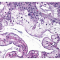
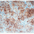
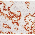
The tumor cells in giant cell carcinoma are discohesive and associated with a marked inflammatory infiltrate, usually composed of neutrophils. Tumor cells have abundant eosinophilic cytoplasm often showing leukocyte emperiopolesis (neutrophils penetrating the cytoplasm of the tumor giant cells), phagocytosed anthracotic pigment, or hyaline globules ( Figs. 14-1,14-2 and 14-3). Nuclei are hyperchromatic to vesicular and contain prominent nucleoli. Pleomorphic giant cells are identified throughout the tumor and are typically very large and often bizarre multinucleated or multilobated cells ( Figs. 14-4,14-5 and 14-6).2,3,4 and 5
Stay updated, free articles. Join our Telegram channel

Full access? Get Clinical Tree


