General Principles and Management


General Principles and Management


Waleed F. Mourad

Kenneth S. Hu

Walter H. Choi

Bruce Culliney

Zujun Li

Adam S. Jacobson

Louis B. Harrison


INTRODUCTION

Oropharynx cancer (OPC) is a common manifestation for squamous cell carcinoma of the head and neck (SCCHN), and most OPC is caused by the human papillomavirus (HPV).1 The estimated incidence of pharyngeal cancers in the United States for 2011 was 13,580 new cases and 2,430 deaths.2 There is increasing incidence of OPC despite the decline in the overall incidence of SCCHN.1 Current estimates suggest that by the year 2020, the annual number of HPV-positive OPC will exceed the annual number of HPV-positive uterine cervix carcinomas and there will be >15,000 cases of HPV-positive OPC per year by 2030.1

OPC typically involves patients in their fifth through seventh decades of life. Due to the increasing association with HPV, younger patients are being diagnosed. Men are afflicted 3 to 5 times more often than women.2 Investigators have reported that the increase in OPC is exclusively in the base of tongue (BOT) and tonsil and that HPV/p16 accounts for approximately 70% to 90% of OPC-SCCHN.1,3,4,5 Furthermore, HPV-positive OPC patients are often middle-aged (40-59 years) white men, of higher socioeconomic status, nonsmokers, and nondrinkers, but with a higher number of oral sexual partners.1,6,7,8,9,10,11,12,13,14,15

A history of tobacco or alcohol use can also be associated with other metachronous or synchronous tumors of the upper aerodigestive tract. Thus, tobacco and alcohol abuse still represent significant risk factors for the development of OPC and other cancers of the head and neck.16,17 Other risk factors for OPC may include18


  • a diet poor in fruits and vegetables19


  • the consumption of maté, a stimulant beverage commonly consumed in South America20


  • the chewing of betel quid, a stimulant preparation commonly used in parts of Asia.21

Historically, the prognosis for OPC depends upon the location of the primary tumor and the stage at presentation. Studies have shown that HPV-positive OPC have more favorable prognosis, with approximately two times better 5- and 10-year overall survival (OS) compared with HPV-negative patients.1 Likewise, recent oncologic and functional outcome data have shown superior results in HPV/p16-positive OPC patients. Of note, the significant improvement in the OS was mainly seen in patients with tonsil and BOT cancer, without obvious survival improvement in other SCCHN sites.22 Interestingly, the survival for African Americans patients with OPC has not shown such improvement. This is attributed to the difference in OPC origin (smoking induced among African Americans vs. HPV induced among Caucasians).10,23

The leading cause of death in OPC and SCCHN patients is loco-regional recurrence (LRR). However, advances in radiation therapy (RT) as well as concomitant chemotherapy and RT (chemo-RT) have improved loco-regional control (LRC) rates, even for advanced-stage disease. There has also been a growing interest in the role of minimally invasive surgery (MIS) including transoral lateral oropharyngectomy (TLO), transoral laser microsurgery (TLM), and transoral robotic surgery (TORS) in the otolaryngology community.24,25,26,27,28,29 Their application to OPC management is being actively explored. Thus, the treatment strategies for these patients are numerous, and advances in organ preservation with attention to quality of life (QOL) remain the major focus of clinical research investigations.


ANATOMY OF THE OROPHARYNX

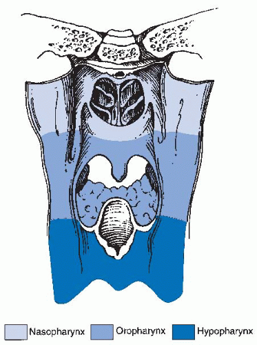
The pharynx is divided into the nasopharynx, the oropharynx, and the hypopharynx (Fig. 17-1). The oropharynx is located between the soft palate superiorly and the hyoid bone
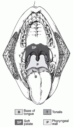
inferiorly. It is continuous with the oral cavity anteriorly and communicates with the nasopharynx above and the supraglottic larynx and the hypopharynx below. Within the oropharynx are four different sites: soft palate, tonsillar region (fossa and pillars), BOT, and posterior and lateral oropharyngeal wall between the nasopharynx and the pharyngoepiglottic fold (Fig. 17-2).


FIGURE 17-1. Regions of the pharynx.


Soft Palate

The soft palate includes the uvula and incompletely separates the oral cavity and oropharynx from the nasopharynx. It is continuous laterally with the tonsillar pillars and attaches anteriorly to the hard palate. It forms both the roof of the oropharynx and the floor of the nasopharynx. The soft palate demarcates the oral cavity from the oropharynx as well as the oropharynx from the nasopharynx. Tumors arising from the oropharyngeal surface are far more common than are those arising from the nasopharyngeal surface. The authors have never actually seen a tumor arising from the nasopharyngeal surface.


Tonsillar Region

The palatine (or faucial) tonsils, located posteriorly on the lateral wall of the oropharynx, are almond-shaped structures of largely lymphoid tissue embedded in a fibrous capsule. The tonsillar fossa, which encases the palatine tonsil, is bounded by an anterior and posterior portion, commonly called the pillars. These contain the palatoglossus and the palatopharyngeus muscles, respectively, and converge superiorly to join with the soft palate. The medial portion of the fossa becomes the glossotonsillar sulcus.


FIGURE 17-2. Topographic surface anatomy of the oropharynx; view from oral cavity.


Base of Tongue

The BOT is the tissue that extends inferiorly from the circumvallate papillae to the vallecula (base of the epiglottis) and encompasses the pharyngoepiglottic and the glossoepiglottic folds. Laterally, it extends to the glossotonsillar sulcus. The tongue musculature is composed of the genioglossus, the styloglossus, the palatoglossus, and the hyoglossus muscles. The blood supply is identical to that of the oral tongue. Motor innervation of the muscles of the tongue is through the hypoglossal nerve (XII), except for the palatoglossal muscle, which is innervated by the pharyngeal branch of the vagus nerve (X). In the BOT, both taste and general sensations are innervated by the glossopharyngeal nerve (IX).


Pharyngeal Wall

The posterior pharyngeal wall starts at the inferior aspect of the nasopharynx, in the region of the soft palate, and extends to the level of the epiglottis inferiorly. It comprises the posterolateral surfaces of the oropharynx. The pharyngeal constrictor muscles constitute the framework of the pharyngeal wall. The wall is related to the second and third cervical vertebrae and contains the mucosa, the submucosa, the pharyngobasilar fascia, the underlying superior constrictor muscle, and the buccopharyngeal fascia. The lateral aspect of the pharyngeal wall is continuous with the pharyngoepiglottic fold and continues into the lateral aspect of the pyriform sinus. Nerve supply is from cranial nerves IX and X. The pharyngeal wall is rich in lymphatics, the primary drainage being directed to the retropharyngeal (RP) nodes and levels II and III.


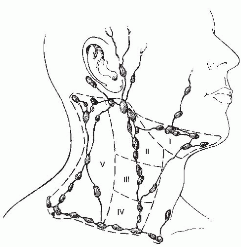
FIGURE 17-3. Anatomic lymph node levels of the neck.


Lymphatics of the Oropharynx

The lymphatic drainage of the neck was described by Rouviere in 1938 and has been refined by others.30,31,32 The lymph node groups are described by clinical levels I to V as depicted in Fig. 17-3.

The primary drainage of the oropharynx is to the jugulodigastric (level II) node(s) located in the upper deep jugular chain. The tonsillar region, pharyngeal portion of the soft palate, the lateral and posterior oropharyngeal walls, and the BOT are also drained by the RP and parapharyngeal nodes. These nodes are located in the RP and parapharyngeal space that is closely related to cranial nerves IX through XII, the internal jugular vein, and the internal carotid artery at the base of skull. The RP lymph nodes are subdivided into lateral (Rouviere) and medial nodal chains.33,34,35 The lateral nodes lie posterolateral to the nasopharynx and the oropharynx, just medial to the internal carotid artery. The parapharyngeal lymph nodes are also known as the junctional nodes, owing to the junction of the spinal accessory (level V) and the upper internal jugular lymphatic chains.

The probability of lymphatic metastasis is related to size and location of the primary tumor within the oropharynx. The order of progression of lymph node metastases usually proceeds superiorly, from the high cervical first-echelon nodes (level II) inferiorly to the midcervical and lower cervical nodes (levels III and IV). Skip metastasis can occur in which a particular lymph node level is bypassed, but this is very unusual. Candela et al. evaluated 333 previously untreated patients with SCC of the oropharynx or the hypopharynx to ascertain the prevalence of neck node metastases by neck level. The patients underwent classic radical neck dissections. Isolated skip metastases outside of level II, III, or IV occurred in only one patient (0.3%). Otherwise, level I or V involvement was always associated with nodal metastases at other levels.36 Metastases to the RP nodes are most commonly associated with cancers of the nasopharynx and the pharyngeal wall,37,38,39,40 but can also occur from other subsites, particularly with advanced disease.40,41,42 Notably, these metastases occur primarily along the lateral RP nodal chains. Involvement of the medial chain is extremely rare.30,38,40

In 1994, Hasegawa and Matsuura reported on a series of 24 patients with advanced carcinoma of the oropharynx and the hypopharynx treated with surgery and RP node dissection up to the base of the skull. This was followed by postoperative RT (PORT) of 50 Gy to RP space if nodes were pathologically involved. A total of 50% (12 of 24) of the patients had positive RP nodes: 1 of 5 (20%) with pathologic N0 neck and 12 of 19 (63%) with pathologic N-positive neck. More specifically, RP node positivity was established histopathologically in 36% (4 of 11 patients) and 61.5% (8 of 13 patients), respectively.43 In a retrospective study of 774 patients with SCC of the nasopharynx, the oropharynx, the hypopharynx, and the supraglottis, using CT scans for RP nodes evaluation, McLaughlin et al. found an overall incidence of radiologically positive RP nodes in 9% of the patients. The highest incidence was seen in patients with cancer of the nasopharynx (74%) and the pharyngeal walls (19%). They also noted that in patients with advanced cancer of the oropharyngeal walls and the hypopharynx, the incidence of radiologically positive RP nodes was higher in patients with positive cervical nodes than in those with an N0 neck (pharyngeal wall: N+ 21%, N0 16%; hypopharynx: N+ 9%, N0 0%).38

Shimizu et al. reported a rate of 13% of RP node metastases for OPC. They found that the risk was higher for patients with posterior and lateral pharyngeal wall cancers (29% for lateral and posterior wall vs. 0% for others sites).44

Dirix et al. reported a rate of RP nodal metastases of 16% in a series of 208 patients with OPC after evaluation of pretreatment CT scans. Patients with RP nodal metastases had significantly more regional recurrences (45% vs. 10%). RP nodal involvement independently predicted the likelihood of regional recurrence in multivariate analysis (p = 0.01). Furthermore, disease-specific survival (DSS) was significantly lower in the RP node-positive group (38% vs. 58%).45

Bussels et al. analyzed the same population of 208 patients with OPC. They reported RP node involvement in 23% of those patients with nodal disease in other neck levels. More frequently, the RP nodes were involved in the case of posterior pharyngeal wall (5 of 13 = 38%) and soft palate primaries (5 of 9 = 56%). Ipsilateral involvement of level II and contralateral involvement of level III predicted for involvement of the ipsilateral RP nodes on multivariate analysis (p < 0.05). A solitary ipsilateral RP node was present in 3 (9%) of 34 patients with RP nodes. Two of these three patients had a primary posterior pharyngeal wall tumor. No patients presented with a solitary contralateral RP node.46

Yoshimoto et al. reported their series of 84 patients with BOT cancer. Two patients had RP node metastases at the time of diagnosis and two more developed recurrent cancer in the RP node. Three of these four patients had a tumor extending to the lateral pharyngeal wall. They found that the incidence of contralateral or RP node metastasis was low if the tumor neither crossed the midline nor infiltrated the lateral wall.47

Chan et al. retrospectively reviewed the 18F-FDG PET-CT studies of 244 patients with SCC of the oro- and the hypopharynx. RP node involvement was identified in 17%.48

It is clear from the data that inclusion of the lateral RP nodes is an important part of the treatment. This is especially true for patients with cervical node metastases, but it is also potentially important for patients with N0 necks.

Tumors located in the midline (the BOT, the soft palate, and the posterior pharyngeal wall) exhibit a higher propensity for bilateral nodal metastases. The probability of cervical node metastases, as demonstrated by clinical and radiological examinations of the soft palate, the tonsillar fossa, the BOT, and the oropharyngeal wall, is shown in Tables 17.1, 17.2 and 17.3.41,49,50


TABLE 17.1 Percentage Incidence of Cervical Lymph Node Metastasis as Determined by Clinical Examination


Location


NO


N1


N2


Oropharyngeal wall


T1


75


0


25


T2


70


10


20


T3


33


23


45


T4


24


24


52


Soft palate


T1


92


0


8


T2


64


12


25


T3


35


26


39


T4


33


11


56


Tonsillar fossa


T1


30


41


30


T2


33


14


54


T3


30


18


52


T4


11


13


77


Base of tongue


T1


30


15


55


T2


29


15


57


T3


26


23


52


T4


16


9


76


Source: From Lindberg R. Distribution of cervical lymph node metastases from squamous cell carcinoma of the upper respiratory and digestive tracts. Cancer. 1972;29:1446-1449, with permission.


TABLE 17.2 Defining the Risk of Involvement for Each Neck Nodal Level in Patients with Early T-Stage/Node-Positive Human Papillomavirus-related Oropharyngeal Carcinoma


Neck Level


Pathologically Involved


Risk of Involvement When Negative on CT


IB


8.4%


2.8%


II


89.4%


72%


III


38.3%


16%


IV


20%


7%


V


2.6%


0.8%


Source: From Sanguineti G, Gunn GB, Parker BC, et al. Weekly dose-volume parameters of mucosa and constrictor muscles predict the use of percutaneous endoscopic gastrostomy during exclusive intensity-modulated radiotherapy for oropharyngeal cancer. Int J Radiat Oncol Biol Phys. 2011;79:52-59, with permission.


TABLE 17.3 Percentage Incidence of Cervical Lymph Node Metastasis in Soft Palate Cancer


Nodal Group


Ipsilateral (%)


Contralateral (%)


I


9


6


II


86


29


III


26


6


IV


6


0


V


0


3


Supraclavicular


0


0


Tonsillar fossa


I


16


3


II


97


13


III


24


3


IV


13


2


V


23


7


Supraclavicular


0


0


Base of tongue


I


8


1


II


88


31


III


40


7


IV


9


3


V


12


2


Supraclavicular


4


1


Source: From Mukherji SK, Armao D, Joshi VM. Cervical nodal metastases in squamous cell carcinoma of the head and neck: what to expect. Head Neck. 2001;23:995-1005, with permission.


PATHOLOGY

More than 90% of tumors of the oropharynx are SCC, the remainder being malignant melanomas, minor salivary gland tumors, sarcomas, plasmacytomas, lymphomas, and other rare tumors.51 Benign and malignant tumors that can be found in the oropharynx are listed in Table 17.4.


Clinical Presentation, Pathogenesis, and Patterns of Spread

Patients with OPC are frequently asymptomatic until their primary tumors reach a significant size or metastasize to a lymph node in the neck. The usual complaints consist of vague discomfort or irritation, or a mass in the neck. The manifestation of symptoms depends upon the location of the primary tumor. Some tumors are visualized easily, but others can be insidious and are only found by careful examination of all the mucosal surfaces of the oropharynx. Pain can be a presenting complaint, which can be attributed either to deep infiltration of tumor or to referred pain. The following sections list some of the possible clinical scenarios based on the site of the primary tumor.


TABLE 17.4 Differential Diagnosis of an Oropharyngeal Mass


Malignant


Benign


Squamous cell carcinoma


Papilloma


Minor salivary gland tumor


Retention cyst


Lymphoma


Fibroma


Sarcoma


Lipoma


Melanoma


Hemangioma


Plasmacytoma


Lymphangioma


Other


Neuroma


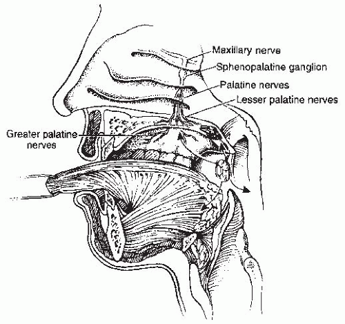
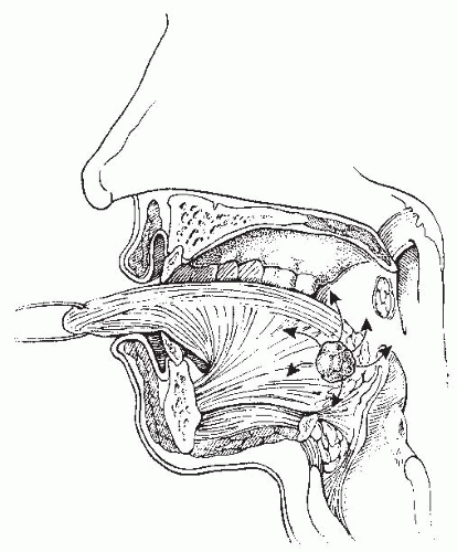
Soft Palate. Tumors of the soft palate are almost exclusively found on the oropharyngeal as opposed to the nasopharyngeal surface (Fig. 17-4). Tumors can extend to involve the tonsillar pillars and the BOT. Occasionally, these lesions may extend laterally and superiorly as far as the nasopharynx. Involvement of the palatine nerve can result in tumor tracking along this pathway, with extension to the skull base. Lymphatic involvement is primarily to level II. Lesions of the midline and the uvula can result in bilateral nodal metastases more frequently than do lateralized lesions. The RP nodes are also at risk.52

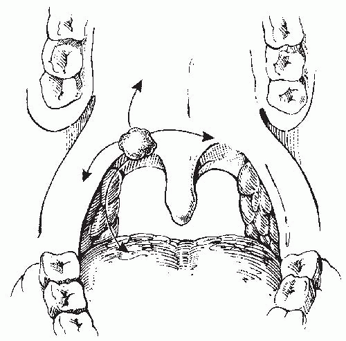
Tonsil. The tonsil is the most common location for a primary tumor of the oropharynx. Common presenting symptoms are ipsilateral referred otalgia, odynophagia, or sensation of a lump or foreign body in the throat. Lesions involving the anterior tonsillar pillar can appear as areas of dysplasia, inflammation, or a superficial spreading or exophytic lesion. Frequently, these lesions become endophytic, ulcerate, and can spread laterally to the buccal mucosa and directly to the retromolar trigone (Fig. 17-5). Inferior extension to the BOT can occur. Perez et al. reported that 60% of tonsil cancers involved the soft palate and 55% extended into the BOT.53 Superior extension can even involve the hard palate. Medial extension can involve the oral tongue. The close proximity of the anterior tonsillar pillar to the mandible places the periosteum and the bone at risk for tumor involvement in advanced cases. Posterior and superior extension with destruction of the tonsillar pillars can lead to involvement of the pterygoid muscles, with subsequent trismus and pain. The lymphatic drainage is primarily to level II but can also involve RP nodes, as previously discussed. Patients with neck node involvement are at higher risk for RP node metastases.38


FIGURE 17-4. Patterns of tumor spread in soft palate.


FIGURE 17-5. Patterns of spread of tonsillar carcinoma.

Tumors of the tonsillar fossa (in contrast to those of the tonsillar pillar) are either exophytic or ulcerative and present in more advanced stages than do tumors of the pillars or soft palate. Approximately 75% of patients will present with stage III or IV disease, for which the patterns of extension are similar to those of the tonsillar pillar. In addition, lateral extension can involve the parapharyngeal space toward the base of skull, causing neurologic signs and symptoms. Tumors of the posterior tonsillar pillar can extend inferiorly and involve the pharyngoepiglottic fold and the posterior aspect of the thyroid cartilage.

The probability of clinical lymph node involvement is greater with tumors of the tonsillar fossa in contrast to that of the tonsillar pillar. The lymphatic drainage depends upon the location of the primary tumor. Lindberg describes nodal metastases in 76% of patients with tonsillar fossa tumors.41 The most common nodal group was level II. Contralateral lymph nodes were detected in 11% of patients. In contrast, tumors of the anterior tonsillar pillar or retromolar trigone region have an incidence of ipsilateral lymph node metastases of 45%, level II being the most common node-bearing region. Contralateral adenopathy was present in only 5% of patients. The issue of ipsilateral versus bilateral neck metastasis is discussed later in this chapter.

Base of Tongue. Squamous cell carcinoma of the BOT is highly insidious. The BOT is almost devoid of pain fibers. Frequently, these tumors are asymptomatic until they have progressed significantly. Patients with cancers of the BOT often present with neck node metastases, only to be found to have a BOT lesion upon full evaluation. Visualization of this area on physical examination can be difficult, and a lesion can be often missed.54
Patients may experience the sensation of a mass or discomfort in the throat, with bleeding and pain at later stages. Patients might also experience difficulty with speech and swallowing. Occasionally, referred otalgia is the first symptom.


FIGURE 17-6. Patterns of spread of base of tongue cancer.

Extension anteriorly can involve the oral tongue, superior and lateral extension can involve the tonsil, and inferior extension can involve the vallecula, the epiglottis, and the preepiglottic space (Fig. 17-6). Locally advanced BOT tumors can infiltrate the deep muscle and cause fixation. Larynx and hypopharynx involvement can occur with advanced, inferior extension.

Lymph node metastasis is common, owing to the rich lymphatic drainage of the BOT. The most common first-echelon nodal region is level II, though involvement of levels III and IV is also seen. Approximately 70% of patients or more will present with ipsilateral metastases, and 10% to 20% will present with bilateral nodal metastases.41

Pharyngeal Wall. Tumors of the pharyngeal wall are generally diagnosed in an advanced stage due to the silent location in which they develop. Symptoms can include pain and bleeding and a mass in the neck. Disease involvement can extend to the nasopharynx superiorly, the prevertebral fascia posteriorly, and the pyriform sinuses and the hypopharyngeal wall inferiorly. Clinically palpable cervical lymph node metastases are present in 25% of patients with T1 lesions, 30% of those with T2 lesions, 66% of patients with T3 lesions, and >75% of patients with T4 disease. Because most pharyngeal wall tumors extend past the midline, bilateral cervical metastases are common. RP nodes are also at great risk.38

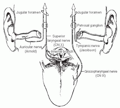
Referred Otalgia. Referred otalgia can be one of the first symptoms that a patient experiences with a mass in the oropharynx. The pathway for this referred pain is mediated by cranial nerves IX and X. The pathophysiology for otalgia is demonstrated in Figure 17-7.


FIGURE 17-7. Neural pathways of referred otalgia. For pain sensed in the front of the helix and tragus, the skin of the anterior wall of the external auditory canal, the tympanic membrane, and the temple, pain is referred through the auriculotemporal nerve, which joins the lingual nerve, where the two become the mandibular nerve and enter the foramen ovale. The sensation of deep ear pain is through the tympanic nerve (Jacobson), which joins the glossopharyngeal nerve (cranial nerve IX) as the two traverse the jugular foramen. These general somatic afferent fibers innervate the base of tongue, the inner surface of the tympanic membrane, and the upper pharynx. For pain sensed in the back of the pinna, the posterior wall of the external auditory canal, and the external surface of the tympanic membrane, pain is referred through the auricular nerve (Arnold), which joins the superior laryngeal nerve (cranial nerve X), which in turn innervates the larynx, the pharynx, and the epiglottis as it traverses the jugular foramen.


Diagnostic Evaluation

History. The history should be part of a comprehensive evaluation of any patient with head and neck cancer. Patients can present with sore throat, odynophagia, otalgia, foreign body sensation in the throat, or a neck mass. If the history strongly reveals tobacco and alcohol use, efforts should be made to determine whether the patient is an alcoholic and continues to smoke. Alcoholic smokers are at risk for other chronic diseases of the heart, lungs, peripheral vascular system, liver, kidneys, and may present with signs of malnutrition. Before the institution of any therapeutic modality, the pathology material should be evaluated for HPV/p16 tumor status. All patients should cease alcohol and tobacco use. It may be necessary to guide the patient toward cessation programs. Clearly, those who discontinue smoking will better tolerate treatment and obtain a better result.55

Physical Examination. The details of the physical examination are discussed in Chapter 1. Specific aspects of the physical examination should include evaluation of the lesion (exophytic or infiltrative), tongue mobility, and palatal motion. Fixation of the tongue will result in incomplete protrusion or deviation of the tongue to the side of tumor involvement. For tumors of
the tonsil or the lateral pharyngeal wall, the examiner should test for anesthesia in the distribution of the ipsilateral mandibular nerve (V3). Any abnormality might suggest involvement of the inferior alveolar nerve in its pathway as it courses through the mandible or the base of skull and may direct the appropriate imaging study.56 Because the tonsil is adjacent to the ascending ramus of the mandible, the BOT, and the parapharyngeal space, tumors may extend into these areas. This can often be detected by palpation. Indirect mirror examination and flexible endoscopy are important parts of the physical examination. It is useful to take a photograph or extensively diagram the physical findings in every patient. This can include a videotape. Such records are an excellent means of documenting the physical findings and of comparing future examination results to the initial presentation. In selected cases, an examination under anesthesia is recommended as a mean of obtaining information that is not completely accessible during office examination.

Initial Workup and Radiographic Evaluation. In addition to a his tory and physical examination, a complete blood cell count and metabolic profile are recommended. Biopsy of a suspicious lesion is necessary to confirm the diagnosis.57 The overwhelming majority of patients will also undergo PET-CT imaging as a key part of the staging evaluation. PET-CT may remove the need for a chest x-ray in many patients. See Chapter 4 for further discussion of general principles of head and neck imaging.


TABLE 17.5 American Joint Committee on Cancer (AJCC) Classification of Oropharyngeal Cancer


Primary tumor (T)


Stage


T1: Tumor ≤2 cm in greatest dimension


Stage 0


Tis


N0


M0


T2: Tumor >2 cm by not >4 cm in greatest dimension


Stage I


T1


N0


M0


T3: Tumor >4 cm in greatest dimension or extension to lingual surface of epiglottis


Stage II


T1


N0


M0


T4A: Moderately advanced local disease; Tumor invades the larynx, extrinsic muscle of tongue, medial pterygoid, hard palate, or mandible*


* Mucosal extension to lingual surface of epiglottis from primary tumors of the base of the tongue and vallecula does not constitute invasion of larynx.


T4B: Very advanced local disease: Tumor invades lateral pterygoid muscle, pterygoid plates, lateral nasopharynx, or skull base or encases carotid artery


Regional lymph nodes (N)


Nx: Regional lymph nodes cannot be assessed


* Metastases at Level VII are considered regional lymph node metastases.


N0: No regional lymph node metastasis


Stage III


T3


N0


M0


N1: Metastasis in a single ipsilateral node, <3 cm


T1-3


N1


M0


N2A: Metastasis in a single ipsilateral node, >3 cm but ≤6 cm


Stage IVA


T4A


N0-2


M0


N2B: Metastasis in multiple ipsilateral nodes, >3 cm but ≤6 cm


T1-3


N2


M0


N2C: Metastasis in bilateral or contralateral lymph nodes, none >6 cm


Stage IVB


Any T


N3


M0


N3: Metastasis in lymph node >6 cm


T4B


Any N


M0


Distant metastasis (M)


M0: No distant metastasis present


M1: Distant metastasis present


Stage IVC


Any T


Any N


M1


Source: From Edge SB, Compton CC. The American Joint Committee on Cancer: the 7th edition of the AJC cancer staging manual and the future of TNM. Ann Surg Oncol. 2010;17:1471-1474, with permission.


Staging. The current staging criteria for tumors of the oropharynx, as defined by the American Joint Committee on Cancer (AJCC),58 are listed in Table 17.5. This is a clinical staging system and not a pathologic staging system. If radiographic information reveals a discrepancy from clinical staging, this should be noted. Current staging allows the radiographic findings to factor into the clinical staging designation.

Dental Evaluation and Prophylaxis. Treatment of OPC can result in a number of temporary and permanent effects on dental hygiene. Radiation-related xerostomia places dentulous patients at increased risk for dental caries, owing to the reduction in salivary flow, pH alteration, and proliferation of bacteria. A complete dental evaluation should be performed before any therapy is undertaken. See Chapter 9 for further discussion of dental oncology and maxillofacial prosthetics.


Prognostic Factors

Traditionally, OPC prognostic factors for LRC, disease-free survival (DFS), or OS included TNM classification, location of the tumor, gender, age, and performance status.59,60 As previously mentioned, in the current era, the impact of smoking and HPV/p16 tumoral positivity on OPC oncologic and functional outcomes has evolved remarkably.61 A recent SEER analysis showed that the overall 5- and 10-year OS were approximately two times
better for those patients with HPV-positive disease regardless of the treatment modality.1 Although HPV/p16-positive patients have better outcomes than HPV/p16-negative patients, this advantage disappears in the HIV-positive population and heavy smokers. Mourad et al. reported on 73 HIV-positive patients with SCCHN, of whom 24 patients had OPC (12 of whom were HPV/p16 positive).62 The outcomes revealed no difference between HPV/p16-positive versus HPV/p16-negative in the setting of HIV-positive patients and no advantage to chemo-RT over RT alone. These data are limited by their retrospective nature, but suggest that HIV status is an important prognostic factor.63 Gillison et al. reported that the risk of OPC progression and death increases directly as a function of tobacco exposure at diagnosis and during therapy and is independent of tumor p16 status and treatment.8 Smoking-induced OPC has been associated with mutated p53, epidermal growth factor receptor (EGFR) overexpression, and lower HPV/p16 expression.64 Thus, given its unique prognostic significance, HPV is reviewed in detail in Chapter 12. In addition, Chapter 3 reviews all other important prognostic factors.

Chan and McBride et al. reported that active smoking during and after RT is predictive for decreased DSS, OS, progressionfree survival (PFS), and distant metastasis-free survival (DMFS) rates.65,66 In addition to smoking, anemia around the time of RT is associated with higher rates of persistent/recurrent disease,60,67,68 and its correction may improve outcome.69

Multiple studies have investigated various molecular markers as potential predictive factors in patients treated definitely for OPC. Single-institution studies have reported that a high intratumoral microvessel density predicts for poorer LC and worse OS after definitive RT.70 High Ki-67 labeling index is associated with local relapse71 and shorter time to relapse72 after definitive treatment. Shoushtari et al. reported that tissue microarray analysis using p16, EGFR, and HIF-1α for OPC could provide prognostic information.73 These markers need validation in larger, multi-institutional trials.

Age is an important factor in the OPC management paradigm. Meta-analysis has shown that the effectiveness of chemo-RT and altered RT fractionation decreases with increasing age. For patients older than 70 years, there was no difference in survival for either chemo-RT or altered fractionation RT over conventional RT alone.74,75,76,77 A similar observation regarding older patients (≥65 years) was also reported by Garden et al.78 Likewise, Bonner et al. reported that the effectiveness of RT plus cetuximab versus RT alone decreases with the increase in age.79,80 Recently, Michal et al. challenged the dogma that elderly patients (≥70) may not benefit from concomitant chemotherapy. They explored two cycles of concomitant cisplatin with RT. Older patients did experience greater myelosuppression and required more supportive care. However, their outcomes were the same as younger patients. Moreover, they recommended that age alone is not a contraindication to primary chemo-RT for locally advanced SCCHN.81


MANAGEMENT STRATEGIES, RESULTS, AND OUTCOMES


General Principles

Treatment decisions are always a balance between tumor control, cost-effectiveness, and QOL. The best decisions are usually made in a multidisciplinary context. It is very important to understand the goals and preferences of the patient before finalizing any recommendation. In general, early-stage disease can be treated by either RT or surgery, whereas more advanced disease introduces the potential use of chemotherapy and multimodality therapies into the discussion. RT has the advantage in many cases because it can be used as a single modality, it comprehensively treats the primary site and the neck, and it provides excellent functional outcomes. The advent of MIS, such as TORS and TLM, has augmented the role of surgery in early-stage diseases and will be discussed. For some patients, the use of TORS or TLM can reduce the need for RT, providing a trade-off, which can be attractive. Chemotherapy is generally reserved for patients with advanced disease. In the following sections, the management guidelines, outcomes, and results of OPC will be reported.


SELECTION OF THERAPEUTIC MODALITY


General Principles

Early-stage Disease. Early-stage OPCs can be treated with primary RT or primary surgery. RT is often used because it comprehensively includes the primary site, neck nodes, RP nodes, lymphatic channels in transit, and produces excellent long-term results. The increasing adoption of intensity-modulated radiotherapy (IMRT) with the addition of image-guided radiotherapy (IGRT) has led to high rates of LRC with low incidence of long-term functional morbidity.

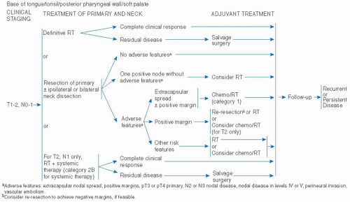
Surgery involves the use of TLM or TORS for the primary site. This needs to be combined with a separate procedure to remove the neck nodes. Surgery may not be the only treatment needed, as patient may require additional treatment with RT, or chemo-RT, depending upon the pathologic findings in the primary tumor and the neck. Moreover, surgery generally does not address the RP nodes, which is an area at risk. Complete and multidisciplinary discussion is essential for every patient so that the best management decisions can be made. A fundamental management principle for early-stage disease is to maximize the likelihood of treatment with single-modality therapy by either RT or surgery alone. The best outcome for the patient is when one treatment modality is used. Multimodality therapy invariably increases morbidity and cost and should be unnecessary in most cases. Figure 17-8 shows the National Comprehensive Cancer Network (NCCN) 2012 guidelines for early-stage OPC treatment.

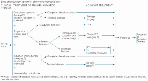
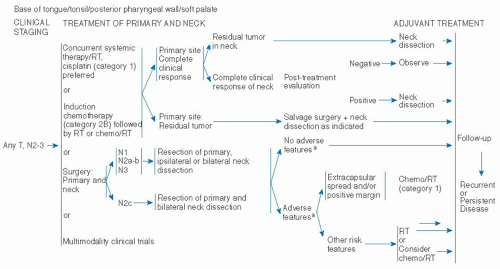
Advanced-stage Disease. For patients with more advanced disease, combined therapy options are generally preferred. However, treatment needs to be individualized. See Figures 17-9 and 17-10 for the algorithm of care for advanced-stage OPC. In general, concomitant chemo-RT is the preferred management for most patients with stages III and IVA/B disease.82,83 RT alone is often considered for certain patients, especially those with low bulk stage III lesions (i.e., T1-2N1M0).63 Similarly, primary surgery may be used for selected patients with low bulk stage III disease (Hurtuk et al., 2012).83a If there are negative margins on the primary site resection, and the neck specimen is negative, surgery alone may be adequate. For other stage III patients, pathologic analysis may reveal positive nodes, inadequate margins, or other adverse features such as perineural invasion or lymphovascular invasion. This would trigger the use of PORT, or even postoperative chemo-RT, especially if the margins are positive or there is extranodal spread.84,85

In selecting stage III patients for surgery, it is important to try to choose patients who are unlikely to have indications for chemotherapy (positive margins or extracapsular spread), to minimize treatment morbidity. If a patient turns out to need postoperative chemo-RT, then it is probably true that the patient could have been treated definitively with primary chemo-RT and the surgery has not added value to the outcome. Indeed, it may have only added cost and toxicity. On the other hand, if the pathologic findings only call for PORT alone, or no adjuvant therapy, then the surgical procedure may have saved the patient from chemotherapy or RT, thus providing a trade-off, which may
be attractive to certain patients. These complex issues require the patient to have a thorough understanding of all the options and management algorithms, so that a personalized choice can be made. Multidisciplinary evaluation and discussion is mandatory.


FIGURE 17-8. National Comprehensive Cancer Network (NCCN) 2012 guidelines for treatment of early-stage cancer of the oropharynx. All recommendations are category 2A unless otherwise indicated. NCCN believes that the best management of any cancer patient is in a clinical trial; participation in clinical trials is especially encouraged. Chemo, chemotherapy; RT, radiation therapy.

Source: Adapted from the NCCN Guidelines.


FIGURE 17-9. National Comprehensive Cancer Network (NCCN) 2012 guidelines for treatment of locally advanced cancer of thvthe best management of any cancer patient is in a clinical trial; participation in clinical trials is especially encouraged. Chemo, chemotherapy; RT, radiation therapy.

Source: Adapted from the NCCN Guidelines.


FIGURE 17-10. National Comprehensive Cancer Network (NCCN) 2012 guidelines for treatment of regionally advanced cancer of the oropharynx. All recommendations are category 2A unless otherwise indicated. NCCN believes that the best management of any cancer patient is in a clinical trial; participation in clinical trials is especially encouraged. Chemo, chemotherapy; RT, radiation therapy. For more information, consult the specific NCCN guidelines at the website ww.nccn.org.

Source: Adapted from the NCCN Guidelines.

For stage IVA/B disease, concomitant chemo-RT is the mainstay of treatment. In general, this involves concomitant cisplatin chemotherapy with IMRT. Full-dose RT is delivered to the primary site and neck. The current paradigm calls for a PET-CT scan to be done as part of the baseline evaluation. Three months after the completion of chemo-RT, a repeat PET-CT scan is done for evaluation of response and disease status. If the scan is negative, no further treatment is needed.86 If the scan shows residual disease in the neck, then a neck dissection (ND) is performed. If the scan is equivocal in the neck, management needs to be individualized. Some patients will get ND.87 Others will have close follow-up to assess the evolution of the findings. Sometimes, a fine-needle aspiration can help determine whether there is residual disease warranting surgery, or whether the findings are likely benign, warranting close follow-up alone, with ND reserved for salvage. Refer to Chapters 4 and 14 for comprehensive discussion on follow-up PET-CT scan and neck management, respectively.

For patients treated with primary surgery, adjuvant RT or chemo-RT will be indicated in the overwhelming majority of cases. Therefore, it is essential to balance any potential benefit of surgery with the morbidity, given the reality that chemo-RT will be needed anyway. In selected circumstances, patients with very advanced disease at either the primary site (e.g., T4) and/or neck (e.g., N3 or low neck disease) have been treated with an induction chemotherapy approach.68,88,89,90,91,92,93 The rationale for this sequence is related to both the aggressive nature of the systemic therapy, as well as the greater risk of distant metastases in this cohort of patients. However, results from recent clinical trials have not shown any clear benefit from induction over concomitant chemo-RT,94,95 thereby questioning the appropriateness of induction chemotherapy outside of a clinical trial.

For stage IVC, management decisions must be individualized and will be determined by the extent of the loco-regional disease, resectability, presence and extent of distant metastases, and the performance status of the patient. Thus, careful balance of morbidity, cost, and oncologic outcome must be considered. As mentioned earlier, these complex issues require the patient to have a thorough understanding of all the options and management algorithms, so a personalized choice can be made. Multidisciplinary evaluation and discussion is mandatory.


SITE-SPECIFIC MANAGEMENT STRATEGIES, RESULTS, AND OUTCOMES


Soft Palate

Early-stage Disease. Treatment of soft palate cancer must address the primary site as well as both sides of the neck including bilateral RP nodes.50,96,97,98

Chera et al. reported on 59 patients with SCC of the soft palate, who presented with early-stage disease. All patients underwent
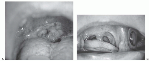
primary RT. Stage I disease included 23 patients (16%) and stage II disease included 36 patients (25%). The 5-year initial and ultimate (i.e., after successful salvage) LRC rates for stage I were 84% and 89%, respectively. The 5-year initial and ultimate LRC rates for stage II were 85% and 88%, respectively. Specifically, the LC rates for both T1 and T2 were 90%. Nodal control rate at 5 years for N0 disease was 90%. The 5-year freedom from distant metastases rate was 95% for stage I and 97% for stage II. The 5-year DSS for stages I and II were 89% and 87%, respectively.97 Figure 17-11 shows a T2 SCC of the soft palate before and after RT.


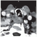
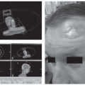
FIGURE 17-11. Patient with T2N0 squamous cell cancer of the soft palate before and after radiation therapy. This patient was treated with external beam radiation therapy to the primary site and both sides of the neck. The patient currently has no evidence of disease, more than 7 years after treatment.

Iyer et al. reported on patients, with SCC of the soft palate, treated with primary surgery. With a median follow-up of 52 months, the 5-year DFS for stages I and II were 70% and 53%, respectively. Specifically, the 5-year LC for stages I and II were 85% and 84%, respectively. The 5-year regional control (RC) for stages I and II were 83% and 74%, respectively. The 5-year distant control (DC) for stages I and II were 96% and 81%, respectively. The 5-year DSS for stages I and II were 87% and 66%, respectively (Table 17.6).98

Advanced-stage Disease. Chera et al. reported on 86 patients presented with advanced disease SCC of the soft palate. All patients underwent primary RT. Stage III disease included 23 patients (16%) and stage IVA/B diseases included 51 (35%) and 12 patients (8%), respectively. The 5-year initial and ultimate LRC rates for stage III were 66% and 96%, respectively. The 5-year initial and ultimate LRC rates for stage IVA were 85% and 88%, respectively, and for stage IVB were 43% for both. Specifically, the LC rate at 5 years was 67% for T3 and 57% for T4. Nodal control rates at 5 years were N1, 82%; N2, 68%; and N3, 71%. The 5-year rates of freedom from distant metastases were stage III, 100%; stage IVA, 93%; stage IVB, 69%; and overall, 94%. The 5-year DSS for stage III, IVA, and IVB were 88%, 57%, and 0%, respectively. In multivariate analysis, overall treatment time significantly affected local and ultimate LRC, and nodal stage significantly affected OS.97

Iyer et al. reported on patients with SCC of the soft palate, 81% underwent primary surgery, of whom 36% underwent surgery + PORT, and 19% underwent primary RT ± chemotherapy. With a median follow-up of 52 months, the 5-year DFS for stages III and IV were 54% and 36%, respectively. Specifically, the 5-year LC for stages III and IV were 88% and 54%, respectively. The 5-year RC for stages III and IV were 77% and 73%, respectively. The 5-year DC for stages III and IV were 72% and 83%, respectively. The 5-year DSS for stages III and IV were 64% and 55%, respectively, and for OS for stages III and IV were 55% and 37%, respectively.98 In total, in the surgical cohort, 43% developed recurrent disease: 19% local recurrences, 20% nodal recurrences, and 12% distant
metastases. Patients with early disease (n = 90) had a DSS of 79%, whereas T1-2 but with neck-positive nodes (n = 22) had a DSS of 56%. For OS and DSS, N classification was predictive of outcome. For LC and DC, margin status was a significant predictor, whereas the T classification was only a significant predictor for LC.98


TABLE 17.6 Five-year Oncologic Outcomes of Primary Surgery ± Postoperative Radiation Therapy in Soft Palate Cancer


5-yr Outcomes (%)


Stage


LC


RC


DC


DFS


OS


DSS


I


85


83


96


70


60


87


II


84


74


81


53


54


66


III


88


77


72


54


55


64


IV


54


73


83


36


37


55


DFS, disease-free survival; LC, local control; RC, regional control; DC, distant control; DSS, disease-specific survival; OS, overall survival.


Source: From Iyer NG, Nixon IJ, Palmer F, et al. Surgical management of squamous cell carcinoma of the soft palate: factors predictive of outcome. Head Neck. 2012;34:1071-1080.


Many single institutional reports have shown relatively similar outcomes.99,100,101,102,103,104,105,106,107,108 In the modern era, results for loco-regionally advanced OPC are often combined for all primary sites, making it very difficult to extract the specific outcomes for each anatomic subsite. Most patients have primary lesions of either the tonsil or BOT, with fewer soft palate and pharyngeal wall lesions. Refer to the sections of this chapter titled “Selection of Therapeutic Modality—General Principles” and “Management of Advanced Stage Disease—Principles of Combining Chemotherapy and Radiation Therapy” for additional data and discussion related to OPC, all sites combined. Chemo-RT is the mainstay of management of loco-regionally advanced OPC, and this is reviewed for OPC sites in the section “Management of Advanced Stage Disease—Principles of Combining Chemotherapy and Radiation Therapy.”


Pharyngeal Wall Tumors

Early-stage Disease. Treatment of pharyngeal wall tumors must address the primary site and both sides of the neck, including bilateral RP nodes. Hull et al. reported on 37 patients with pharyngeal wall tumors treated with primary RT. Five-year LC rates for T1 and T2 were 93% and 82%, respectively. On multivariate analysis, factors associated with improved LRC included twice-daily fractionation (p = 0.0009), stages I to II disease (p = 0.0051), and oropharyngeal primary site (p = 0.0193). The same group previously reported the effect of the use of once-daily versus twice-daily fractionation on LC. The LC rates for patients treated with once-daily versus twice-daily fractionation were as follows: T1—100% versus 100% and T2—67% versus 92% (p = 0.0009). These investigators also examined their outcomes as a function of RT technique. They found superior outcomes when using a more modern technique where there was more generous anatomic coverage of the prevertebral area and adjacent vertebral body. The LC rate for T1 was 100%. The LC rate for T2 was 57% using older techniques versus 100% for the modern approach, emphasizing the importance of including the entire prevertebral space into the target volume.109,110

Mendenhall et al. recently reported the results of primary RT. The 5-year LC and ultimate LC were: T1—93% and 93%, respectively; T2—84% and 91%, respectively. Multivariate analysis revealed stages I to II tumors, female gender, and altered fractionation were associated with improved LRC. The 5-year DSS and OS were as follows: stage I—88% and 50%, respectively; stage II—89% and 57%, respectively.111

Spiro et al. reported on 78 patients with posterior pharyngeal wall tumors treated with primary surgery (n = 60) or primary RT (n = 18). Survival was greater in those with stage I and stage II tumors, but the difference was significant only in comparison to those with stage IV disease. Local recurrence occurred in 16% and 21% for stages I and II, respectively.112

Advanced-stage Disease. Hull et al. reported the outcomes of primary RT. The 5-year LC rates for T3 and T4 tumors were 59% and 50%, respectively. The LC rates for patients treated with once-daily versus twice-daily fractionation were as follows: T3—43% versus 80%; and T4—17% versus 50% (p = 0.0009). These investigators also examined their outcomes as a function of RT technique. Similar to their finding for T2 disease, they found superior outcomes when using a more modern technique where there was more generous anatomic coverage of the prevertebral area and adjacent vertebral body. The LC rate for T3 was 46% for the older technique versus 73% for the modern approach. The LC rate for T4 was 29% for the older technique versus 75% for the modern approach, emphasizing the importance of including the entire prevertebral space into the target volume.109,110 The current practice would employ IMRT, and issues of target coverage and spinal cord protection become easier to assure.

Mendenhall et al. recently reported the results of primary RT. The 5-year LC for T3 was 60% and T4 was 44%. Multivariate analysis revealed female gender, and altered fractionation were associated with improved LRC. The 5-year DSS and OS were the following: stage III—49% and 31%, respectively, and stage IV—35% and 21%, respectively. Fatal complications occurred in nine patients (5%).113

Spiro et al. reported on 78 patients with posterior pharyngeal wall tumors treated with primary surgery (n = 60) or primary RT (n = 18). Local recurrence occurred in 37% and 63% for stage III and IV, respectively. Neck failure was noted in 21% of patients. Distant metastasis was documented in 8%.112

Julieron et al. reported the outcomes for primary surgery ± RT. In their hands, primary surgery followed by RT produced the best outcomes. For combined therapy, the rate of local failure (LF) was 11% and the 5-year survival rate was 35%. In the group of patients who developed LF post primary RT, the 5-year survival rate was 16%.114 In the modern era, results for loco-regionally advanced OPC are often combined for all primary sites, making it very difficult to extract the specific outcomes for each anatomic subsite. Refer to the sections of this chapter titled “Selection of Therapeutic Modality—General Principles” and “Management of Advanced Stage Disease—Principles of Combining Chemotherapy and Radiation Therapy” for additional data and discussion related to OPC, all sites combined. Chemo-RT is the mainstay of management of loco-regionally advanced OPC, and this is reviewed for OPC sites in the section “Management of Advanced Stage Disease—Principles of Combining Chemotherapy and Radiation Therapy.”


Tonsillar Region

Early-stage Disease. Treatment of tonsil cancer must address the primary site as well as the regions of the neck at risk. There is a growing body of evidence to support the strategy of unilateral treatment to the ipsilateral neck alone, including the ipsilateral, lateral RP nodes.115,116,117 Unilateral treatment provides the patient with a significant QOL advantage, both with respect to acute toxicity and late functional outcomes. Lesions that approach the midline or involve the tongue base or glossotonsillar sulcus are not good candidates for unilateral treatment and should be treated to both sides of the neck. Treatment planning must ensure protection of critical normal structures, which is easily achieved with image-guided IMRT. Treatment technique will be further described in Chapter 17, Part C.

Eisbruch et al. have demonstrated that patients treated unilaterally report less xerostomia and better QOL compared to those patients treated bilaterally, even with IMRT, which spares the contralateral parotid gland.118 However, careful patient selection is required to minimize the risk for contralateral neck failure. Multiple reports document excellent outcomes using ipsilateral neck RT.

Jackson et al. report on 178 patients receiving ipsilateral treatment for tonsillar cancers. Patients presented primarily with T1-2 (66%) and N0 (57%) disease. The rates of LRC and contralateral neck recurrence by stage were as follows: stage I—91% and 0%, respectively; stage II—74% and 2%, respectively. None of the patients with N0 disease had experienced contralateral failure. The LC rate was 84% for T1-2.115

O’Sullivan et al. reported on 228 tonsillar carcinomas treated with ipsilateral RT. Eighty-four percent of patients had
T1-2 tumors of which 70% with N0. At a median follow-up of 5.7 years, none of the patients with T1 or N0 lesions had experienced contralateral neck failure. The median 3-year LC rate for stages I and II were 91% and 76%, respectively. The 3-year median ipsilateral RC for stages I and II were 98% and 91%, respectively. For the whole cohort, the 3-year cause-specific survival was 76%. The only patients subsets with significant risk of contralateral neck failure (>10%) were those with tumor involvement of the medial one-third of palate or BOT.116

Kagei et al. reported 32 patients with SCC of the tonsil and soft palate treated unilaterally. Each tumor did not cross midline and demonstrated no obvious contralateral disease. With a median follow-up of 3.7 years, there were no contralateral neck failures.119

Levendag et al. reported on 190 patients with T1-3 N0-3 SCC of the tonsil and soft palate. All patients underwent primary RT followed by surgery or brachytherapy boost (BT). The tumor control rates after BT versus surgery at 5 years were 88% versus 88% for LC; 93% versus 85% for RC; 57% versus 52% for DFS; 67% versus 57% for OS; and 5% versus 6% for RF. They concluded that there was no need to treat the contralateral neck unless the tumor extends beyond the midline of the soft palate (uvula) or beyond the lateral one-third of the ipsilateral BOT.120

Hu et al. performed one of the few prospective trials assessing the safety of unilateral neck treatment.117 The eligibility criteria required patients to have primary lesions >1 cm from midline, no involvement of the BOT, and PET-CT documentation of the absence of contralateral neck metastasis. With a median follow-up of 16 months, there were no failures in the contralateral neck. These criteria seem reasonable to use in making the decision to exclude the contralateral neck.

The oncologic outcomes for early-stage tonsil cancer with primary RT have been excellent. Mendenhall reported on 503 patients with tonsillar cancer, who underwent definitive RT. Five-year rates of LC were as follows: T1—88% and T2—84%. The ultimate LC rates (after salvage surgery) at 5 years were T1—94% and T2—91%. Five-year cause-specific survivals were as follows: stage I—100% and stage II—86%.121

It is important to recognize that the shift of patient characteristics toward younger, healthier, nonsmoker, more favorable prognosis of the HPV-related cancer, as well as the major advances in RT delivery, have had a major impact on outcomes and QOL in the modern era. Thus, current outcomes are much better than the prior era.61,63,122,123,124,125,126

Mourad et al. reported on 79 patients with SCC of the tonsil who underwent primary RT.127 After a median follow-up of 56 months, the 5-year local control (LC), RC, DC, and OS for stages I/II combined were all 100%.

Garden et al. reported on 777 patients with OPC. The median follow-up was 54 months. The 5-year LRC, DFS, and OS rates were 90%, 82%, and 84%, respectively. Five-year DFS was 90% in never smokers with T1-2 disease. The 5-year DFS for N0 was 92%.128 Table 17.7 shows the excellent outcomes of OPC as reported in the English literature. It is clear that the overwhelming majority of patients with early-stage tonsil cancer achieve excellent LRC with primary RT.

Primary surgical treatment is also an option for early-stage tonsillar cancer. Most surgical series report outcomes by T and N classification and is not always possible to determine the outcomes by overall stage. Thus, there are some limitations in the interpretation of the surgical literature related to this point.

The use of primary surgery, via conventional approach, as definitive treatment for early tonsillar disease is not reported frequently. However, excellent LC rates ranging from 80% to 90% have been reported.

Moncrieff et al. retrospectively reported on 92 patients with early-stage OPC, of whom 54% had tonsillar cancer. All patients underwent primary surgery, 76% underwent ND, and 62% underwent PORT or chemo-RT. Twenty-eight percent had T1 lesions, 72% had T2 lesions, and 52% were N0. The 5-year DSS was 83% and LC rate was 87%.129

Walvekar et al. reported on 49 patients with early-stage OPC, of whom 53% had tonsillar cancer. All patients underwent primary surgery, 94% underwent ND, and 33% underwent PORT or chemo-RT (8%). Thirty-five percent had T1 lesions, 65% had T2 lesions, and 42% were N0. The 3-year DFS and 5-year OS was 85% and 83%, respectively.130 Table 17.8 shows the oncologic and functional outcomes of OPC status postsurgery and microinvasive surgery (MIS), including TLO, TLM, and TORS. The vast majority of patients underwent primary surgery and ND followed by adjuvant RT or chemo-RT based upon the pathological findings. If surgery is selected as the primary treatment, MIS is the preferred surgical approach.

Laccourreye et al. reported on 166 patients with SCC of the tonsil who underwent TLO. The 5-year LC rate was 89% for T1 and 82% for T2 lesions, respectively (p = 0.02).25

There has also been a growing interest in the role of TLM and TORS in the management of OPC.28,29,131,132

Grant et al. reported on 69 patients with OPC, of whom 41% were tonsil cancer. All underwent primary TLM, 86% underwent ND, and 26% had adjuvant RT. With a mean follow-up of 44 months, the 5-year LRC estimate for stages I and II disease were 90% and 73%, respectively. The 5-year OS estimate was 86%.133

Moore et al. reported on 66 patients who underwent TORS with a minimum of 2-year follow-up. Long-term gastrostomy tube use was required in 4.5% and long-term tracheotomy in 1.5%. Three-year estimated LC and RC were 97% and 94%, respectively. Two-year DSS and DFS were 95% and 92%, respectively.134

One of the strongest arguments for performing TLM or TORS is the potential to avoid RT and chemotherapy in properly selected, favorable patients. If negative margins are achieved and no other poor prognostic factors are noted in the specimen (lymphovascular invasion, perineural invasion, multiple positive lymph nodes, extracapsular extension), then adjuvant therapy can potentially be deintensified or eliminated. Thus, the patient may have the personal choice of selecting an algorithm that uses surgery to avoid chemotherapy or RT, or uses RT to avoid surgery. However, it must be pointed out that surgery alone generally does not address the RP nodes, which are at risk.

It is clear that excellent outcomes should be achieved with either primary RT or primary surgery for patients with early-tonsil cancer. In the best of circumstances, patients should be treated with single modality. Because there are options, and because these options require the patient to understand their risks and benefits, multidisciplinary evaluation is mandatory.

Advanced-stage Disease. In the current era, patients with loco-regionally advanced disease are still highly curable. Excellent outcomes can be achieved in stages III, IVA, and IVB.63,126 Most patients will require multimodality therapy. Concomitant cisplatin-based chemotherapy and RT (chemo-RT) has been the mainstay of treatment.61,77,82,83,135,136 Primary surgical treatment is also an option, but will usually require additional treatment with RT ± chemotherapy.85,137 Patients with stage III disease can be managed by single-modality therapy of primary RT or primary surgery, but the treatment of these patients must be individualized.

Treatment of loco-regionally advanced tonsil cancer involves management of the primary site and the regions of the neck at risk. As is the case with early-stage disease, unilateral treatment may still be appropriate, even in patients with ipsilateral nodal metastases.116 O’Sullivan et al. report the results of 228 tonsillar carcinomas. Patients with stages III and IVA/B were included.
At a median follow-up of 5.7 years, the median 3-year LC for stages III and IVA/B were 79% and 70%, respectively. The 3-year median RC for stages III and IVA/B were 75% and 60%, respectively. Contralateral neck failure occurred in 3%.116


TABLE 17.7 Oncologic and Functional Outcomes of OPC, Tonsil, and Base of Tongue, Treated by Primary RT


Study


Number of Patients


Site


RTb


Median Follow-up (m)


Stage III—IV (%)


Oncologic Outcome


OS%


PEG


Mourad (2012)127


79


Tonsil


Daily (37%, 70 Gy), 14% CRT, 49% CRT + ND


56


92


5-yr LRC: 95%


5-yr LRC for stages I/II, III/IVA, and IVB: 100%, 95%, 100%


5-yr DM for stages I/II, III/IVA, and IVB: 0%, 7%, 33%


80%


4%


Setton (2012)126


442


Tonsil, 50%


BOT, 46%


PPW, 3%


Soft palate, 2%


IMRT, 70, 59.4, 54 Gy


37


94


3-yr LC: 95%; RC: 94%


85%


4%


Eisbruch (2010)280


69


Tonsil 49%


BOT 39%


Soft palate 12%


IMRT 66/2.2, 54/1.8 Gy


34


0


2-yr LRC: 91%


DFS 82%


OS 95.5%


0


Mendenhall (2006)121


503


Tonsil


Daily (25%, 70 Gy) or BID (75%, 76.8 Gy or DCB 72 Gy) N + CRT 18%


24


47


5-yr LC: T1, 88%; T2, 84%; T3, 78%; T4, 61%


RC: N0, 95%; N1, 93%; N2a, 89%;


N2b, 84%; N2c, 77%; N3, 66%


RC: 97% contralateral neck post URT


DSS:


Stage I, 100%


Stage II, 86%


Stage III, 84%


Stage IVA, 73%


Stage IVB, 46%


3.6%


Mendenhall (2006)281


333


BOT


Daily (25%, 70 Gy) or BID (75%, 76.8 Gy or DCB 72 Gy) N + CRT 18%


79


50


5-yr LC: T1, 98%; T2, 92%; T3, 82%; and T4, 53%


LRC: Stages I-II, 100%; III, 82%; IVA,


87%; and IVB, 58%


5-yr OS and DSS:


Stages I—II, 67%, 91%


Stage III, 66%, 77%


Stage IVA, 67%, 84%


Stage IVB, 33%, 45%


6.3%


Garden (2004)78


299


Tonsil, 47%


BOT, 40%


Soft palate, 7%


PP wall, 6%


Daily RT, 51%, 70 Gy


DCB, 40%, 72 Gy


XHF, 9%, 81.4 Gy


82


100


5-yr LRC: 85%, DFS: 71%, DM: 19%


2-, 5-, 10-yr OS: 80%, 64%, 50%


NR


Rusthoven (2009)124


20


Tonsil


URT, 70 Gy primary CRT, 60-66 for PORT


19


100


2-yr LRC: 100%


2-yr DMFS and DFS: 87% and 80%


80%


0%


Chronowski (2011)125


102


Tonsil


URT


39


65


5-yr ipsilateral LRC: 100%, 2% contralateral metastasis


95%


0%


O’Sullivan (2001)116


228


Tonsil


URT


68


0%


3-yr actuarial LC: 77%, 3.5% contralateral metastasis


3-yr DSS: 76%


0%


Jackson (1999)115


178


Tonsil


URT


NR


63%


5-yr LRC:


Stage I: 91%


Stage II: 74% and after salvage 81%


Stage III: 51% and after salvage 71%


Stage IV: 53% and after salvage 70%


5-yr DSS: 69%


OS: 56%


0%


Kagei (2000)119


30


Tonsil


URT, 65 Gy/26 fx, ±5-15 Gy boost


44


NR


5-yr LC: 74%


RC: 81%


No contralateral neck failure


5-yr OS: 64%


DSS: 79%


NR


Hu (2011)117


22


Tonsil


URT IMRT, 70, 63, 54 Gy


16


100


1.5-yr LC 100%, ipsilateral RC 93%, 0% contralateral metastasis


88


0%


Chao (2004)282


74


OPC


IMRT, 70 Gy


33


93


4-yr LRC: 87%


87


NR


Selek (2004)283


175


Tonsil, 34%


Soft palate, 31%


BOT, 24%


PPW, 11%


Median, 66 Gy; CF: 49%;


DCB: 42%, 10% XHF or BT boost


76


0


5-yr LRC: 81%


DFS: 77%


5-yr ultimate LRC: 87%


5- and 10-yr actuarial OS: 70% and 43%


5- and 10-yr actuarial DSS: 85% and 79%


0%


de Arruda (2006)185


50


OPC


IMRT, 70, 59.4, 54 Gy


18


92


2-yr LC: 98% RC: 88%


98


12%


Yao (2006)284


66


OPC, 11%


Tonsil, 47%


BOT, 39%


Soft palate, 1%


PPW, 2%


IMRT 70-74, 60, and 54 Gy


27


92


3-yr LRC: 99%


OS: 78%, DFS: 64%


15%


Cmelak (2007)122


69


OPC


aIC-CCRT, IMRT 70 Gy


37


100


2-yr LRC: 84%


83


3%


Garden (2007)285


51


Tonsil, 65%


BOT, 31%


OPC, 4%


IMRT 66 and 54 Gy


45


84


2-yr LRC: 94%


94


8%


Lawson (2008)157


34


BOT


CCRT-IMRT 70 (2.13/fx) 63(1.9/fx), 57 (1.75 Gy/fx)


20


94


2-yr LC: 92%


RC: 97%


90


9%


Sanguineti (2008)286


50


Tonsil, 68%


BOT, 16%


PPW, 4%


Soft palate, 12%


IMRT: CH, hypofx, AHF


33


88


3-yr LC: 94%


RC:85%


NR


NR


Huang (2008)269


71


OPC


IMRT-CCRT


70 at 2.12 Gy/fx


59.4 at 1.8


54 at 1.64


33


100


3-yr LRC: 94%


83


NR


Fahkry (2008)123


62


OPC


aIC-CCRT, IMRT 70 Gy


39


100


2-yr LRC: 95% HPV positive


2-yr LRC: 67% HPV negative


2-yr LRC: 81% whole cohort


95 HPV +ve


62 HPV -ve


79 All patients


NR


Ang (2010)61


433


OPC


58


100


3-yr LRC: 86% HPV positive


3-yr LRC: 65% HPV negative


3-yr LRC: 76% whole cohort


82 HPV +ve


57 HPV -ve


70 All patients


NR


Daly (2010)287


107


21% S+


RT


Tonsil, 44%


BOT, 50%


PPW, 4%


Soft palate, 3%


IMRT 66 at 2.2 Gy/fx


27


96


3-yr LRC: 92%


83


3%


Garden (2011)128


777


OPC


IMRT


54


89


5-yr LRC: 90%


84


NR


Palta (2011)288


204


OPC


CCRT, HF (64%), CF (29%), r AXF (2%)


56


100


10- and 15-yr LRC: 80%, 70%


DFS: 72%, 63%


DMFS: 84%, 84%


OS: 47%, 26%


<10%


Koyfman (2011)138


82


BOT, 51%


Tonsil, 46%


OPC, 3%


75% HPV +ve


3DCRT 70-74 Gy -CCRT


26


100


NR


2-yr OS 97%


13%


Greskovich (2011)289


30


OPC


IMRT-CCRT


21


100


LRC: 97%, 100% after salvage


100%


NR


Chan (2011)66


132


OPC 92% HPV +ve


42% IMRT


48


100


3-yr DMFS: 82%, LRC: 95%


DSS: 90%


PFS: 81%


OS: 84%


NR


McBride (2011)290


DFS, disease-free survival; OS, overall survival; URT, unilateral radiotherapy; OPC, oropharyngeal cancer; LC, local control; RC, regional control; LRC, loco-regional control; HPV, human papilloma virus.


a 2 cycles of paclitaxel 175 mg/m2 followed by CCRT paclitaxel 30 mg/m2 IV, IMRT 70 Gy/35 fx/7 weeks, 2 Gy/fx.

b Doses are stated as either PTV or as a dose per fraction.

Only gold members can continue reading. Log In or Register to continue

Stay updated, free articles. Join our Telegram channel

Jun 21, 2016 | Posted by in ONCOLOGY | Comments Off on General Principles and Management

Full access? Get Clinical Tree
Get Clinical Tree app for offline access