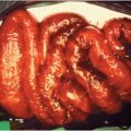
| Intrarenal abscesses Renal cortical abscesses Renal corticomedullary abscesses Acute focal bacterial nephritis Acute multifocal bacterial nephritis Xanthogranulomatous pyelonephritis |
| Perinephric abscesses |
Renal cortical abscess
A renal cortical abscess results from hematogenous spread of bacteria from a primary focus of infection outside the kidney, often the skin. The most common causative agent is Staphylococcus aureus (90%). Predisposing conditions include entities associated with an increased risk for staphylococcal bacteremia, such as hemodialysis, diabetes mellitus, and injection drug use. The primary focus of infection may not be apparent in up to one-third of cases. Ascending infection is an infrequent cause of renal cortical abscess formation. Ten percent of renal cortical abscesses rupture through the renal capsule, forming a perinephric abscess.
Patients present with chills, fever, and back or abdominal pain, with few or no localizing signs (Table 67.2). Most patients do not have urinary symptoms as the process does not generally communicate with the excretory passages. Physical examination may reveal costovertebral angle tenderness and involuntary guarding in the upper lumbar and abdominal musculature. A flank mass or bulge in the lumbar region with loss of lumbar lordosis may be present.
| Renal cortical abscess | Renal corticomedullary abscess | Perinephric abscess | |
|---|---|---|---|
| Epidemiology | Males 3× > females, 2nd–4th decades, hematogenous seeding of kidneys | Males = females (females > males in xanthogranulomatous pyelonephritis), incidence increases with age, associated with underlying abnormality of the urinary tract | Males = females, 25% of patients are diabetic, rupture of intrarenal suppurative focus into perinephric space |
| Clinical presentation | Chills, fever, localized back or abdominal pain | Chills, fever, flank or abdominal pain, nausea and vomiting (65%) | Insidious onset over 2–3 weeks: fever (early), flank pain (late) |
| Urinary symptoms | Nonea | Dysuria/other urinary tract symptoms variably present | Dysuria 40% |
| Physical exam | Flank mass | Flank mass in 60%, hepatomegaly in 30% | Flank or abdominal mass in ≤50%, 60% have abdominal tenderness |
| Organisms | S. aureus | Enteric aerobic gram-negative rods (Escherichia coli, Klebsiella species, Proteus mirabilis) | Enteric aerobic gram-negative rods and S. aureus; occasionally Pseudomonas species, gram-positive bacteria, obligate anaerobic bacteria, fungi, mycobacteria; 25% polymicrobial |
| Urinalysis | Normala | Abnormal in 70% | Abnormal in 70% |
| Urine cultures | Negativea | Generally positive | Positive in 60% |
| Blood cultures | Often negative | Often positive | Positive in 40% |
a If there is no communication between the abscess and the collecting system.
Laboratory data vary, though leukocytosis is common. Radiologic techniques can establish the diagnosis. Ultrasonography is useful diagnostically and may guide percutaneous drainage of the abscess and follow response to therapy. Computed tomography (CT) is the most precise noninvasive diagnostic technique and may guide percutaneous aspiration. Magnetic resonance imaging (MRI) with gadolinium may help diagnose renal abscess and define its extent, with accuracy comparable to CT but without exposure to radiation and ionizing contrast.
Renal cortical abscesses often respond to antibiotics alone without surgical intervention. If the diagnosis is suspected and bacteriologic evaluation of the urine or aspirated abscess fluid reveals large, gram-positive cocci or no bacteria, antistaphylococcal therapy should be started promptly (Table 67.3). The choice of empiric therapy depends on the susceptibility patterns of S. aureus in the community. If methicillin-susceptible S. aureus (MSSA) is suspected a semisynthetic penicillin (oxacillin or nafcillin) is appropriate empiric therapy. For penicillin-allergic patients, a first-generation cephalosporin, such as cefazolin, may be used. Vancomycin should be used for patients with a severe immediate β-lactam allergy. If the prevalence of methicillin-resistant S. aureus (MRSA) is high, empiric therapy with vancomycin should be initiated. In the absence of occult bacteremia, parenteral antibiotics are administered for 10 days to 2 weeks, followed by oral antistaphylococcal therapy for at least 2 to 4 more weeks. Fever generally resolves after 5 to 6 days of antimicrobial therapy. If there is no response to therapy in 48 hours, percutaneous aspiration should be considered, and, if unsuccessful, open drainage should be undertaken. The prognosis is good if the diagnosis is made promptly and effective therapy is instituted immediately.
| Empiric therapya | Durationa | Drainage | Surgery | |
|---|---|---|---|---|
| Renal cortical abscess | Antistaphylococcal penicillin: oxacillin or nafcillin (1–2 g IV every 4–6 h) Penicillin allergy: First-generation cephalosporin: cefazolin (2 g IV every 8 h) or vancomycin (15 mg/kg IV every 12 h) if severe immediate β-lactam allergy or if MRSA is suspected | Intravenous antibiotics for 10 days to 2 weeks followed by 2–4 weeks oral antistaphylococcal antibiotic depending on antibiotic susceptibility testing | If no response to treatment after 48 h → percutaneous drainage followed by open drainage if no response | |
| Renal corticomedullary abscess | ||||
| Acute focal bacterial nephritis | Extended-spectrum penicillin (piperacillin–tazobactam 3.375 g IV every 6 h), extended-spectrum cephalosporin (ceftriaxone 1 g IV every 24 h, cefotaxime 1 g every 8 h), fluoroquinolone (ciprofloxacin 200–400 mg IV every 12 h), ampicillin (1 g IV every 4–6 h) with gentamicin or cefazolin (1 g every 8 h) with gentamicin | |||


