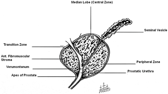
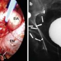
Fig. 1.1
Prostate and its anatomic relationships. All illustrations by Forrestall O. Dorsett Jr.
Anatomy
The prostate can be separated into four regions; anterior, peripheral, central, and transitional zones (Fig. 1.2). These zones are important in that they each can be the site of distinct pathology. The anterior fibromuscular zone which represents 30 % of the prostate mass contains virtually no glandular elements and is primarily smooth muscle. The peripheral zone is the largest zone, comprising 75 % of prostate glandular elements. It is of significant clinical importance as it is the primary site of prostatic carcinomas. The central zone has roughly 20–25 % of prostate glandular elements and surrounds ejaculatory ducts while the transitional zone contains up to 5 % of prostate glandular elements, is the site of BPH, and represents approximately 15–30 % of prostate volume.
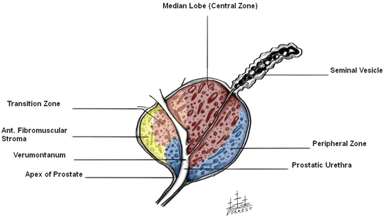

Fig. 1.2
Zones of the prostate
The prostate is the only solid organ in the pelvis and is typically easily located on digital rectal exam (DRE) especially when enlarged. It has been classically described as being about the size of a walnut with a texture not unlike the tip of the nose when palpated. It is oriented with its base angled posteriorly toward the bladder neck and its apex is angled anteriorly in continuity with the urethra. At the apex of the prostate we have the formation of the striated external urethral sphincter (Fig. 1.3). This sphincter is a vertically oriented tubular sheath that surrounds the membranous urethra and prostate. The gland is also suspended by the pubo-rectal portion of the levator ani muscle (Fig. 1.4). The prostate possesses an outer capsule which is made up of collagen, smooth muscle, and elastin. The longitudinal fibers of the detrusor muscle mesh with the fibro-muscular tissue of the capsule. While contributing to the rubbery external texture of the prostate and some protective benefit, the capsule also serves as scaffolding for adjacent support structures besides the levator ani. Additional support is provided by three distinct yet intertwining layers of fascia found on the anterior, lateral, and posterior aspects of the prostate. The anterior and antero-lateral fascia is in direct continuity of the capsule. This is where the deep dorsal vein of the penis and its tributaries are found. Laterally the levator fascia fuses with the anterior and antero-lateral fascia. The recto-vesical or Denonvilliers fascia is a connective tissue layer located between the anterior wall of the rectum and posterior aspect of the prostate. This fascial layer covers the prostate and seminal vesicles posteriorly and extends caudally to terminate as a fibrous plate just below the urethra at the level of the external urethral sphincter near the apex of the prostate. This median fibrous plate or raphe has a distal extension to the level of the central tendon of the perineum.
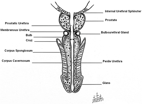
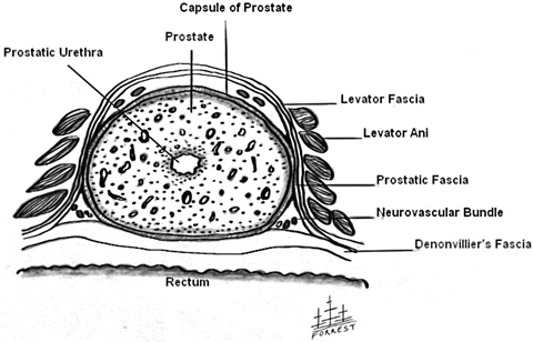

Fig. 1.3
Coronal view of penis and prostate

Fig. 1.4
Axial view of prostate showing muscle fibers of levator ani
Seminal Vesicles
The seminal vesicles are two saccular glands that pair with its corresponding ductus to form the ejaculatory ducts that enter into the prostate. The seminal vesicles are located superior to the prostate under the base of the bladder and are approximately 6 cm in length (Fig. 1.5). Seminal vesicles are derived from the wolffian ducts through testosterone stimulation. Together with the prostate the seminal vesicles create seminal fluid that facilitates sperm transport for fertilization.

