Endocrine Cell (Carcinoid) Tumors of the Vermiform Appendix
Rhonda K. Yantiss
Avariety of neoplasms develop in the appendix, many of which are incidentally discovered tumors encountered in appendectomy specimens obtained for other reasons. Of these, endocrine (carcinoid) tumors are, by far, most common. Endocrine neoplasms are recognized by their histologic patterns, cytoplasmic argentaffinity or argyrophilia, and immunohistochemical staining for cytosolic markers (e.g., neuron-specific enolase and CD56), vesicles (e.g., synaptophysin), or secretory granules (e.g., chromogranin). Most endocrine tumors of the appendix are composed of enterochromaffin cells similar to those of tumors involving the distal small intestine and have been termed “classic” carcinoid tumors. These lesions should be distinguished from goblet cell carcinoid tumors, as discussed in Chapter 20.
CLINICAL FEATURES OF ENDOCRINE (CARCINOID) TUMORS
Endocrine tumors account for slightly more than 50% of all appendiceal neoplasms and nearly 20% of all gastrointestinal endocrine tumors. They are identified in less than 1% of patients undergoing appendectomy.1 Most tumors are identified in appendices obtained from adults but tend to occur in a slightly younger age group than endocrine tumors elsewhere in the gastrointestinal tract. Females are affected more often than males, although some of the difference may reflect the greater number of incidental appendectomies performed on women undergoing gynecologic surgery.2 Appendiceal endocrine tumors are usually located in the distal appendix and, thus, are asymptomatic. Some tumors that develop in the mid or proximal appendix are associated with appendicitis, presumably due to obstruction of the lumen.
PATHOLOGIC FEATURES OF ENDOCRINE TUMORS
Enterochromaffin Cell Endocrine Tumors (Classic Carcinoid Tumors)
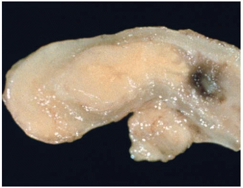
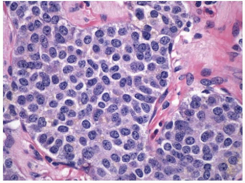
Appendiceal endocrine tumors appear as yellow, or white, firm nodules in the appendiceal tip, and most are composed of enterochromaffin cells (Figure 18.1). These serotonin-producing tumors are usually low grade and contain cells that are argentaffin and argyrophil positive. The tumors are well circumscribed, but unencapsulated, and consist of tightly packed nests and acini containing neoplastic cells that fill the mucosa and variably extend into the appendiceal wall (Figure 18.2). Acinar structures may harbor luminal material that is positive for periodic acid-Schiff stain but lack mucin production. The tumor cells contain abundant amphophilic or eosinophilic granular cytoplasm with round, smooth nuclei and coarse “salt and pepper” chromatin and rare mitoses (Figure 18.3). Some may contain spindle cells, whereas others show nuclear pleomorphism and multinucleation. Enterochromaffin cells show strong immunohistochemical staining for
chromogranin A, synaptophysin, serotonin, and substance P. Tumor cell nests are surrounded by sustentacular cells that are immunopositive for S-100 protein. More than 70% of serotoninproducing endocrine tumors of the appendix span less than 1 cm in diameter and show very low proliferation indices by Ki-67 immunolabeling.3
chromogranin A, synaptophysin, serotonin, and substance P. Tumor cell nests are surrounded by sustentacular cells that are immunopositive for S-100 protein. More than 70% of serotoninproducing endocrine tumors of the appendix span less than 1 cm in diameter and show very low proliferation indices by Ki-67 immunolabeling.3
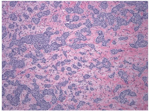 FIGURE 18.2: Enterochromaffin cell endocrine tumors resemble lesions of the distal jejunum and ileum. They contain solid cell nests and trabeculae enmeshed in a collagenous stroma. |
Enteroglucagon (L) Cell Endocrine Tumors
Stay updated, free articles. Join our Telegram channel

Full access? Get Clinical Tree


