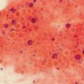
Fig. 4.1
Budding cell of Blastomyces dermatitidis. Note that the daughter cell is almost as large as the mother cell and still connected by a pore. H&E hematoxylin and eosin. (From MB Smith, MR McGinnis. Diagnostic histopathology. In Diagnosis and Treatment of Human Mycoses, DR Hospenthal, MG Rinaldi, eds. Humana Press. Totowa, NJ. 2008. Reprinted with permission from Springer)
Candidiasis
Pseudohyphae and yeast cells are seen in invasive candidiasis. Pseudohyphae differ from true hyphae in having constrictions at the septae and showing branching only at points of septation. Yeast cells, also called blastospores or blastoconidia, may bud off pseudohyphae at septae (Fig. 4.2). Yeast cells are also seen separately in the tissue, along wih pseudohyphae. Yeast cells are round or elliptical, smaller than Blastomyces, and may show budding or even chains of budding cells. Pores between mother and daughter yeast cells are too small to be seen readily. Yeast cells are rarely seen without pseudohyphae, with the exception that Candida glabrata has only yeast cells (Fig. 4.3). If patients are not granulocytopenic, the predominant inflammation is neutrophilic. Blastoschizomyces capitatus and Trichosporon asahii share similar features with candida in tissue.
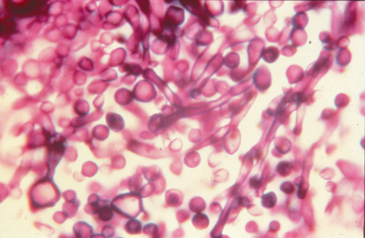
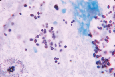
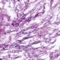
Fig. 4.2
Candida albicans pseudohyphae and blastospores. Note constrictions along the pseudohyphae. GMS Gomori methenamine silver
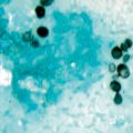
Fig. 4.3
Candida glabrata in a cardiac valve vegetation. Note the absence of pseudohyphae. GMS Gomori methenamine silver
Chromoblastomycosis
The most distinctive structures are thick-walled golden hued cells in the dermis, called “copper penny” cells, often surrounded by neutrophils and a granulomatous response (Fig. 4.4). The hyperplastic epidermis sometimes has short, wavy “fumigoid” hyphae.
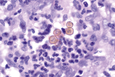
Fig. 4.4
Brown thick-walled “copper penny” cells of chromoblastomycosis in the dermis. H&E hematoxylin and eosin
Coccidioidomycosis
Spherules are all round, nonbudding, and range in size from that of Blastomyces or Cryptococcus to several times that large. Rounded endospores may be seen inside the larger spherules (Fig. 4.5). Neutrophils clusters around endospores released from ruptured spherules. Large pockets of pus may form in the tissue. Granulomatous inflammation or pyogranulomas are usual, similar to blastomycosis. Tissue eosinophilia is often present on H&E stain but easily missed. Caseous necrosis may be seen in chronic lesions, resembling tuberculosis, but calcification is not seen. Short hyphae are occasionally seen in Coccidioides lesions. Spherule-like structures, larger than Coccidioides and filled with numerous endospores, can be seen with Rhinosporidium seeberi, a protistan parasite of mucous membranes (Fig. 4.6), and with the Emmonsia species causing adiaspiromycosis of the lung.
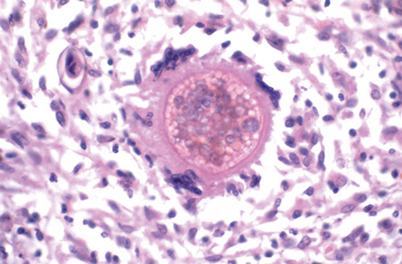
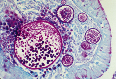


Fig. 4.5
Large spherule of Coccidioides species containing endospores. H&E hematoxylin and eosin. (From MB Smith, MR McGinnis. Diagnostic histopathology. In Diagnosis and Treatment of Human Mycoses, DR Hospenthal, MG Rinaldi, eds. Humana Press. Totowa, NJ. 2008. Reprinted with permission from Springer)
Fig. 4.6
Large cyst of Rhinosporidium seeberi containing spores. PAS Periodic acid Schiff. (From MB Smith, MR McGinnis. Diagnostic histopathology. In Diagnosis and Treatment of Human Mycoses, DR Hospenthal, MG Rinaldi, eds. Humana Press. Totowa, NJ. 2008. Reprinted with permission from Springer)
Related posts:

Stay updated, free articles. Join our Telegram channel
Full access? Get Clinical Tree
