Figure 10.1 Oblique section of top of neck. Note contiguity of spaces and resultant potential for spread. Adapted with permission from Hollingshead WH. Anatomy for Surgeons. Vol. 1: The Head and Neck, 2nd edn. New York, NY: Harper & Row; 1968.
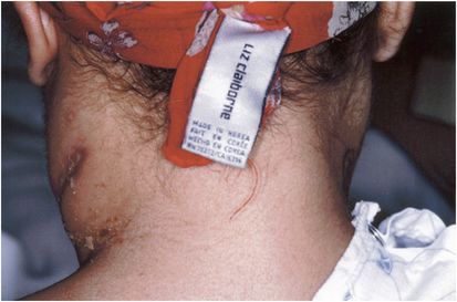
Patients with deep neck infections are frequently either diabetic, human immunodeficiency virus (HIV)-infected (irrespective of antiretroviral therapy status), or immunocompromised in some other way. An example of a deep neck infection of the lateral pharyngeal space in an HIV-infected patient is shown in Figure 10.2. Nocardia asteroides grew from the deep neck cultures. In addition, a history of injection drug use (IDU), neutropenia, or exogenous steroid therapy is common. A dental source is frequently present. On rare occasions, deep neck space infection may complicate head and neck cancers. Cases complicating traumatic airway intubation have also been reported.

Figure 10.2 Recurring deep neck abscess of lateral pharyngeal space caused by Nocardia asteroides in an HIV-infected patient with CD4 count of 86/mm3.
Management of deep neck infections is very costly. A recent analysis of 71 such patients treated in the United States found that the total cost of care exceeded $1.1 million.
Deep neck infections are usually polymicrobial, reflecting the oral cavity source of most of these infections. When associated with IDU, community-acquired methicillin-resistant Staphylococcus aureus (MRSA) is the most likely organism to be encountered. The microbiology of these infections is shown in Table 10.1. The increasing prevalence of multidrug-resistant gram-negative rods complicates therapy. Wound botulism as a complication of deep neck infection may occur in IDUs.
| Common Viridans and other streptococci Staphylococcus aureus (including MRSA) Prevotella, Fusobacterium, Bacteroides, Porphyromonas |
| Rare Moraxella Haemophilus species Pseudomonas species Actinomyces species |
Relevant anatomy of the deep neck spaces
The deep neck spaces are bound by a variety of anatomic landmarks and are anatomically distinct. However, all have the potential for communicating with each other, and thus infection may spread from one region to another during the course of the illness. It is important to delineate the various spaces clinically and radiologically so that appropriate management can be provided. The main spaces are as follows:
- The submandibular space (composed of two spaces separated by the mylohyoid muscle: the sublingual space, which is superior, and the submaxillary space, which is inferior).
- The lateral pharyngeal spaces (anterior and posterior)
- The retropharyngeal spaces (including the retropharynx, the prevertebral space, and the “danger space”).
Radiologic testing in deep neck infections
Computed tomography (CT) and magnetic resonance imaging (MRI) are excellent diagnostic tools for deep neck infections. However, it must be appreciated that these patients are critically ill with a risk for acute airway obstruction at any time in their clinical course. Thus, appropriately trained personnel must accompany these patients when they go for such scans. Equipment must be brought along to allow immediate airway protection should acute airway obstruction develop in (or on the way to) the radiology department.
In the postoperative setting, rim enhancement (>50%) of fluid collections in the neck on CT scanning correlates with abscess formation.
Submandibular space infections (“Ludwig’s angina”)
This bilateral infection of the submandibular space most commonly arises from infection of the posterior two molar teeth. There is the rapid onset of fever, mouth pain, and drooling of oral secretions. Soft-tissue spread of infection causes woody induration of the submandibular space and a stiff neck. Because the tongue may be displaced upward and backward, acute airway obstruction may develop. Tracheal compression due to surrounding edema is another cause of airway failure in this infection. Management is outlined in Tables 10.2 and 10.3 and complications in Table 10.4.
| NEVER LEAVE THE PATIENT UNATTENDED out of a monitored unit |
| Protect the airway – experienced otolaryngology evaluation is essential |
| CT or MRI of mouth, neck, mediastinum to evaluate for drainable collections, airway compression, vascular complications, or mediastinal involvement |
| Dentist/oral surgery evaluation for a (removable) dental source of infection |
| Correct underlying medical issues – maximize glycemic control, correct neutropenia, taper steroids (if feasible), etc. |
Stay updated, free articles. Join our Telegram channel

Full access? Get Clinical Tree


