Current Imaging Techniques for the Diagnosis and Staging of Lung Cancer
Philip T. Cagle
Ronald E. Fisher
INTRODUCTION
Radiology and nuclear medicine play a vital role in the diagnosis, staging, and treatment of lung cancer and typically provide the basis for obtaining specimens for the surgical pathologist. Computerized tomography (CT), magnetic resonance imaging (MRI), and positron emission tomography (PET) have been in use for decades as imaging techniques for many purposes, including detection and evaluation of primary lung tumors and metastases.1,2,3 and 4 The frequent use of CT and high resolution CT (HRCT) in the diagnosis of chest diseases, including interstitial lung diseases, has resulted in the increased detection of incidental lung nodules, including clinically unsuspected lung cancers which, in turn, has led to renewed interest in the possibilities of lung cancer screening.5 Currently, several imaging techniques are popular for the diagnosis and staging of primary lung cancers and their metastases. These include PET/CT co-registration for diagnosis, staging, planning therapy and evaluating response to therapy,1,2,3 and 4,6,7,8,9,10 and 11 and endobronchial ultrasound (EBUS)-guided transbronchial needle aspiration and transesophageal endoscopic ultrasound (EUS)-guided fine needle aspiration primarily for diagnosing mediastinal lymph node metastases.12,13,14,15,16,17,18,19 and 20
CT AND MRI
CT and MRI have been in routine clinical use for anatomic imaging for decades and the images they produce should be very familiar to surgical pathologists. CT scanners, using x-rays, and MRI scanners, using radio frequency, produce multiple two-dimensional cross-sectional images or “slices” of tissue that can be reconstructed in three dimensions. Both may be enhanced by the use of contrast agents. MRI provides images in any plane. Although earlier CT scanners could only provide images in an axial or near-axial plane (computed axial tomography or CAT scanners), current multidetector CT (MDCT) scanners generate data that subsequently can be reconstructed in any plane. There are advantages and disadvantages to both techniques, but CT is preferred for solid thoracic tumors such as lung cancer in most situations.1,2
PET AND PET/CT
PET is a nuclear medicine technique in which a radioactively labeled compound or radiotracer is injected into the patient.3,4,6,7,8,9,10 and 11 Fluorine-18-fluorodeoxyglucose (FDG) is the radiotracer most often used to detect cancers. A positron-emitting radionuclide with a short half-life (fluorine-18
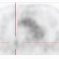
has a half-life slightly <2 hours) is incorporated into a metabolically active compound (glucose) to produce the radiotracer FDG. The FDG is injected into the patient’s bloodstream and concentrates in normal body tissues, inflammatory lesions, and tumors in proportion to the cells’ glucose metabolic rate that use glucose at a high rate, including brain and liver, and in some inflammatory lesions. Most cancers have an increased rate of glucose metabolism due to their high reproductive rates and their inefficient use of glucose—poorly differentiated tumors cannot undergo aerobic metabolism even if sufficient oxygen is present. As a result, most cancers have to transport a large amount of glucose across their cell membranes in order to generate a sufficient amount of ATP. Once in the cell, FDG is phosphorylated to FDG-6-phosphate by hexokinase. FDG-6-phosphate, however, cannot be metabolized further because an oxygen atom has been replaced with fluorine. And because mammalian cells lack significant levels of the phosphatase needed to remove the phosphate group, the molecule is trapped inside cells. The more glucose (and thus FDG) a cell transports in, the more FDG is trapped inside it and the “hotter” it will appear on an FDG PET scan. Most cancers are hot on PET, with poorly differentiated, high-grade tumors being extremely hot (Fig. 18-1).3,4,6,7,8,9,10 and 11
has a half-life slightly <2 hours) is incorporated into a metabolically active compound (glucose) to produce the radiotracer FDG. The FDG is injected into the patient’s bloodstream and concentrates in normal body tissues, inflammatory lesions, and tumors in proportion to the cells’ glucose metabolic rate that use glucose at a high rate, including brain and liver, and in some inflammatory lesions. Most cancers have an increased rate of glucose metabolism due to their high reproductive rates and their inefficient use of glucose—poorly differentiated tumors cannot undergo aerobic metabolism even if sufficient oxygen is present. As a result, most cancers have to transport a large amount of glucose across their cell membranes in order to generate a sufficient amount of ATP. Once in the cell, FDG is phosphorylated to FDG-6-phosphate by hexokinase. FDG-6-phosphate, however, cannot be metabolized further because an oxygen atom has been replaced with fluorine. And because mammalian cells lack significant levels of the phosphatase needed to remove the phosphate group, the molecule is trapped inside cells. The more glucose (and thus FDG) a cell transports in, the more FDG is trapped inside it and the “hotter” it will appear on an FDG PET scan. Most cancers are hot on PET, with poorly differentiated, high-grade tumors being extremely hot (Fig. 18-1).3,4,6,7,8,9,10 and 11
Stay updated, free articles. Join our Telegram channel

Full access? Get Clinical Tree


