VI.B.001 Acute Mast Cell Leukemia. Blood and Marrow
VI.B.001
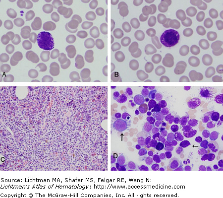
Acute mast cell leukemia. (A and B) Blood film. Occasional blast cells (no granules) and frequent mast cells at diagnosis as shown here. Blast cells increased in later blood films. (C) Marrow biopsy. Diffusely infiltrated with mast cells that also formed focal aggregates. The marrow biopsy was hypercellular and contained 29% of cells with mast cell granules and 7% blast cells (no granules). Vertical arrow indicates one of several blast cells. (D) The mast cells vary in maturity from early myelocytes (often with nucleoli and immature chromatin patterns) but with a few large mast cell granules to mature mast cells (horizontal arrow). Many of the mast cells have multilobed nuclei and atypical granules. The leukemic mast cells were positive for CD117, CD25, and tryptase. The flow cytometric phenotype of the blast cells was slightly different from the mast cells in that it CD34-positive but CD25-negative, whereas the mast cells were CD34-negative and CD25-positive. CD117 was much stronger in the mast cells than in the blast population.
VI.B.002 Acute Mast Cell Leukemia. Marrow Immunocytostains
VI.B.002
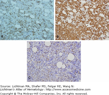
Acute mast cell leukemia: Marrow immunocytostains. Marrow biopsy. The marrow aspirate is hypercellular and contains 29% cells with mast cell granules and 7% blasts (no granules). The mast cells vary in maturity from early myelocytes (often with nucleoli and immature chromatin patterns) but with a few large mast cell granules through to mature mast cells. The brown cytoplasmic staining of marrow cells indicates a positive immunological reaction for (A) CD25, (B) CD117, and (C) tryptase. The latter is a specific marker for mast cells granules.
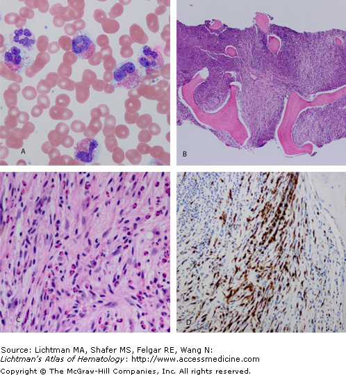
VI.B.003. Mastocytosis with eosinophilia.
VI.B.003
Related posts:

Stay updated, free articles. Join our Telegram channel

Full access? Get Clinical Tree


