Chapter 34 Clinical Manifestations, Staging, and Treatment of Follicular Lymphoma

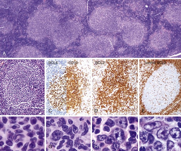
Figure 34-1 FOLLICULAR LYMPHOMA: MORPHOLOGIC AND IMMUNOPHENOTYPIC FINDINGS.
A, A low-power photomicrograph illustrates a lymph node involved by follicular lymphoma. The lymphoma cells grow in nodules or follicles that resemble the normal lymphoid follicles of a reactive lymph node. However, in the lymphomatous growth, the follicles are crowded, show back-to-back localization, and lack many of the features of their reactive counterparts. At higher power (B), the neoplastic follicles lack mantle zones and the normal polarization of small and large germinal center cells (centrocytes and centroblasts, respectively), which occurs due to the cellular response to antigenic stimulation as it sweeps thorough the follicle. The neoplastic follicles stain for the germinal center marker, BCL6 and CD10 (C); however, they overexpress BCL2 (D) as a result of the associated translocation t(14;18), involving the IgH gene and BCL2. BCL2 is not much expressed in normal germinal center B cells (E, control for comparison). Follicular lymphoma is graded by the number of large neoplastic cells (centroblasts) present among the smaller neoplastic cells (centrocytes) (F to I). The grading system is not entirely accurate, but it provides some framework for subclassifying cases morphologically. Grade 1 is 0 to 5 centroblasts per average 40x field (F); between 6 and 15 is grade 2 (G); and more than 15 is grade 3A (H). Grades 1 and 2 are now considered together. When most of the cells in the neoplastic follicles are centroblasts without centrocytes, the case is considered grade 3B (I).
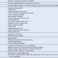
Table 34-1 Initial Evaluation of Follicular Lymphoma
Stage | Criteria |
---|---|
I | Involvement of 1 lymph node (I) or 1 extralymphatic organ or site (IE) |
II | Involvement of 2 or more lymph nodes on same side of diaphragm (II) or localized extralymphatic organ or site and 1 or more involved lymph node on same side of diaphragm (IIE) |
III | Involvement of lymph nodes on both sides of diaphragm (III) or same side with localized involvement of extralymphatic site (IIIE), spleen (IIIS), or both (IIIS+E) |
IV | Diffuse or disseminated involvement of extralymphatic organ or tissues with or without lymph node enlargement |
Table 34-3 Factors Having Prognostic Significance in the FLIPI* and FLIPI2†
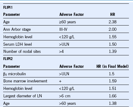
FLIP, Follicular lymphoma international prognostic index; HR, hazard ratio; LDH, lactate dehydrogenase, ULN, upper limit of normal.
† From Federico M, Bellei M, Marcheselli L, et al: Follicular lymphoma international prognostic index 2: A new prognostic index for follicular lymphoma developed by the international follicular lymphoma prognostic factor project. J Clin Oncol 27:4555, 2009.

Data from Federico M, Bellei M, Marcheselli L, et al: Follicular lymphoma international prognostic index 2: A new prognostic index for follicular lymphoma developed by the international follicular lymphoma prognostic factor project. J Clin Oncol 27:4555, 2009.

Stay updated, free articles. Join our Telegram channel

Full access? Get Clinical Tree