GENERAL APPROACH TO THE HEAD AND NECK CANCER PATIENT
Clinical evaluation of the patient with head and neck cancer entails multiple challenges for the physician. In the context of an initial clinical encounter, the caregiver needs to collect the findings that will guide diagnosis, while also establishing a constructive therapeutic relationship with the patient and family members, addressing acute intervention needs, and outlining and initiating a plan for further diagnostic workup and treatment. The imperatives that the physician must address in the evaluation of the head and neck cancer patient may be considered as 12
Key Tasks, as outlined in
Table 2.1. An accurate and comprehensive examination must be performed. A complete medical history should be undertaken, with special attention paid to key risk factors, including prior cancer history, smoking history, alcohol intake, extent of sun exposure, reflux, industrial or occupational exposures, and immunosuppression. Cardinal signs and symptoms associated with head and neck cancer include lesions or masses, persistent hoarseness, mouth or throat pain, dysphagia, odynophagia, hoarseness or dysphonia, dyspnea, stridor, unilateral otalgia or hearing loss, aspiration, weight loss, or cranial nerve deficits. Assessment and documentation must be performed of the patient’s overall medical and functional status, which may influence the suitability of specific treatments such as surgical resection or radiotherapy. Critical information regarding previous treatments must be collected, including previous oncologic surgeries, chemotherapy treatments, or prior radiotherapy. If the patient has had prior radiotherapy, key elements of therapy must be documented, including treatment portals, dosages, fractionation, treatment duration and compliance, and specifics regarding prior surgical or chemotherapeutic interventions obtained.
A provisional diagnosis or differential should be established with rapid formulation of a plan of care and clear delineation of the next steps required. At the same time, the treating physician must be vigilant in assessing areas in which immediate intervention may be necessary, such as treating pain, airway management, or addressing severe nutritional deficiencies. Assessment and treatment of pain is an important aspect of the clinical evaluation of the head and neck cancer patient, although insufficient attention is often paid to this aspect of care. In a recent meta-analysis of studies on pain among patients with cancer, den Beuken and colleagues found that >50% of patients had inadequately treated pain; when considered by cancer type, patients with head and neck cancer had the highest prevalence of insufficient pain management at 70%.
1A comprehensive evaluation of the head and neck cancer patient typically involves adjunctive measures such as radiographic imaging and cytopathologic tissue analysis. However, physical examination of the head and neck and history-taking by the evaluating clinician retain a central role in shaping further assessment and treatment. For example, patient age is a key factor to be considered in evaluating the patient with a solitary neck mass. Whereas the overwhelming majority of neck masses in young patients are either infectious or inflammatory in origin, most neck masses in adults involve neoplasia. Although benign pathologies are common in young patients with cystic neck masses, >80% of patients over the age of 40 who present with such findings are ultimately found to have carcinoma.
2 The clinician should be highly suspicious of the diagnosis of branchial cleft cyst or thyroglossal duct cyst in patients older than 40 with no prior history of symptoms; >30% of patients over 40 initially diagnosed with branchial cleft cyst subsequently prove to have cystic metastases of squamous cell carcinoma.
3As part of the initial visit, head and neck cancer patients should be engaged with a team of professionals dedicated to providing guidance and support through their course of care, including an oncology nurse, nurse practitioner or physician assistant, and patient navigator. Such multilayered staffing in the management of head and neck cancer patients improves treatment compliance and overall satisfaction with care.
4 If the patient is a smoker, provision of smoking counseling and offering treatment or referral to smoking cessation resources is necessary at the initial visit, as persistent smoking during treatment is an independent risk factor for poor treatment outcome.
5In many cases, patients will present having already been told that they have a head and neck cancer or they may have a strong suspicion of such a diagnosis. There is wide variation regarding patients’ desires regarding the detail in which they want cancer diagnoses described and the degree of specificity with which they want prognostic information delivered.
6 Although legal and ethical standards in the United States regarding informed consent demand full disclosure, it is incumbent upon the clinician to attempt to furnish such information in an optimal manner for individual patients. The obligation to full disclosure does not obviate the responsibility of the provider to attempt to tailor the delivery in accordance with patient preferences and sensitivities.
7,8
PHYSICAL EXAMINATION
Thorough physical examination is critical in the evaluation of the patient with possible head and neck cancer. A complete physical examination of the entire head and neck is essential, regardless of suspected primary site. Appropriate steps should be taken prior to the initiation of the physical exam in order to develop and enhance patient comfort and rapport. Appropriate hand washing should precede examination, and adherence to universal precautions should be maintained. These actions promote trust between patient and physician. Physical examination should begin with attention to the general appearance of the patient. Information regarding a patient’s overall health, mood and affect, hygiene, and smoke exposure may be gained with simple observation.
In addition to visualization of anatomic structures of the head and neck, manual palpation is a critical tool for assessment of the head and neck. Palpation should routinely be performed of the neck and oral cavity. Bimanual palpation is necessary for comprehensive evaluation of the floor of mouth and submandibular areas. Palpation techniques can yield critical information regarding submucosal extension, fixation, and tumor thickness. Any abnormal or concerning findings should be described in relation to key landmarks and anatomic subsites, so as to facilitate accurate communication with other providers and appropriately document changes over time.
Examination of the Skin
Examination should begin with assessment of facial structures; any gross asymmetry or facial swelling should be noted. Thorough external inspection of the patient’s skin for any suspicious lesions is performed. Particular attention should be given to sunexposed areas, including the external auricles and the posterior neck. The scalp and other hair-bearing areas should be closely inspected, as lesions in these areas often escape notice. All pigmented lesions and nevi should be examined closely for color variation, border quality, asymmetry, and ulceration.
Examination of the Ear
Examination of the ear and temporal bone is a required portion of the assessment. The skin overlying the external ear and mastoid region is inspected and palpated when appropriate. Following inspection of the external ear, otoscopic examination should be performed to assess the external auditory canal, tympanic membrane, and middle ear space. The presence and quality of any otorrhea or evidence of effusion is noted. Subjective pain associated with aural discharge is particularly concerning for underlying malignancy. In an adult presenting with unexplained unilateral or bilateral serous otitis media, fiberoptic endoscopic assessment should be performed to rule out obstruction of the Eustachian tube by a nasopharyngeal mass.
Examination of the Nose
Anterior rhinoscopy is performed under direct visualization with a nasal speculum and a headlight or head mirror. With exposure enhanced by appropriate speculum technique, the anterior nasal cavity and its contents are visualized, including the vestibule, anterior septum, floor of the nasal cavity, inferior turbinate, middle turbinate, and middle meatus. The presence of secretions, masses, obstruction, scabbing, or active bleeding should raise suspicion for sinonasal pathology.
Examination of the Oral Cavity
The oral cavity begins anteriorly at the skin/mucosal junction of the vermillion border of the lips. The hard/soft palate junction, the anterior tonsillar pillar, and circumvallate papillae form a plane posteriorly, which separates the oral cavity from the oropharynx. The oral cavity is divided into the following subsites: the lips, buccal mucosa, floor of mouth, retromolar trigone, oral tongue, hard palate, and alveoli. Oral cavity examination is best performed with two tongue blades and a head light or head mirror. The use of a handheld light source is discouraged, as it prevents the examiner from using both hands to manipulate oral cavity contents.
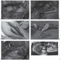
Examination should begin with external inspection of the lips and then proceed to thorough intraoral examination. When asking the patient to open their mouth, the presence of trismus should be noted, as it suggests pterygoid muscle involvement. The buccal mucosa, gingiva, gingivobuccal sulci, the retromolar trigone, and overall state of dentition are then assessed. The tongue is then grasped with a gauze pad and gently manipulated to allow full visualization of the ventral, dorsal, and lateral surfaces. The lateral surfaces should be inspected for lesions and palpated to assess for induration. With the tongue pulled superiorly, the floor of the mouth should be inspected and manually palpated; submucosal masses in the floor of the mouth should raise suspicion for minor salivary gland tumors. Midline protrusion and lateral excursion of the tongue should be tested; atrophy of one side or asymmetric protrusion raises suspicion for tongue invasion or cranial nerve XII involvement. Inspection and palpation of the hard palate is performed.
Oral cavity examination requires assessment of all mucosal surface changes suggestive of potential or
premalignancy: leukoplakia, erythroplakia, lichen planus, oral submucosal fibrosis, discoid lupus erythematosus, and dyskeratosis.
9 The most common sites for premalignant lesions include the buccal mucosa, lower gingiva, tongue, and floor of mouth.
10 Adjunctive screening methods including toludine blue application, fluorescence, and brush biopsy have not proven to be of unequivocal benefit in enhancing detection of oral malignancy,
11 but early findings regarding the potential application of optical coherence tomography in early detection are promising.
Examination of the Oropharynx
The oropharynx extends from the level of the soft palate to the hyoid bone and has four distinct subsites: soft palate, tonsillar region, base of tongue, and posterior and lateral oropharyngeal wall. The soft palate should be examined for mucosal lesions and submucosal fullness or swelling. Elevation of the soft palate should be symmetric and can be assessed by asking the patient to say “ah”; asymmetric elevation should raise suspicion for CN X involvement. The tonsillar region consists of anterior and posterior tonsillar pillars encompassing the palatine tonsils. The anterior tonsillar pillar and tonsil represent the most common sites for primary tumors of the oropharynx. Asymmetrically hypertrophied lymphoid tissue, particularly in the palatine tonsils, should raise suspicion. The lateral and posterior pharyngeal walls are inspected; medialization of the lateral oropharynx may represent mass effect from an entity in the parapharyngeal space, which lies posterolateral to the tonsil. Palpation of the base of tongue is paramount as direct examination of this area is limited. Submucosal lesions may be missed on indirect visualization, but can often be palpated with good manual examination techniques. The base of tongue, pharyngoepiglottic folds, and vallecula should be inspected during indirect laryngoscopy with either mirror examination or fiberoptic endoscopy, as discussed below.
Examination of the Neck and the Parotid Region
Examination of the neck is paramount in the evaluation of head and neck cancer. All levels of the neck, including the supraclavicular region, should be palpated to assess for the presence of lymphadenopathy. The anatomy of the neck is divided into six levels, as described in
Table 2.2.
12 Size, mobility, and location of suspected malignant lymph nodes should be carefully documented in relation to these levels. The thyroid should be palpated for the presence of distinct nodules or gross enlargement. Standing behind the patient and having them swallow facilitates identification of irregularities of the thyroid lobes. The parotid glands, the preauricular lymph nodes, and the postauricular lymph nodes should be palpated. A mass in the parotid may represent primary neoplasm, metastatic lymph node, a cyst, or an inflammatory process. If the patient presents with a neck mass without an obvious primary, the index lesion can be identified in most cases on the basis of a comprehensive examination. In cases where the primary lesion is not discovered on initial physical exam, further workup with adjunctive imaging (CT, MRI, or PET/PET-CT), tissue sampling, operative endoscopy, and immunohistochemical or molecular studies as indicated will facilitate ultimate discovery of the primary lesion in 97% of patients.
13
Neurologic Examination
Complete neurologic examination should be performed with a focus on the cranial nerves (CN). Cranial nerve deficits may be indicators of underlying neoplastic processes and require further workup. Any paresis of the facial nerve (CN VII) must be characterized in accordance with the House-Brackmann grading scale. The House-Brackmann scale (
Table 2.3) has been adopted by the American Academy of Otolaryngology-Head and Neck Surgery as the universal standard for the clinical assessment of facial nerve function.
14 Characterization of facial nerve deficits using the House-Brackmann scale facilitates accurate communication between providers and clear documentation of symptom progression. However, it should be noted that the House-Brackmann scale was developed and tested in neuro-otologic setting and has only limited usefulness in assessing tumors of the head and neck. Nevertheless, it is a common grading system that is now universally known and it does help with communication to other health professionals.
Fiberoptic Endoscopy
The nasopharynx, portions of the oropharynx, larynx, and hypopharynx cannot be assessed by unaided physical examination. These areas can be visualized using either mirror techniques or
fiberoptic endoscopy. Fiberoptic endoscopy provides the most accurate visualization of these areas, allowing for assessment of primary lesions and evaluation of airway status. The nose is typically anesthesized with topical anesthetic spray and decongested with a vasoconstrictive spray. A flexible laryngoscope is passed through both nasal cavities and into the nasopharynx.
The nasopharynx extends from the skull base superiorly to the soft palate inferiorly and communicates with the nasal cavity through the choanae. Midline posterior pharyngeal mucosa and the nasopharyngeal lymphoid tissue should be assessed. Posterior pharyngeal wall fullness raises suspicion for retropharyngeal pathology. The scope is then angled to view the lateral wall and its contents including epithelial mucosa, the Eustachian tube orifice, the posterior lip of the opening of the Eustachian tube or
torus tubarius, and the fossa of Rosenmüller lying behind the
torus tubarius. The lateral wall must be carefully inspected with particular attention given to the fossa of Rosenmüller, as nasopharyngeal tumors have a predilection for this site.
15 The scope should then be angled inferiorly, and the base of tongue and the vallecula inspected. Protrusion of the tongue facilitates complete examination of the vallecula and the base of tongue. Advancing the tip of the scope until it is just above the epiglottis allows for visualization of the larynx and the hypopharynx.
The larynx is divided into three subsites: the supraglottis, glottis, and the subglottis. The supraglottic region includes the epiglottis, the false vocal cords, the ventricles, the arytenoid cartilages, and the aryepiglottic folds. The glottis consists of the true vocal cords and anterior commissure. The supraglottic and glottic larynx are examined for any mucosal lesions or submucosal masses. It is vital to fully visualize the anterior cords and anterior commissure. Bilateral cord involvement, cord mobility, and involvement of surrounding subsites should all be documented. True vocal fold mobility is assessed by having the patient phonate, pant, and breathe quietly. Although clinical staging is partially based on cord mobility, in the setting of impaired cord movement, it is critically important to attempt assessment of the cricoarytenoid unit, which may represent a more specific involvement, rather than a mass effect related to deep muscle invasion. The cricoarytenoid is the basic functional unit of the hemilarynx, and mobility of the cricoarytenoid is often the critical factor determining patient eligibility for organ-preserving laryngeal surgery.
16 Unfortunately, discrimination of vocal cord movement restriction due to paraglottic space involvement or mass effect of bulky lesions from true cricoarytenoid fixation based upon endoscopic examination is difficult, and evaluation in the operating room is often required.
The subglottis begins 5 to 10 mm below the free edge of the true vocal folds. Primary tumors of the subglottis are rare. However, transglottic extension of superior lesions has important staging and prognostic implications. Visualization of the subglottis may be obscured by glottic pathology, and comprehensive evaluation of the subglottis is not possible in the office examination.
The hypopharynx extends from the hyoid bone superiorly to the esophageal introitus inferiorly. It is continuous with the oropharnyx and sits just behind the larynx. The hypopharynx is divided into three distinct subsites: the piriform sinus, posterior pharyngeal wall, and postcricoid region. Adequate visualization of the hypopharynx in the office setting may be difficult. Having the patient perform a Valsalva maneuver may enhance visualization due to clearance of pooled secretions. The majority of tumors that arise in the hypopharynx are SCC or an SCC-variant.
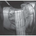
17The larynx and hypopharynx may alternatively be visualized with rigid endoscopy. Patients with sensitive gag reflexes may need to be sprayed with topical anesthesic intraorally. Rigid nasal endoscopy with angled scopes allows for optimum visualization of the nasal cavity and should be performed in any patient with suspected skull-base or sinonasal tumors. Stroboscopic examination of the glottis allows for enhanced visualization of true vocal cords, and subtle changes in mucosal wave dynamics may be detected; it is especially valuable in judging the depth of glottic lesions.
Physical examination findings, considered in the context of patient’s symptomatology and risk factor profile, form the basis for further workup. In most cases, comprehensive evaluation of suspicious findings will require adjunctive imaging. Prior to treatment, definitive tissue diagnosis must be obtained by fine needle aspiration (FNA), core needle biopsy, or open biopsy. Operative endoscopy with biopsy may also assist in securing definitive tissue diagnosis and achieving accurate staging.
IMAGING MODALITIES
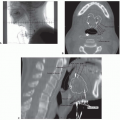
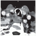
Prior to therapeutic intervention, precise tumor staging is critical. Accurate staging is generally best achieved with a combination of clinical evaluation and radiologic imaging. Pretreatment imaging assists in staging by helping to delineate tumor origin, size, extension, and presence of locoregional metastases. Imaging facilitates preplanning by defining the relationship between masses and nearby vital structures such as blood vessels and cranial nerves, and may also detect possible signs of malignancy not readily appreciable by physical examination such as ill-defined tumor borders, cartilage invasion, bone erosion, perineural spread, and multifocality. Traditionally, computed tomography (CT) and magnetic resonance imaging (MRI) have been the standard imaging modalities employed in the workup of head and neck cancer patients. More recently, positron emission tomography (PET) has emerged as a powerful tool in the diagnosis and management of these patients. Ultrasound (US) is radiation sparing and useful in the performance of directed biopsies, and is currently the first-line imaging modality for most thyroid and salivary lesions. Scintigraphy currently has a limited role in the evaluation of thyroid nodules. Emerging technologies in biologic neuroimaging, such as proton MR spectroscopy, CT-perfusion, perfusion-weighted MRI, and diffusion-weighted imaging, utilize biologic behavior patterns such as tumor cellularity and perfusion to depict additional imaging dimensions.
