Classification of Crushed Stone-like Calcifications Produced by Malignant Processes
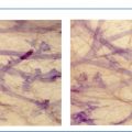
Correlation of subgross, 3D histological and mammographic images provides a basis for the following classification of crushed stone-like calcifications produced by malignant processes.
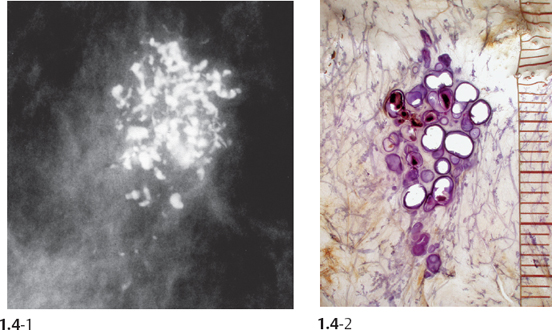
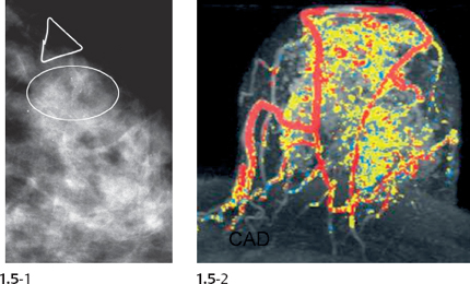
Group 1
Mammograms showing one or two clusters of crushed stone-like calcifications that represent:
Group 1A. The main morphological finding. In these cases there is concordance between mammography and the underlying histology. The temporal changes may be deceptively gradual. The outcome is excellent, even when invasion develops, provided the tumor is surgically removed when it is still in the 1-14 mm size range. The role of MRI in detecting these cases has yet to be defined.
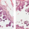
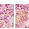
Group 1B. The subtle mammographic finding may represent only the “tip of the iceberg.” In these cases the true extent of the disease is seriously underestimated by mammography, although it may be adequately outlined by functional imaging methods such as MRI, since the disease involves numerous TDLUs, ducts and neoducts with little or no intervening normal tissue. The unexpectedly large tumor burden may result in a poor outcome, which can only become worse when underestimation of the disease leads to delayed or inadequate therapy.
Related posts:
 Overview of the Diagnostic and Management Problems Encountered When Malignant Type Calcifications Are Detected on the Mammogram
 Group 1A: Example 2.1
 Group 1A: Example 2.2
 14 Urothelial Cancer and Transitional Cell Cancer
 7 Colorectal Hepatic Metastatic Disease: Ablation
 13 Emerging Technologies in Interventional Oncology
Stay updated, free articles. Join our Telegram channel

Full access? Get Clinical Tree


