Chemokines


Chemokines


Philip M. Murphy


INTRODUCTION

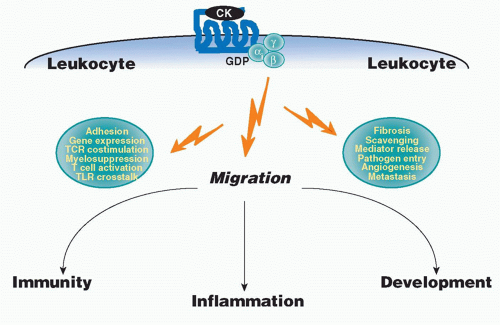
Chemokines constitute a large specialized family of cytokines that regulate immune responses by activating specific G protein-coupled receptors (GPCRs) on leukocytes.1,2,3,4,5,6,7,8,9,10,11,12,13,14 Chemokines may act at multiple levels, including immune system development,15 leukocyte transendothelial migration,15,16,17,18 leukocyte positioning within microenvironments,19,20,21,22,23 and both phagocyte and lymphocyte activation.24,25,26 They may function beneficially, for example in the setting of antimicrobial host defense and tissue repair, or harmfully, for example in the setting of cancer, chronic inflammation, autoimmunity, and infectious disease (Fig. 28.1). Many pathogens produce broad-spectrum chemokine blocking agents, attesting to the importance of chemokines in host defense,27,28,29 and some pathogens, most notably human immunodeficiency virus (HIV) and Plasmodium vivax, exploit host chemokine receptors as essential cell entry factors.30,31,32 Chemokines may also have nonimmunologic functions, including regulation of organ development.33,34,35 Together, these attributes have suggested potential chemokine-based therapeutic opportunities, and there are now two drugs targeting chemokine receptors approved by the U.S. Food and Drug Administration (FDA), the CCR5 antagonist maraviroc (Pfizer) in HIV,36,37 and the CXCR4 antagonist plerixafor (Sanofi-Aventis) in hematopoietic stem cell mobilization for transplantation in cancer.38

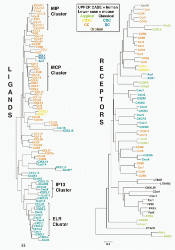
To date,48 chemokines and 23 chemokine receptors have been discovered in man. The goal of this chapter is to delineate their molecular properties and provide an overview of current knowledge of how they coordinate innate and adaptive immune responses, focusing on mouse and human. In this regard, it is important to note that the chemokine system may differ substantially among species, even at the genomic level.39,40,41 In addition, despite adopting a standard nomenclature in the year 2000,42,43,44 competing nonstandard aliases continue to be used in the primary literature, confusing even experts in the field. To address these challenges to clear communication, three illustrations are provided. The first defines the chemokine system repertoire at the genomic level in mouse and human, allowing comprehensive and facile identification of orthologues and species-specific elements (Fig. 28.2); the second is a dendrogram depicting the evolutionary relationship of chemokine and chemokine receptor protein sequences in mouse and man (Fig. 28.3); and the third provides the standard nomenclatures approved by the International Union of Immunological Societies for chemokine proteins and by the International Union of Pharmacology for chemokine receptors, linked to nonstandard aliases and protein sequence accession numbers (Tables 28.1 and 28.2). The names assigned by the HUGO Gene Nomenclature Committee are used for the corresponding chemokine system genes (see Fig. 28.2). In most cases, but not all, the approved gene and protein names are the same.


MOLECULAR ORGANIZATION OF THE CHEMOKINE SYSTEM


Chemokine Structure and Classification

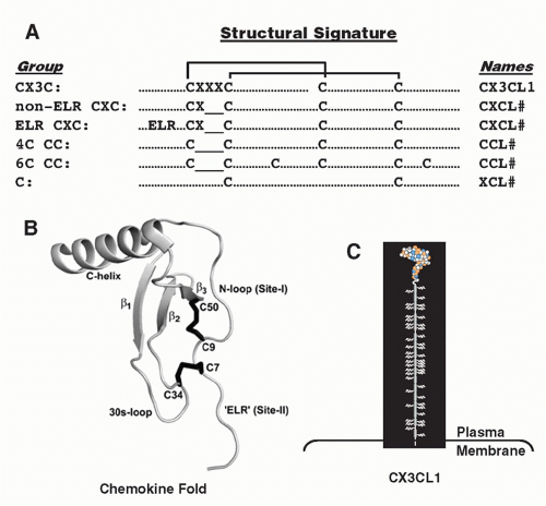
The word “chemokine” is a contraction of “ chemotactic cytokine,” which conveys the main function possessed by members of the family. Nevertheless, chemokines are defined by structure, not function, and may have other functions besides inducing cell movement. At the level of primary structure, chemokines occupy a common sector of sequence space bounded by ˜20% identity for any pairwise comparison, and after processing most are 66 to 111 amino acids in length. At the level of tertiary structure, chemokines fold in a highly conserved manner constrained in part by conservatively spaced disulfide-bonded cysteines.45 The family is subclassified based on the number and location of these cysteines. All chemokines have at least two cysteines, and all but two have at least four cysteines (Fig. 28.4A). In the four cysteine group, the first two are either adjacent (CC motif) or separated by either one (CXC motif) or three (CX3C motif) amino acids. C chemokines have only two cysteines, corresponding to the location of Cys-2 and Cys-4 in the other groups. Most chemokines are members of either the CC or CXC group; there is only one CX3C chemokine and one (mouse) or two (human) C chemokines. The group motifs are used as roots followed by the letter “L” (for “ligand”) and a number to create standard chemokine names (see Tables 28.1 and 28.2, and Fig. 28.2).44 In human names, all letters are capitalized; in the corresponding mouse name, only the first letter is capitalized. Amino acid sequence identity is < 30% between members of the four major chemokine groups, but ranges from ˜30% to 99% among members of the same group, indicating separate evolutionary histories. Although chemokine genes are located on 10 different human chromosomes, most are found in two large clusters, one for CXC chemokine genes on chromosome 4q21 and one for CC chemokine genes on chromosome 17q12 (see Fig. 28.2). These two clusters contain most of the chemokines that mediate inflammatory immune responses, whereas other chemokines, scattered throughout the genome either alone or in
small clusters on chromosomes 1, 2, 5, 7, 9, 10, 16, 17, and 19, mainly subserve homeostatic immune functions. The strongest functional correlate of the four major chemokine groups is receptor specificity: that is, most chemokine receptors bind more than one chemokine but are restricted by group (Table 28.3). With regard to leukocyte specificity, most neutrophil-targeted chemokines are in the CXC group and most of the monocyte-/macrophage-targeted chemokines are in the CC group. Major T and B cell-targeted chemokines can be found in both groups. The leukocyte target specificity of a chemokine may be narrow or broad and is defined by the expression pattern of its cognate receptor(s) (Table 28.4).


FIG. 28.1. Chemokines Coordinate Immune System Development and Immune Responses in Large Part by Inducing Leukocyte Migration Through Activation of G Protein-Coupled Receptors. Other functions induced by chemokines are listed in the green spaces. CK, chemokine.

CC and CXC chemokines can be further subclassified. The ELR subgroup of CXC chemokines is defined by the amino acid motif glu-leu-arg N-terminal to Cys-1 and is encoded by a cluster of genes on human chromosome 4q21 (see Fig. 28.2). A major species difference is the presence of the major neutrophil-targeted chemokine CXCL8 in human, but not in mouse. ELR is required for function of these chemokines, which are all > 40% identical at the amino acid level (see Fig. 28.3), attract neutrophils, bind the same receptor CXCR2, and are angiogenic.46,47 Of the ELR-negative CXC chemokines, only CXCL12 is angiogenic and attracts neutrophils, but by activating CXCR4.48 The so-called IP-10 subgroup, comprised of CXCL9-11, is encoded by a distinct cluster of related genes on human chromosome 4q21. Members share the receptor CXCR3, but are angiostatic, not angiogenic.46,49

The CC group is comprised of multiple subgroups. The two largest are the monocyte chemoattractant protein and macrophage inflammatory protein (MIP) clusters on human chromosome 17q12, which typically chemoattract monocytes/macrophages, plus other differential cell targets50 (see Fig. 28.3). CC chemokines may also be subclassified by the presence or absence of additional cysteines (see Fig. 28.4). Although in separate groups as defined by cysteine motifs, CXCL16 and CX3CL1 also form a unique multimodular subgroup51,52 (see Fig. 28.4). In addition to a typical chemokine domain, both these chemokines have a mucin-like stalk, a transmembrane domain, and a C-terminal cytoplasmic module, and can exist as a membrane-bound or shed form, mediating direct G protein-independent cell-cell adhesion and chemotaxis, respectively.53 The discovery of CXCL17 by sequence threading techniques, not by primary sequence homology, provides proof of principle that the boundaries of chemokine sequence space could expand substantially as protein structures are determined or modeled.54

All chemokines fold into a highly conserved and compact three-dimensional structure. Disulfide bonds link Cys-1 to Cys-3 and Cys-2 to Cys-4.45,55 The core, which contains three β sheets arranged in the shape of a Greek key, is overlaid by a C-terminal α-helical domain and is flanked by an N-terminal domain that lacks order (see Fig. 28.4). Forced chemokine monomers are active,56,57 but dimer and tetramer structures may occur, and complex quaternary structures bound to glycosaminoglycans (GAGs) on the surface of cells may be important for function in vivo.4,58,59,60,61,62 Location of the GAG binding domains is variable and depends in part on the highly basic nature typical of most chemokines. Chemokine heterodimers have been described, both CC/CC and CC/CXC, and some may form preferentially over homodimers in a GAG-dependent manner; native heterodimers have also been identified.63,64,65,66,67,68


Chemokine Receptor Structure and Classification

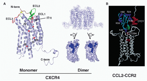
Chemokine receptors are defined as mediators that activate cellular responses upon binding chemokines. All 23 known subtypes, together with receptors for the classical lipid and peptide chemoattractant receptors, are members of the rhodopsin-like seven-transmembrane (7TM) domain superfamily of GPCRs8,42,43 (see Fig. 28.3; www.gpcr.org/7tm). The leukocyte chemoattractant receptors comprise the second largest subfamily of GPCRs (see Fig. 28.3). Chemokine binding, membrane anchoring, and signaling domains are formed from a single polypeptide chain ˜340 to 370 aa long. CXCR4 is the only chemokine receptor, and one of only four 7TM receptors, whose structure has been determined. When bound to small molecule and cyclic
peptide antagonists, it is a dimer, in general agreement with previous biochemical data describing ligand-independent CXCR4 dimerization69,70 (Fig. 28.5). Viral GPCRs have been proposed to hijack human receptors such as CXCR4 by heterodimerization.71 Evidence for homo- and heterodimerization has been reported for other chemokine receptors, such as CCR2, CCR5, and CX3CR1, but the precise physiologic forms remain undefined.72,73,74
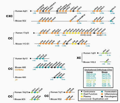


FIG. 28.2. Chemokine Gene Repertoires in Mouse and Human. Data are from www.ncbi.nlm.nih.gov/genome Builds 37.2 for Homo sapiens and Mus musculus. Brackets pair regions of conserved synteny between the indicated mouse and human chromosomes. Bracket labels denote chemokine groups (see text). Gene names are from the HUGO Gene Nomenclature Committee assignments. Protein names that differ from gene names are given in parentheses. Arrows, gene location and orientation relative to centromere; Mb, megabases; plasma, chemokines found at high concentrations constitutively in plasma. Other codes are in box at lower right. The figure layout is based on a previously published design and helpful advice from Hisayuki Nomiyama of Kumamoto University.

Standard receptor names are based on the property of chemokine group restriction, as follows: receptor name = ligand group root + R (for “receptor”) + number, in order of discovery. An exception is the C chemokine receptor XCR1, where “X” distinguishes it from CR1, the previously assigned name for complement receptor 1. For consistency, the XCR1 ligands are named XCL1 and XCL2. All but six chemokines have had their receptors identified (see Table 28.3). Chemokines have unique receptor specificity profiles, and chemokine receptors have unique chemokine specificity profiles. Ligands that share a common receptor interact with the same receptor chain, in contrast to many other types of cytokines that share receptors via specialized subunits.75 Pharmacologically, almost all chemokines are agonists, but a few are agonists at one receptor and antagonists at another (see Table 28.3).


FIG. 28.3. Chemokine and Chemokine Receptor Family Trees in Mouse and Human. Branch distances are calibrated by the bar at the bottom of each tree. The dendrograms were created in collaboration with Steven Tsang, National Institute for Allergy and Infectious Diseases.


Atypical Chemokine System Components

There are at least six categories of atypical chemokine system components. The first includes the chemokines and 7TM chemokine receptors encoded by poxviruses and herpesviruses, the probable result of “molecular piracy” of their hosts.29,71,76,77 This group includes chemokine scavengers and chemokine receptor antagonists thought to function in immune evasion; additional functions include modulation of host cell proliferation and movement. The second is a heterogeneous group of host 7TM chemokine-binding proteins that signal anomalously or not at all.78,79,80,81 Included in this group are Duffy antigen receptor for chemokines (DARC), CXCR7, CCBP2 (also known as D6), CCRL1, and CCRL2. CXCR7, DARC, and CCBP2 are the best studied. CXCR7 has been included in the standard nomenclature system due to evidence of β-arrestin- but not G-protein-mediated signaling.82 Its functions will be discussed in a later section with CXCR4, with which it shares a common ligand, CXCL12. DARC is nonsignaling and is expressed on red cells, lymphatic endothelium, and cerebellar Purkinje cells, but not


leukocytes. It is an exception to the ligand rule as it binds many, but not all, CC and CXC chemokines.83 DARC mediates transcytosis of chemokines across endothelial cells84 and modulates inflammatory responses.85 CCBP2 is expressed on leukocytes but at low levels. More prominently expressed on placental trophoblasts86 and lymphatic endothelium,87 CCBP2 scavenges inflammatory CC chemokines by a process of rapid internalization and recycling to plasma membrane,88 and affects embryo survival, inflammatory responses, immune activation, and antimicrobial resistance in infection and cancer models.89 One theory is that nonsignaling chemokine binding proteins function as antiinflammatory chemokine buffers, and accordingly they have been referred to as chemokine scavengers, interceptors, and silent or decoy receptors. A third category includes structurally unique broad-spectrum anti-inflammatory chemokine
binding proteins encoded by microbes, including several herpesviruses and poxviruses,90 as well as Schistosoma mansoni91 and ectoparasitic ticks.92


TABLE 28.1 Chemokine Nomenclature


Accession Number


Standard Name


Common Aliases


Other Names


Human


Mouse


CXCL1


GROα, MGSA Mouse: KC


SCYB1, NAP-3, FSP, GRO1, N51


P09341


P12850


CXCL2


Groβ; MIP-2α Mouse: MIP-2


SCYB2, CINC-2a, GRO2, MGSA-β


P19875


P10889


CXCL3


Groγ, MIP-2β,


CINC-2b, GRO3, SCYB3 Mouse: Dcip1


P19876


Q6W5C0


CXCL4


Platelet factor-4


SCYB4


P02776


Q9Z126


CXCL4L1


PF4V1


CXCL4V1,SCYB4V1,


P10720


CXCL5


ENA-78 Mouse: LIX


SCYB5


P42830


P50228


CXCL6


GCP-2


SCYB6, CKA-3


P80162


NA


CXCL7


NAP-2


THBGB1; PBP => CTAP-III => β-TG => NAP-2a


SCYB7, β-TG1, Beta-TG, CTAP3, LA-PF4, LDGF, MDGF


P02775


Q9EQI5


CXCL8


IL-8


3-10C, AMCF-I, β-ENAP, GCP-1, K60, LECT, LUCT, MDNCF, MONAP, NAF, NAP-1, SCYB8, TSG-1


P10145


NA


CXCL9


Mig


SCYB9, crg-10, Humig Mouse: CRG-1


Q07325


P18340


CXCL10


γIP-10


SCYB10, C7, crg-2, IFI10, mob-1 Mouse: CRG-2


P02778


P17515


CXCL11


I-TAC


IP9, H174, SCYB11


O14625


Q8R392


CXCL12


SDF-1α


SDF-1βb, PBSF, SCYB12, TPAR1, TLSF


P48061


P40224


CXCL13


BLC


BCA-1, SCYB13, ANGIE, ANGIE2, BLR1L


O43927


O55038


CXCL14


BRAK


bolekine, SCYB14, BMAC, Kec, KS1, MIP-2γ, NJAC


O95715


Q6AXC2


Cxcl15


Lungkine


NA


Q9WVL7


CXCL16


SR-PSOX


Q9H2A7


Q8BSU2


CXCL17


Dcip1, DMC, UNQ473, VCC1


Q6UXB2


Q8R3U6


CCL1


I-309


P500, SISε Mouse: TCA-3


P22362


P10146


CCL2


MCP-1 Mouse: JE


MCAF, HC11, SCYA2, SMC-CF, GDCF-2


P13500


P10148


CCL3


MIP-1α


MIP-1αS, LD78α,


GOS19-1; PAT 464.1; TY-5; SISα


P10147


P10855


CCL3L1


MIP-1αP, LD78β SCYA3L, SCYA3L1, G0S19-2; PAT 464.2


P16619


P10855


CCL3L3


MGC12815


P16619


CCL4


MIP-1β


ACT-2, PAT 744, H400; SIS-γ, LAG-1, HC21, G-26, MAD-5, AT744.1


P13236


P14097


CCL4L1


AT744.2, LAG-1


Q8NHW4


NA


CCL4L2


Q8NHW4


NA


CCL5


RANTES


SIS-δ, MGC17164, p288, TCP228


P13501


P30882


Ccl6


C10, MRP-1


NA


P27784


CCL7


MCP-3


NC28, FIC Mouse: MARC


P80098


Q03366


CCL8


MCP-2


HC14


P80075


Q9Z121


Ccl9


MRP-2, MIP-1γ


CCF18


NA


P51670


CCL10 (reserved)


NA


NA


CCL11


Eotaxin


SCYA11, eotaxin-1, MGC22554


P51671


P48298


Ccl12


Mcp-5


NA


Q62401


CCL13


MCP-4


Ckβ10; NCC-1, MGC17134, SCYL1, SCYA13


Q99616


NA


CCL14


HCC-1


CC-1, NCC-2, CCCK-1/CCCK-3, Ckβ1, MCIF, HCC-3, SCYL2, SCYA14


Q16627


NA


CCL15


HCC-2


Leukotactin-1 (Lkn-1), MIP-5, CC-2, NCC-3, MIP-1δ, HMRP-2B, SCYL3, SCYA15


Q16663


NA


CCL16


HCC-4


LEC, NCC-4, LMC, monotactin-1 (Mtn-1), LCC-1, ILINCK, CKβ12, SCYL4, SCYA16


O15467


NA


CCL17


TARC


STCP-1, SCYA17


Q92583


Q9WUZ6


CCL18


PARC


DC-CK-1, MIP-4, AMAC-1, ckβ7, SCYA18


P55774


NA


CCL19


ELC


MIP-3β, Exodus-3, ckβ11, SCYA19


Q99731


O70460


CCL20


MIP-3α, LARC


Exodus-1, CKβ4, SCYA20 Mouse: ST38


P78556


O89093


CCL21


SLC


6Ckine, exodus-2, TCA4, ckβ9, ECL, SCYA21


O00585


P84444


CCL22


MDC


STCP-1, SCYA22 Mouse: abcd-1, dc/b-ck


O00626


O88430


CCL23


MPIF-1


MIP-3, ckβ8-1, SCYA23


P55773


NA


CCL24


Eotaxin-2


MPIF-2, ckβ6, SCYA24


O00175


Q9JKC0


CCL25


TECK


ckβ15, SCYA25


O15444


O35903


CCL26


Eotaxin-3


MIP-4α, IMAC, N1, TSC-1, SCYA26


Q9Y258


Q5C9Q0


CCL27


CTACK


ILC, SCYA27, ESkine, PESKY, skinkine


mouse: ALP


Q9Y4X3


Q9Z1X0


CCL28


MEC


SCYA28, CCK1


Q9NRJ3


Q9JIL2


XCL1


Lymphotactin α


SCM-1α, ATACa, SCYC1, LPTN


P47992


P47993


XCL2


Lymphotactin β


SCM-1β, ATAC, SCYC2


Q9UBD3


NA


CX3CL1


Fractalkine


SCYD1, ABCD-3, C3Xkine, CXC3, CXC3C,


Mouse: neurotactin


P78423


O35188


6Ckine, chemokine with 6 cysteines; AMAC, alternative macrophage activation-associated CC-chemokine; ATAC, activation-induced, chemokine-related molecule exclusively expressed in CD8+ T lymphocytes; BCA-1, B cell activating chemokine-1; BLC, B lymphocyte chemoattractant; BRAK, Breast and kidney chemokine; CC-#, CC chemokine-#; ckb#, CC chemokine #; CD, cluster of differentiation; CTACK, cutaneous T cell attracting chemokine; CTAP, connective tissue activating peptide; DC, dendritic cell; dc/b-ck, dendritic cell beta chemokine-1; ELC, Epstein-Barr virus-induced receptor ligand chemokine; ENA-78, epithelial cell-derived neutrophil-activating factor, 78 amino acids; FIC, fibroblast inducible cytokine; GCP-#, granulocyte chemoattractant protein-#; Gro, growth-related oncogene; HCC-#, hemofiltrate CC chemokine-#; IL-8, interleukin-8; IP-10, interferon-inducible protein-10; I-TAC, IFN-inducible T cell alpha chemoattractant; LARC, liver and activation-related chemokine; LCC, liver CC chemokine; LEC, liver expressed chemokine; LMC, lymphocyte and monocyte chemoattractant CC chemokine; LYNAP, lymphocyte-derived neutrophil-activating peptide; MARC, mast cell activation related chemokine; MCAF, monocyte chemoattractant and activating factor; MCIF, macrophage colony inhibitory factor; MCP, monocyte chemoattractant protein; MDC, macrophagederived chemokine; MDNCF, monocyte-derived neutrophil chemotactic factor; MEC, mucosa-associated epithelial cell chemokine; MGSA, melanoma growth-stimulatory activity; Mig, monokine induced by gamma interferon; MIP, macrophage inflammatory protein; MPIF-#, myeloid progenitor inhibitory factor-#; MRP-#, MIP-related protein-#; Mtn-1, monotactin-1; NAF, neutrophil-activating factor; NAP-#, neutrophil-activating protein-#; NCC-#, novel CC chemokine-#; PARC, pulmonary and activation-regulated chemokine; PBP, platelet basic protein; PBSF, pre B cell stimulatory factor; PF4, platelet factor 4; RANTES, regulated upon activation normal T cell-expressed and secreted; SCM, single cysteine motif-1; SCY#, small cytokine #; SDF-1, stromal cell-derived factor-1; SIS-#, small inducible secreted protein-#; SLC, secondary lymphoid tissue chemokine; SR-PSOX, scavenger receptor for phosphatidylserine and oxidized lipoprotein; TARC, thymus and activation-related chemokine; TCA-#, T-cell activation protein-#; TECK, thymus expressed chemokine; TG, thromboglobulin; TLSF, thymic lymphoma cell stimulating factor; TPAR, TPA repressed protein.


aSequential N-terminal truncation of PBP produces the chemokines shown. Only NAP-2 has leukocyte chemoattractant activity, specifically for neutrophils.


bSDF-1α and β are splice variants of the same human gene.


TABLE 28.2 Chemokine Receptor Nomenclature


Accession Number


Name


CD#


Previous Names


Human


Mouse


CXCR1


CD181


IL8RA, IL-8R-I, CDw128a, CKR-1, IL-8Rα


P25024


Q810W6


CXCR2


CD182


IL8RB, IL-8R-II, CMKAR2, IL-8Rβ


P25025


P35343


CXCR3


CD183


IP10/Mig R, GPR9, CKR-L2, CMKAR3, IP10-R, MigR


P49682


O88410


CXCR4


CD184


HUMSTSR, LESTR, fusin, HM89, LCR1, NPYR, D2S201E, D2S201E, fusin, HSY3RR, NPY3R,


P61073


P70658


CXCR5


CD185


BLR-1, MDR15


P32302


Q04683


CXCR6


CD186


BONZO, STRL33, TYMSTR


O00574


Q9EQ16


CXCR7


RDC1,GPR159, CMKOR1


P25106


P56485


CCR1


CD191


CKR1, CC CKR1, MIP-1α/RANTES, CMKBR1


P32246


P51675


CCR2


CD192


CKR2, CC CK2, CC CKR2, MCP-1, CMKBR2


CC-CKR-2, FLJ78302, MCP-1-R


P41597


P51683


CCR3


CD193


CKR3, CC CKR3, Eotaxin receptor, CMKBR3


P51677


P51678


CCR4


CD194


CKR4, CC CKR4, K5-5, CMKBR4, CHEMR1


P51679


P51680


CCR5


CD195


CKR5, CC CKR5, ChemR13, CMKBR5, IDDM22


P51681


P51682


CCR6


CD196


GPR-CY4, CKR-L3, STRL-22, DRY-6, DCR2, BN-1, GPR29, CMKBR6


P51684


O54689


CCR7


CD197


EBI-1, BLR-2, CMKBR7


P32248


P47774


CCR8


CDw198


TER1, CKR-L1, GPR-CY6, CMKBR8, CKR-L1


P51685


P56484


CCR9


CDw199


GPR28, GPR 9-6


P51686


Q9WUT7


CCR10


GPR2


P46092


Q9JL21


XCR1


GPR5


P46094


Q9R0M1


CX3CR1


Fractalkine receptor, GPR13, V28, CMKBRL1, CCRL1, CMKDR1


P49238


Q9Z0D9


DARC


CD234


Duffy; glycoprotein D, CCBP1, Dfy, GPD, FY


Q16570


Q9QUI6


CCBP2


D6, CCR9 (unofficial), CCR10 (unofficial)


O00590


Y12879


CCRL1


CCX-CKR, CCBP2, CCR11, PPR1, VSHK1


Q9NPB9


Q924I3


CCRL2


CKRX, CRAM-A, L-CCR, CRAM-B, HCR, CCR11 (unofficial)


O00421


O35457


FIG. 28.4. Chemokine Classification, Nomenclature, and Structure. A: Chemokine groups are defined by the number and arrangement of conserved cysteines, as shown. Brackets link cysteines that form disulfide bonds. ELR, glu-leu-arg; X, an amino acid other than cysteine. The underscore is a spacer used to optimize the alignment. The N- and C– termini can vary considerably in length (not illustrated). For the molecules with four cysteines, there are approximately 24 amino acids between Cys-2 and Cys-3 and 15 amino acids between Cys-3 and Cys-4. At right is listed the nomenclature system. B: The chemokine fold illustrated by monomeric CXCL8. C: Model of transmembrane chemokine CX3CL1. See text for details.


TABLE 28.3 Chemokine Specificities for Human Seven-Transmembrane Chemokine Receptors


CXC


CC


XC


CX3C


Atypical


CXCR1


CXCR2


CXCR3


CXCR4


CXCR5


CXCR6


CCR1


CCR2


CCR3


CCR4


CCR5


CCR6


CCR7


CCR8


CCR9


CCR10


XCR1


CX3CR1


DARC


CCBP2


CCRL1


CCRL2


CXCR7


CXC


CXCL1/Groα


+


+


CXCL2/Groβ


+


CXCL3/Groγ


+


CXCL4/PF-4


CXCL4L1


CXCL5/ENA-78


+


+


CXCL6/GCP-2


+


+


CXCL7/NAP-2


+


+


CXCL8/IL-8


+


+


+


CXCL9/Mig


+


A


CXCL10/γIP-10


+


A


CXCL11/I-TAC


+


A


+


CXCL12/SDF-1


+


+


CXCL13/BCA1


+


+


CXCL14/BRAK


*Cxcl15/lungkine


CXCL16


+


CXCL17


CC


CCL1/I-309


+


CCL2/MCP-1


+


+


+


+


CCL3/MIP-1α


+


+


CCL4/MIP-1β


A


+


+


CCL5/RANTES


+


+


+


+


+


*Ccl6/MRP-1


+


CCL7/MCP-3


+


+


+


A


+


CCL8/MCP-2


+


+


+


*Ccl9/MRP-2


+


CCL11/eotaxin


A


A


+


+


+


*Ccl12/MCP-5


+


CCL13/MCP-4


+


+


+


+


CCL14a/HCC-1


+


+


+


CCL14b/HCC-3


CCL15/HCC-2


+


+


CCL16/HCC-4


+


+


+


CCL17/TARC


+


CCL18/PARC


CCL19/ELC


+


+


+


CCL20/LARC


+


CCL21/SLC


+


+


+


CCL22/MDC


+


CCL23/MPIF-1


+


CCL24/eotaxin-2


+


CCL25/TECK


+


+


CCL26/eotaxin-3


+


CCL27/CTACK


+


CCL28/MEC


+


+


XC


XCL1/lymphotactin α


+


XCL2/lymphotactin β


+


CX3C


CX3CL1/fractalkine


+


MCP, monocyte chemoattractant protein; NAP, neutrophil-activating protein.


TABLE 28.4 Chemokine Receptor Distribution on Some Hematopoietic Cells


CXC


CC


XC


CX3C


Atypical


CXCR1


CXCR2


CXCR3


CXCR4


CXCR5


CXCR6


CCR1


CCR2


CCR3


CCR4


CCR5


CCR6


CCR7


CCR8


CCR9


CCR10


XCR1


CX3CR1


DARC


CCBP2


CCRL1


CCRL2


CXCR7


CD34+ HSC


+


Erythrocytes


+


Megakaryocytes


+


Platelets


+


+


+


+


+


Granulocytes


Neutrophils


+


+


+


+


+


Eosinophils


+


+


+


+


Basophils


+


+


++


+


+


Mononuclear Phagocytes


CD14+ Monocytes


+


+


+


+


+


+


CD16+ Monocytes


+


Immature DC


+


+


+


+


+


+


+


+


Mature DC


+


+


++


+


NK Cells


CD56dim CD16+ NK


+


+


+


+


+


+


+


+


+


+


+


CD56bright CD16- NK


+


+


+


+


+


+


+


+


+


+


NK-T cells


CD4 NK-T


+


+


+


+


+


CD8 NK-T


+


+


+


+


+


+


+


CD4-CD8- NK-T


+


+


+


+


+


+


+


B cells


Marginal Zone B Cells


+


Plasma cells


+


+


+


+


IgA Ab-secreting cells


+


+


+


T cells


Naïve T cells


+


+


Follicular help T cells


+


Central memory T cells


+


+


+


+


Effector memory T cells


+


+


+


Th1 Effector T cells


+


+


+


+


+


+


+


Th2 Effector T cells


+


+


+


+


+


α4β7+ Gut-homing memory T cells


+


+


+


+


CLA+ Skin-homing memory T cells


+


+


+


+


+


CD4+ CD25+Foxp3 + Regulatory T cells


+


+


+


+


CD, cluster of differentiation; DC, dendritic cell; Ig, immunoglobulin; HSC, hematopoietic stem cell.


FIG. 28.5. Chemokine Receptor Structure. A: Structure of CXCR4 bound to ligand IT1t, at left70; CXCR4 dimer, at right, illustrating the dimer contact surface at the bottom right.70 B: CCL2 monomer docking to model of CCR2.135

The fourth group of atypical components includes endogenous nonchemokine ligands that bind chemokine receptors. Examples include aminoacyl transfer ribonucleic acid synthetases, some of which function as autoantigens in autoimmune disorders93,94; the antimicrobial peptide β defensin 2, which activates CCR6 on immature dendritic cells (DCs), possibly linking innate to adaptive immunity95; and cluster of differentiation (CD)82 (KAI1), a tetraspanin expressed on leukocytes and cancer cells that binds to endothelial cell-expressed DARC, triggering senescence in tumor cells and suppression of metastasis.96 The fifth group includes chemokines that bind scavenger receptor ligands, such as oxidized LDL, through their receptor binding domains.97 The prototype is CXCL16, which was originally called scavenger receptor for phosphatidyl serine and oxidized LDL, or SR-PSOX. CXCL16 has also been shown to function as a transporter for CpG dinucleotides across the plasma membrane for action at toll-like receptor 9, as well as to mediate bacterial phagocytosis by antigen-presenting cells.98

In contrast, a truncated form of CCL23, unlike full-length CCL23, has not been shown to activate any chemokine receptors, yet is an agonist at FPR2, a member of the classical chemoattractant fMet-Leu-Phe receptor subfamily.99 Perhaps the most surprising example of an alternative chemokine receptor agonist is the tripeptide Pro-Gly-Pro, a breakdown product of collagen from extracellular matrix that is found in bronchoalveolar lavage samples from patients with chronic obstructive pulmonary disease.100 This peptide shares structural homology with a domain in CXC chemokines and has been shown to recruit neutrophils via CXCR2 activation in a mouse model of lung inflammation. It has been reported to be a chemotactic agonist at CXCR2, but this has not been confirmed.101

A final category of atypical components includes nonchemokine ligands for chemokine receptors (agonists or antagonists) produced by pathogens. For example, HIV Tat is an antagonist at CXCR4,102 and CCR5 can be activated by viral (HIV gp120), bacterial (Mycobacterium tuberculosis Hsp70), and protozoan (Toxoplasma gondii immunophilin) factors.103,104,105,106 M. tuberculosis Hsp70 activation of CCR5 induces DC aggregation, T-DC immune synapse formation, and effector immune responses.


Chemokine System Genes and Evolution

The chemokine system appears to have originated in teleost fish (for updates and phylogenies, see the Cytokine Family cDNA database at http://cytokine.medic.kumamoto-u.ac.jp). Comparative whole genome analysis has now shown that the chemokine repertoire differs dramatically in size throughout phylogeny with zebrafish having 63 chemokine genes compared to 48 in human.40,41,107,108 Only human CXCL12 and CXCL14 have unambiguous orthologues in fish, and none of the other mammalian CXC chemokines has clear orthologues in any other vertebrate class, including birds. In addition, gene copy number may differ among closely related species (see Fig. 28.2 for human and mouse) and among individuals of the same species (eg, the triplicated MIP cluster in humans). A model of chemokine system evolution has been proposed involving lineage-specific en bloc and tandem duplications with expansion and functional specialization accompanying the origin of adaptive immunity.107

Chemokine genes typically are ˜4 kb long and usually have four exons in the case of CXC chemokines and three exons in the case of CC chemokines. The promoter is immediately upstream of exon 1, which encodes a leader sequence and a few amino acids of the mature peptide. Alternative
splicing has been reported for several chemokines, but the significance is not well defined. CCL14 and CCL15 are an exceptional example of neighboring genes that give rise to a family of mono- and bicistronic transcripts.109

The 23 chemokine receptor genes can be divided by chromosomal location into three groups: a large cluster on human chromosome 3p21-23 including multiple CCRs, CX3CR1, XCR1, and CXCR6, plus CCBP2 and CCRL1; CXCR1, CXCR2, and one receptor pseudogene clustered on 2q34-q35; and the rest unclustered. With the exception of CCR9, genes in the two clustered groups lack introns in the open reading frame (ORF) but have at least one and as many as 10 introns separating the promoter from the ORF. In contrast, an intron divides the N-terminus of the majority of unclustered receptor genes (CXCR3, 4, and 5; CCR6 and 10). Several of these undergo alternative splicing but the products appear to function similarly. CCR2 undergoes alternative splicing of a virtual intron in the C-terminal region of the ORF, but the two products have similar function and CCR2B appears to be the major expressed form.

With regard to horizontal evolution, variation in gene copy number among individuals has been observed for only a few chemokines. CCL3 is an example of this in human,110 and the plt locus (paucity of lymph node T cells), which will be discussed in a later section, is an immunologically important example in mouse111 (see Fig. 28.2). In contrast, variation in gene sequence is common among individuals for most chemokine and chemokine receptors. However, the degree of polymorphism varies greatly among different genes. The most extreme and important example is the CCR2-CCR5 locus in human, in which combinations of common dimorphic single nucleotide polymorphisms in the CCR5 promoter with a single nucleotide polymorphism in the ORF of CCR2 named CCR2 V64I and a 32 base pair deletion in the ORF of CCR5 named CCR5Δ32 together form at least eight distinct haplotypes and in some cases affect HIV disease susceptibility.112,113,114


TABLE 28.5 Sources and Main Immunologic Functions of Human CXC, CX3C, and C Chemokines


Chemokine


Main Source


Main Immunologic Roles


CXCL1


Inducible in most hematopoietic and tissue cells


Neutrophil trafficking


CXCL2


Many tumors Neutrophil


trafficking


CXCL3


Neutrophil trafficking


CXCL4


Preformed in platelets


Procoagulant


CXCL4L1


Preformed in platelets


ND


CXCL5


Induced in epithelial cells of gut and lung; N, Mo, Plts, EC


Neutrophil trafficking


CXCL6


Induced in lung microvascular EC; Mo; alveolar epithelial cells, mesothelial cells, EC and MΦ


Neutrophil trafficking


CXCL7


Preformed in platelets


Neutrophil trafficking


CXCL8


Induced in most cell types


Neutrophil trafficking


CXCL9


Induced in PMN, MΦ, T cells, astrocytes, microglial cells, hepatocytes, EC, fibroblasts, keratinocytes, thymic stromal cells


Th1 response


CXCL10


Induced in ECs, Mo, keratinocytes, respiratory & intestinal epithelial cells, astrocytes, microglia, mesangial cells, smooth muscle cells


Th1 response


CXCL11


ECs, Mo


Th1 response


CXCL12


Constitutive in bone marrow stromal cells; most tissues


Myelopoiesis


HPC, neutrophil homing to BM


B lymphopoiesis


CXCL13


Constitutive in follicular HEV of secondary lymphoid tissue


Naïve B- and T-cell homing to follicles


B1 cell homing to peritoneum


Natural Ab production


CXCL14


Constitutive in most tissues, breast and kidney tumors


Macrophage migration


(CXCL15)


Constitutive in lung epithelial cells


Neutrophil trafficking


CXCL16


Constitutive in spleen; DCs of the T zone


T-cell, NK and DC trafficking


CX3CL1


EC, neurons, Mo, DC


NK, Monocyte, MΦ, and Th1 cell migration; neuroprotection


XCL1


γδ epidermal T cells, NK, NK-T, activated CD8+ and Th1 CD4+ T cells


Cross presentation of antigen by XCR1+ CD8+ DC to CD8+ T cells


XCL2


Immunologic Classification of the Chemokine System

Chemokines and chemokine receptors have differential leukocyte specificity but together regulate all leukocyte subsets (Tables 28.5 and 28.6). As such, they can be loosely divided into two main functional systems, homeostatic and

inflammatory. Homeostatic chemokines are differentially and constitutively expressed primarily in specific microenvironments of primary and secondary immune organs, and coordinate migration of hematopoietic precursor cells, mature DCs, and naïve and central memory lymphocyte subsets via constitutively expressed receptors.115,116,117,118,119 Inflammatory chemokines are induced by noxious stimuli in tissue cells and leukocytes. Inflammatory chemokine receptors tend to be constitutively expressed on myeloid cells, natural killer (NK) cells, and effector but not naïve lymphocytes.120,121 Dynamic shifts in receptor expression occur during leukocyte differentiation, maturation, and activation.115,122,123,124,125 This is not an absolute classification, because constitutive chemokines may be further induced, and chemokines that are highly inducible in some cell types may be constitutively expressed in others. In addition, there may be differences between mouse and human leukocyte subsets. Table 28.4 condenses a small portion of this vast subject available from the literature. Additional information can be obtained from the Immunologic Genome Project (www.immgen.org), which is a consortium systematically evaluating surface molecule expression in over 250 leukocyte subsets.


TABLE 28.6 Sources and Main Immunologic Functions of Human CC Chemokines

Only gold members can continue reading. Log In or Register to continue

Stay updated, free articles. Join our Telegram channel

Aug 29, 2016 | Posted by in IMMUNOLOGY | Comments Off on Chemokines

Full access? Get Clinical Tree

Get Clinical Tree app for offline access