V.A.001 Aplastic
V.A.001
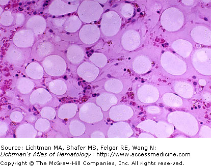
Aplastic. Marrow biopsy. Fat cells are the clear ovoid or circular spaces and compose about three-quarters of the area of this section. Most of the remaining area is a clear pink matrix. No megakaryocytes are evident. Hematopoietic cells are profoundly decreased in number. In the aplastic marrow most of the cells are lymphocytes and occasional plasma cells. Rarely, “hot spots” are seen in aplastic marrow. These are very infrequent areas of residual hematopoietic cells.
V.A.002 Aplastic
V.A.002
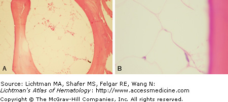
Aplastic. Marrow biopsy. (A) Lower power view. Absence of marrow hematopoietic cells between bone trabeculae, characteristic of severe aplasia, as might be seen after irradiation or other profound marrow injury. Marrow totally replaced by fat cells. (B) Higher power view. Total replacement of hematopoietic marrow by fat cells. Each space represents fat dissolved during preparation. Contrast to V.A.001 in which some residual lymphocytes and plasma cells are present characteristic of autoimmune (autoreactive T cell-induced) aplastic anemia.


