The classification applies only to carcinomas. There should be histological confirmation of the disease. See Head and Neck Tumours. See Head and Neck Tumours. The pT and pN categories correspond to the T and N categories.
NASAL CAVITY AND PARANASAL SINUSES (ICD‐O C30.0, 31.0, 1)
Rules for Classification
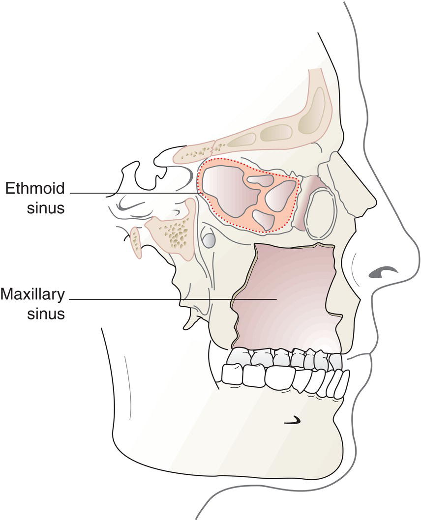
Anatomical Sites and Subsites
Regional Lymph Nodes
TN Clinical Classification
T – Primary Tumour
TX
Primary tumour cannot be assessed
T0
No evidence of primary tumour
Tis
Carcinoma in situ 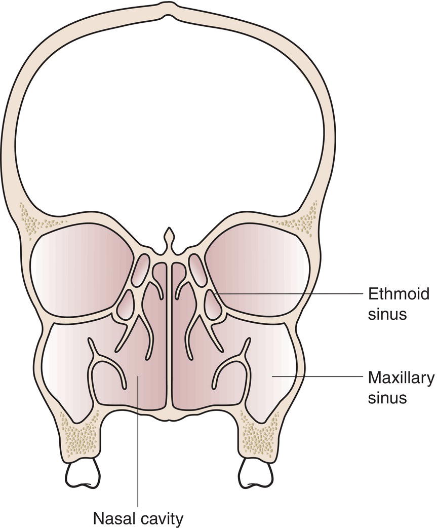
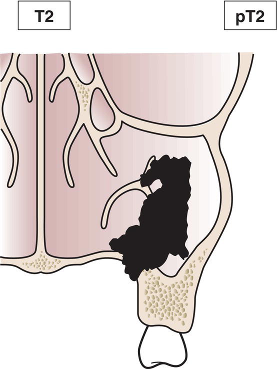
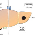
Maxillary Sinus
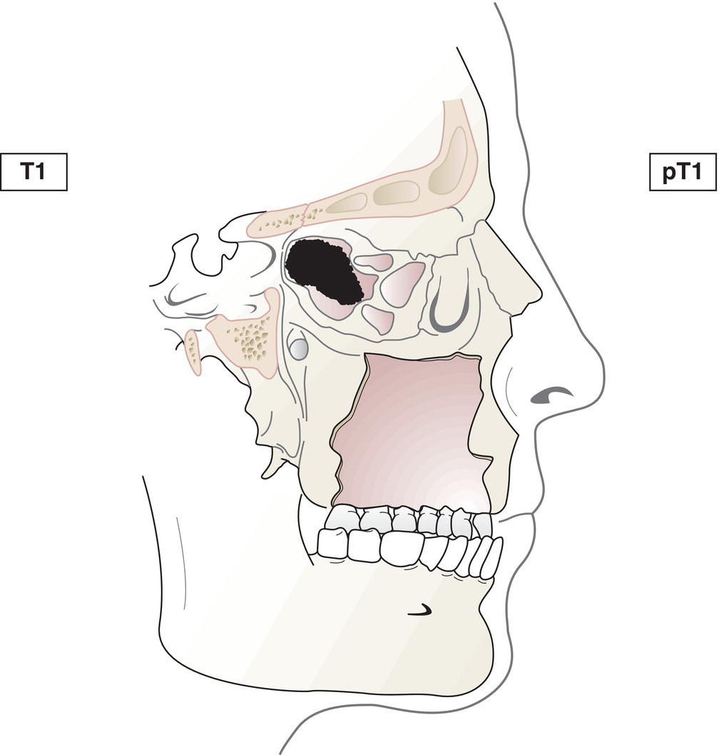
T1
Tumour limited to the mucosa with no erosion or destruction of bone (Fig. 99)
T2
Tumour causing bone erosion or destruction, including extension into the hard palate and/or middle nasal meatus, except extension to posterior wall of maxillary sinus and pterygoid plates (Fig. 100)
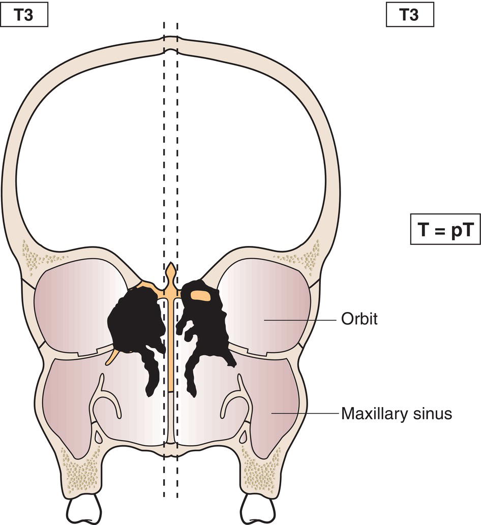
T3
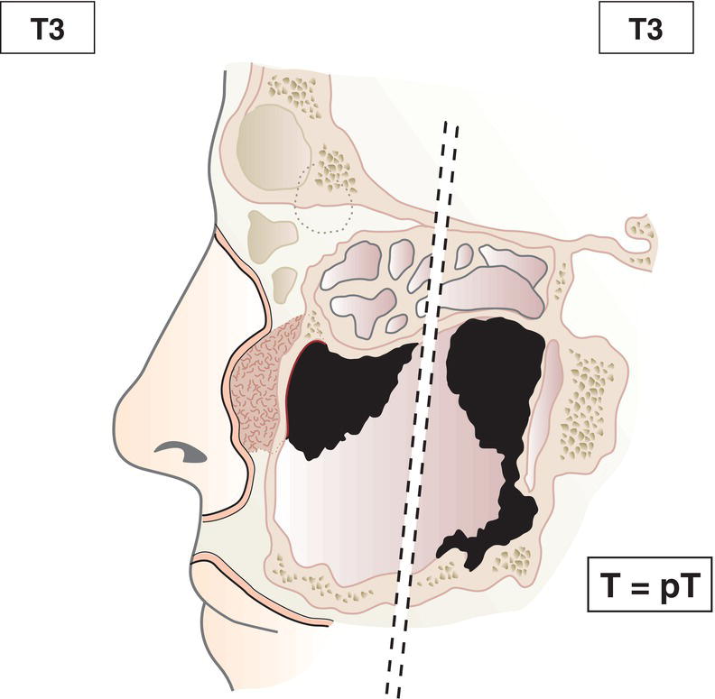
Tumour invades any of the following: bone of posterior wall of maxillary sinus, subcutaneous tissues, floor or medial wall of orbit, pterygoid fossa, ethmoid sinuses (Figs. 101, 102)
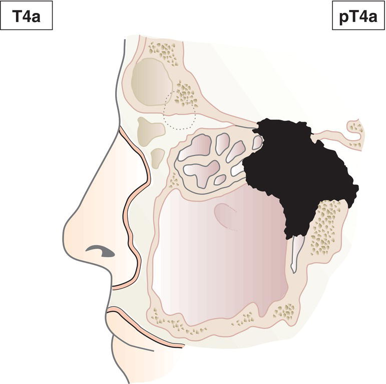
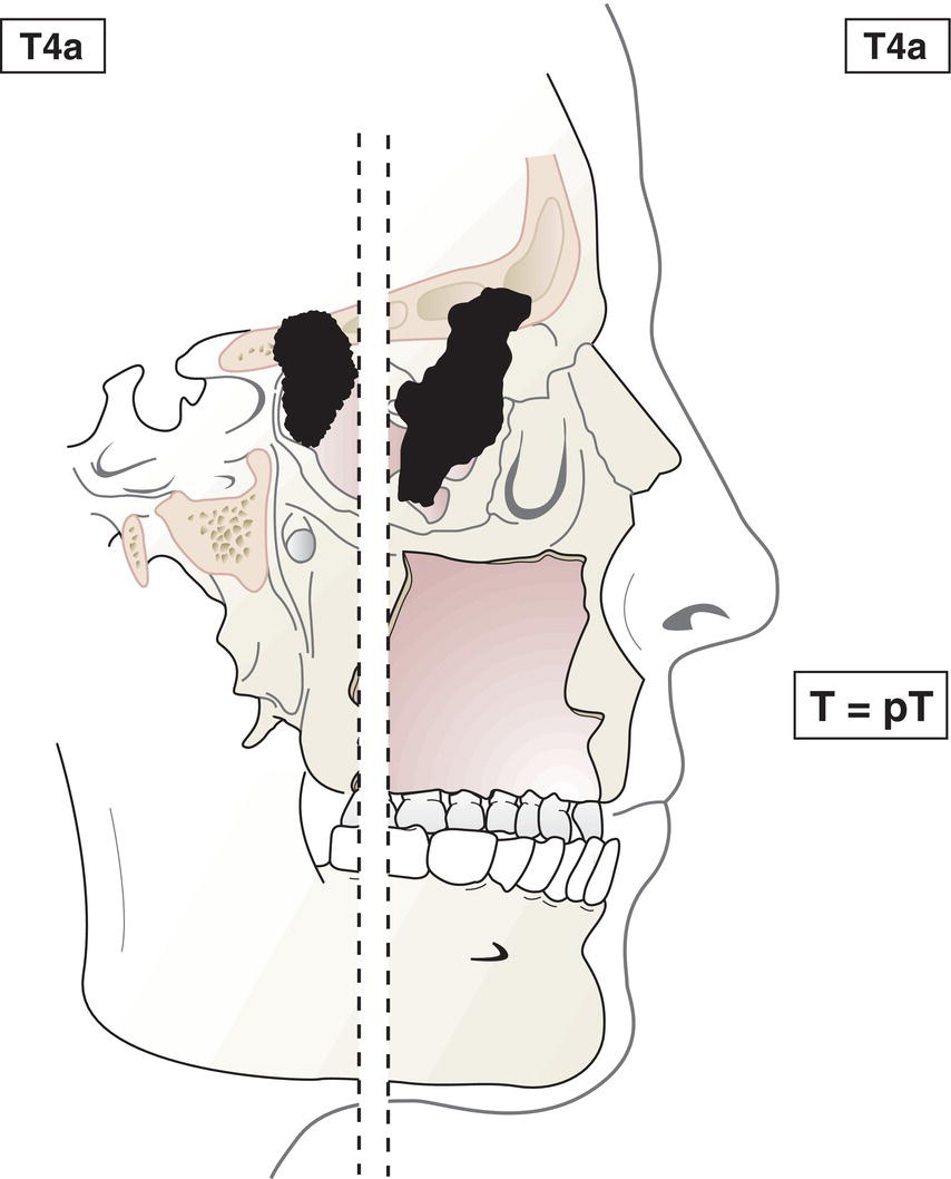
T4a
Tumour invades any of the following: anterior orbital contents, skin of cheek, pterygoid plates, infratemporal fossa, cribriform plate, sphenoid or frontal sinuses (Figs. 103, 104)
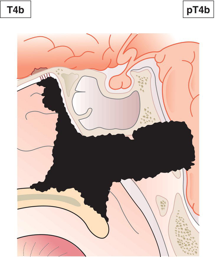
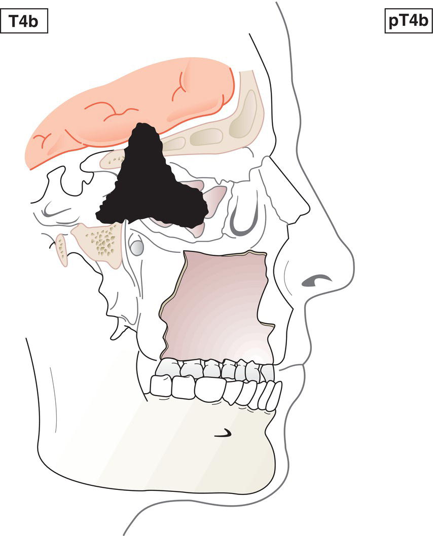
T4b
Tumour invades any of the following: orbital apex, dura, brain, middle cranial fossa, cranial nerves other than maxillary division of trigeminal nerve (V2), nasopharynx or clivus (Fig. 105) 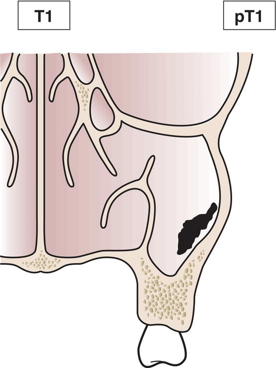
Nasal Cavity and Ethmoid Sinus
T1
Tumour restricted to one subsite of nasal cavity or ethmoid sinus, with or without bony invasion (Figs. 106, 107)
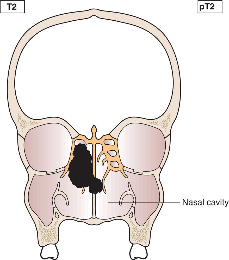
T2
Tumour involves two subsites in a single site or extends to involve an adjacent site within the nasoethmoidal complex, with or without bony invasion (Fig. 108)
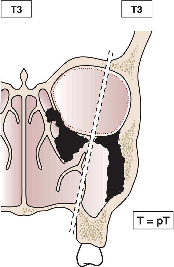
T3
Tumour extends to invade the medial wall or floor of the orbit, maxillary sinus, palate, or cribriform plate (Fig. 109)
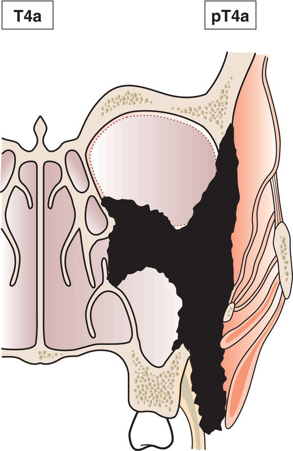
T4a
Tumour invades any of the following: anterior orbital contents, skin of nose or cheek, minimal extension to anterior cranial fossa, pterygoid plates, sphenoid or frontal sinuses (Fig. 110)
T4b
Tumour invades any of the following: orbital apex, dura, brain, middle cranial fossa, cranial nerves other than V2, nasopharynx or clivus (Fig. 111) 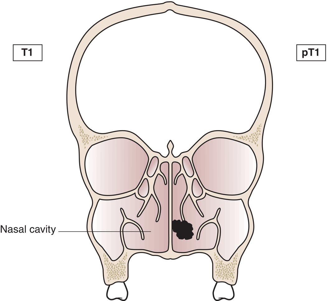
Regional Lymph Nodes
pTN Pathological Classification
Summary
Related posts:


Stay updated, free articles. Join our Telegram channel

Full access? Get Clinical Tree


