The Candida genus represents a group of more than 200 species present in both human flora and the environment. Of these, approximately 20 have been implicated in human disease. Typically, the pathogens from this genus are divided into C. albicans and non- albicans Candida species. The epidemiologic distribution of Candida spp. in children differs somewhat from that of adults. C. albicans is the most common species, and contemporary studies suggest that this species accounts for 30% to 35% of all invasive candidiasis (IC) cases. C. parapsilosis represents the second most common species, causing approximately 15% of IC events, followed by C. glabrata , C. tropicalis , and C. krusei . This is in contrast to the epidemiology of adult patients, in which C. glabrata is the second most common species. Collectively, these five species account for more than 90% of all identified isolates of candidemia. The remaining 10% of IC cases are caused by a variety of less commonly identified species, such as C. lusitaniae , C. dubliniensis , C. guilliermondii , C. stellatoidea , C. keyfr , C. pseudotropicalis , and C. intermedia . More recently, significant attention has been focused on C. auris. This species is still relatively uncommon but is important as it has the potential to harbor multidrug resistance.
Candida spp. are often a component of the normal commensal flora in children, but they can cause both superficial and invasive disease in specific clinical settings. Children with malignancy and hematopoietic stem cell transplant (HSCT) and solid organ transplant (SOT) recipients in whom IC develops often have at least one predisposing risk factor for candidiasis beyond their underlying condition.
Understanding the epidemiology of Candida spp. in these high-risk children is important because treatment strategies are dependent on the Candida spp., location of disease, and immune status. Although the epidemiology and prophylactic strategies for Candida spp. infections differ among clinical manifestations, diagnostic and treatment strategies remain similar.
Epidemiology and risk factors
The rate of IC in hospitalized children has been decreasing. This decline is likely multifactorial and been attributed to improvements in infection control measures, such as managing central venous catheters (CVCs) and prophylaxis in high-risk patient groups. Although these efforts have helped to reduce the frequency of IC, IC persists as the most common form of invasive fungal disease (IFD) for children receiving chemotherapy for malignancy and those undergoing transplantation, resulting in significant morbidity and mortality. A number of factors can predispose children to candidiasis, including compromise of anatomic barriers (e.g., the presence of CVCs or peritoneal catheters and recent surgery or trauma); disturbance of the normal flora with gastrointestinal insufficiency or after antimicrobial exposures; compromise of the immune system in the setting of primary or acquired immunodeficiencies; and iatrogenic compromise of the immune system secondary to receipt of chemotherapy or immune-modulating agents. The epidemiology and risk factors specific to oncology, pediatric HSCT, and SOT populations are discussed in the following sections.
Hematopoietic stem cell transplant
Candida infections account for nearly 10% of all infections and 60% of all IFDs in the first year after HSCT in children. IC has been reported as either the most common or second most common source of IFDs in pediatric allogeneic HSCT patients. The epidemiology of the different species has varied over time. From 1991 to 1996, C. albicans and C. parapsilosis dominated as the cause of IC in pediatric HSCT patients. However, rates of C. glabrata and C. tropicalis have increased in more recent years, exceeding rates of C. albicans and C. parapsilosis in some cohorts. The timing of IC can differ by HSCT-related factors, but typically these infections occur early with a median onset of 20 days after allogeneic HSCT. Rates of IC are higher in allogeneic versus autologous (8% vs. 5%) transplant recipients, likely related to immunosuppression and graft-versus-host disease (GVHD). The presence of GVHD can prolong the risk period, with some cases of IC presenting 8 months after transplantation in the setting of chronic GVHD. Other studies describe the majority of Candida infections occurring within the first 30 days after transplant. In pediatric recipients of an autologous or matched sibling allogeneic HSCT, IC tends to develop early after HSCT, compared with children undergoing haploidentical, umbilical cord blood, or matched unrelated transplants presumably owing to the reduced risk of GVHD in the former patient groups.
Solid organ transplant
Recent publications have begun to better define the epidemiology of IC in each respective SOT groups. IC appears to have the most significant burden in children undergoing small bowel transplant. In a cohort of 98 small bowel recipients, 25 (25.5%) patients had 59 episodes of IC. Candidemia was the most common presentation, but patients also had urinary tract and abdominal involvement, with presentations including abscess or peritonitis. In small bowel recipients, non- albicans Candida species predominate, with C. albicans accounting for only 25% to 38% of the events. Risk factors for IC after small bowel transplantation include administration of total parenteral nutrition and receipt of antibiotics during the 7 days preceding infection.
Although less common than in small bowel transplant recipients, IC is still relatively common after pediatric liver transplantation, occurring in 2.5% to 7% of all liver recipients. The majority of IC in this organ group occurs in the first 3 weeks after transplant. Interestingly, C. albicans is the most common species, accounting for more than 50% of IC events. Candidemia is the most common presentation of IC, although hepatic abscesses and surgical site infections have also been described. Intensive care unit admission before liver transplant and prolonged operative time have been associated with IC in univariate analyses, with intensive care unit admission remaining associated in multivariate modeling.
Pulmonary fungal disease has been reported in approximately 10% of pediatric lung transplant recipients. Aspergillus species are the most common pathogen identified, followed by Candida species, with C. albicans as the predominant Candida species isolated. However, identification of Candida from a sputum or bronchoalveolar specimen may represent colonization and not true infection, and thus clinicians must take caution in ascribing Candida as the true pathogen of pulmonary disease. Candida species do not typically cause a primary pulmonary process but can result in lung parenchymal involvement upon dissemination from the bloodstream.
In a pediatric cardiac transplant cohort of 1854 patients, the rate of all IFDs was estimated at 5.9%. Candida species were the most common etiology of IFD in this cohort, accounting for more than 75% of IFD events. Of note, the majority of Candida species isolated in patients designated as IC were from nonsterile sites, which calls into question whether these were true IC events versus colonization. Thus the frequency of IC in pediatric cardiac transplant recipients may be overestimated from this cohort. Similar to the epidemiology of liver and pulmonary transplant recipients, C. albicans (55%) was the most common detected species. The majority of IC infections occurred early after transplant, within the first 2 months.
Candida infections in pediatric renal transplant patients are uncommon. A 13-year retrospective review of 234 pediatric kidney transplants revealed no cases of IC. In a mixed, but predominantly adult cohort of renal transplant patients, 11% of subjects had candiduria. However, the majority (54%) were asymptomatic, and only 5% had concomitant candidemia.
Oncology
IC is a leading cause of IFD and infection-related mortality in pediatric oncology patients. The frequency of IC can vary by malignancy type, which is likely a function of the variation in the intensity of chemotherapy regimens across cancer types. When considering a cohort inclusive of all pediatric cancers, the incidence of IFD was 4.9% and Candida spp. accounted for 69% of these events. The overall case-fatality rate in the children with IC was 63%, which rivaled the rate of 85% in children with invasive aspergillosis. In children with acute myelogenous leukemia (AML), the incidence of IC has ranged from 6% to 13%, with incidence varying based on intensity of treatment. The overall case-fatality rate of these children with AML and IC was 17.6%. In cohorts of pediatric acute lymphoblastic leukemia, lower rates (3.5%) of IC have been described during the induction chemotherapy period. The distribution of Candida species varies by study and malignancy and may be a function of the local hospital epidemiology. Although some studies suggest that C. albicans accounts for the majority of cases, other studies have found a predominance of C. parapsilosis.
Data on risk factors for IC within cohorts restricted to oncology patients are limited. Generally, IC is more common in children with a hematologic malignancy compared with those with a solid tumor. In a study of children in an intensive care unit, the presence of a malignancy increased the odds of candidemia by 4-fold. The presence of a CVC, recent receipt of vancomycin, or recent receipt of anaerobic antimicrobials, all common in children with malignancy, further increased the risk of candidemia. However, predictive models have been difficult to validate.
As chemotherapy protocols and SOT immunosuppressive induction and maintenance regimens are adapted to include novel immunosuppressive agents, and as supportive care measures are optimized (e.g., administration of antifungal prophylaxis and more judicious use of antibiotics), the rates of IC in children with a specific SOT or malignancy will continue to vary. Recommendations on certain supportive care measures are provided in later text, but clinicians need to remain vigilant to changes in care protocols that will affect the risk of IC in these patient groups.
Clinical manifestations
Candida species can cause superficial and invasive infections. The former are often identified at mucosal surfaces. The latter can involve the bloodstream (i.e., candidemia), be limited to one organ (i.e., focal candidiasis), or have multiple foci (i.e., disseminated candidiasis). These clinical entities have signs and symptoms that are often similar across children with malignancy or transplant recipients.
Superficial and mucosal infections
Dermatitis.
Candida dermatitis occurs in both oncology and transplant patients, similar to non-immunocompromised children. It commonly occurs in the distribution of a diaper and presents as an erythematous rash with or without satellite lesions. Use of broad-spectrum antibiotics, either as prophylaxis or treatment, is often a predisposing factor. Other locations of infection include areas of skin overlap (e.g., inguinal and axillary regions or areas of skin breakdown or breech, particularly at surgical incision or CVC sites). Compared with immunocompetent children, these children are at higher risk to develop secondary IC from a superficial Candida dermatitis infection.
Mucosal infections.
Infection of the oropharynx, esophagus, or vulvovaginal mucosa typically occurs secondary to changes in normal flora after exposure to broad-spectrum antibiotics and/or after receipt of chemotherapeutic or conditioning agents that can also result in dysbiosis or breakdown of the mucosa. Oropharyngeal and esophageal candidiasis can occur in isolation or concurrently. Oropharyngeal candidiasis (thrush) is characterized by a white pseudomembrane on the oral mucosa, including buccal regions, gums, tongue, palate, tonsils, and uvula. Children typically present with pain and dysphagia, leading to decreased oral intake. Rarely, lesions are reported to be painless. The lesions are apparent on physical examination and can be removed by scraping. Underlying mucosa may be erythematous or normal in appearance.
Candidal esophagitis typically presents with dysphagia or odynophagia. In patients without oropharyngeal candidiasis, the diagnosis often requires invasive testing to confirm candidiasis or to evaluate for other infectious causes of esophagitis (e.g., cytomegalovirus or herpes simplex virus), which can present similarly or in tandem with candidal esophagitis. Gross inspection on endoscopy may reveal erythematous, edematous, and ulcerated mucosa as well as white plaques. Rarely, strictures have been reported. In cases of severe disease, mucosal changes may be seen on barium esophogram.
Similar to other mucosal areas of involvement, candidal vulvovaginitis may be asymptomatic, pruritic, or painful. Dysuria and vaginal discharge may also occur. White plaquelike lesions are often present on examination.
Suspicion for mucosal candidiasis is often based on direct visualization of the affected area along with reported symptoms. However, isolation of a Candida spp. may be desired to confirm the diagnosis or to differentiate from other possible processes on the differential (e.g., herpes simplex virus esophagitis). Culture of mucosal surface scrapings can be performed, but it must be recognized that isolation of Candida from these cultures may represent only colonization rather than definitive evidence of infection. Nonetheless, isolation of a Candida spp. may be helpful in directing therapy, especially if a patient is not responding to initial empiric antifungal therapy. C. albicans is the most commonly identified species in the setting of mucosal disease, although children receiving antifungal prophylaxis may present with a non- albicans Candida species.
Invasive candidiasis
IC most commonly presents as a bloodstream infection (i.e., candidemia) with potential for dissemination to multiple organs, often referred to as disseminated candidiasis. It is also possible to have IC of a single organ system or body compartment that may result from local inoculation of Candida, such as with intraabdominal candidiasis in the setting of a peritoneal catheter.
Candidemia.
Candidemia can present with nonspecific findings that mimic the clinical presentation of bacteremia. Signs and symptoms range from fever without localizing symptoms to septic shock. Focal symptoms (e.g., dyspnea, abdominal pain) may be present in cases with dissemination to end organs. This would include extension to the skin and soft tissues that may be clinically evident as erythematous macular, papular, or pustular lesions. Laboratory findings such as hyperglycemia and thrombocytopenia may be present but are not sensitive or specific. The presence of a CVC is common and is often considered as the portal of entry. However, translocation from the commensal flora across a compromised alimentary track without mucosal infection is also possible.
Genitourinary and renal candidiasis.
Candida spp. can result in both lower and upper genitourinary tract involvement. The presence of Candida in the bladder, candiduria, can be either asymptomatic or symptomatic. Asymptomatic candiduria often occurs in association with an indwelling bladder catheter in contrast to symptomatic candiduria, or cystitis, in which children may report dysuria. Candida can infrequently ascend the genitourinary tract to involve the renal parenchyma, similar to bacterial pyelonephritis. In this clinical scenario, signs and symptoms are similar to that of a bacterial pyelonephritis, including fever, vomiting, dysuria, and flank discomfort. Alternatively, Candida spp. can hematogenously disseminate to the kidneys, resulting in renal parenchymal involvement. For this process, patients may be asymptomatic but may also have signs and symptoms consistent with pyelonephritis, including fever and flank pain, in addition to hypertension and an elevated creatinine level. Radiographically, disseminated candidiasis that involves the kidney appears as numerous small nodular lesions, similar to that of hepatosplenic candidiasis (see next section). Less commonly, a mycelial mass (fungal ball) may be present and cause symptoms of urinary obstruction.
Hepatosplenic (chronic disseminated) candidiasis.
Hepatosplenic candidiasis primarily occurs in neutropenic patients. It can present with gastrointestinal symptoms, such as abdominal pain, nausea, and/or vomiting. However, neutropenic patients may be asymptomatic or present with only fever. Fever often persists until and even well beyond neutrophil recovery, at which time abdominal symptoms may develop. Disease may be identified in patients with candidemia undergoing abdominal imaging for evaluation of disseminated disease; however, the majority (>80%) of children with hepatosplenic disease do not have Candida detected in the bloodstream by culture. It has been postulated that in the setting of neutropenia, Candida spreads to the liver and spleen from the gastrointestinal tract via the hepatoportal circulation. Thus hepatosplenic disease is not dependent on antecedent candidemia. This would explain the rarity of positive blood culture results for Candida spp. in these patients. Laboratory findings and imaging may be unrevealing during the neutropenic period. After neutrophil recovery, elevations in the alkaline phosphatase level and abnormalities on imaging (e.g., computed tomography [CT] or ultrasound) can be identified. The majority of patients have lesions identified in both the liver and spleen, although isolation to one organ is possible. Classic CT findings include multiple small lesions that may appear as a “wheel within a wheel” or bull’s-eye pattern. This radiographic appearance correlates with a histologic inner circle of necrotic fungal elements, a middle circle of inflammatory cells, and an outer circle of fibrosis. Other imaging findings include hypoechoic lesions, or later in the course of disease, echogenic foci, which may be due to calcifications or fibrosis.
Intraabdominal candidiasis.
Intraabdominal candidiasis is a distinct entity from hepatosplenic candidiasis. Intraabdominal disease is typically related to surgery (e.g., anastomotic leaks after liver transplantation or bowel perforation) or the presence of a foreign body (e.g., peritoneal dialysis catheter or drain). Peritonitis and/or intraabdominal abscess may develop. Both Candida peritonitis and intraabdominal abscess are clinically similar to their bacterial counterparts; fever and abdominal pain are common. Intraabdominal abscesses may be polymicrobial. In patients with postsurgical infections, the surgical site may become erythematous or tender, and purulent drainage may be present. Alternatively, a nonhealing wound or wound dehiscence should raise suspicion for Candida infection in high-risk patients. Identification of Candida from a peritoneal catheter or drain culture may represent colonization rather than true infection. Thus the diagnosis is made from aspiration of peritoneal fluid or intraabdominal abscess in conjunction with clinical signs and symptoms consistent with intraabdominal candidiasis. Similarly, surgical sites may also be colonized with Candida, so the presence on a surgical site swab does not confirm Candida as a pathogen and clinical correlation is important.
Osteoarticular candidiasis.
Candida osteoarticular disease presents with signs and symptoms similar to those in bacterial osteoarticular disease. Disease is typically secondary to hematogenous dissemination, and the long bones are the most commonly involved foci. Focal findings of bone pain and/or limited joint range of motion or pain are often present. Candida osteoarticular disease more frequently involves multiple bones compared with bacterial disease. Additionally, because Candida osteoarticular disease often presents in an immunocompromised host, symptoms may be less apparent than bacterial osteoarticular disease. This is of particular concern in the neutropenic patient, who may have absence of significant bone discomfort or micromotion tenderness typically associated with osteomyelitis or septic arthritis. However, with neutrophil count recovery, these signs or symptoms may become more apparent.
Endovascular candidiasis.
Endovascular disease, including endocarditis and suppurative thrombophlebitis, most commonly occurs in the presence of persistent candidemia. Children presenting with endocarditis may have a history of predisposing cardiac lesions, cardiac surgery, or prosthetic cardiac material. Persistent fever is common but is not absolute. Fever may be the only sign of endovascular infection. Other signs or symptoms of endovascular disease include chest pain, dyspnea, evidence of embolic phenomena (e.g., wedge-shaped lesions on imaging of other organs, nodular lesions in distal extremities or skin), new heart murmur, or signs of heart failure. Notably, these signs and symptoms are not specific for Candida endovascular disease versus other infectious causes of endocarditis. Suppurative thrombophlebitis may present with focal tenderness, overlying erythema, or a palpable cord at a previous or current CVC site.
Ocular candidiasis.
Candida endophthalmitis is typically endogenous, secondary to candidemia, although traumatic exogenous disease can occur. Endogenous endophthalmitis is often asymptomatic and is recognized only upon a dedicated ophthalmologic examination. Symptoms may present as the disease progresses and include unilateral or bilateral vision loss. Pain is uncommon. In patients with neutropenia, examination findings, such as chorioretinal lesions, may be present only after neutrophil recovery. Chorioretinitis presents with focal, white lesions on the retina. In cases of vitreal involvement, fluffy white balls in the vitreous can be seen. Anterior chamber involvement, sometimes as a hypopyon, occurs with significant inflammation.
Central nervous system candidiasis.
Central nervous system (CNS) disease typically results from hematogenous dissemination in the presence of candidemia. CNS disease after hematogenous spread may be asymptomatic, but it also can be associated with altered mental status, meningismus, seizures, or headache. The size and location of the lesions influence clinical manifestations. The radiographic appearance of CNS candidiasis can be quite varied and include meningeal enhancement, vasculitis, and/or parenchymal foci that are nodular in appearance. The latter can range from multiple small lesions to a single large mass. Histopathologically, these nodular lesions can appear as granulomas or abscesses. Pediatric patients requiring surgical interventions, such as resection of a tumor or placement of a shunt, are at risk of Candida surgical site or device-related infections.
Pulmonary candidiasis.
Candida species do not typically cause primary pneumonia. In fact, identification of Candida spp. from a respiratory tract specimen (e.g., sputum culture or bronchoalveolar lavage) almost always represents airway colonization and not true disease. However, pulmonary candidiasis can result from hematogenous spread to the lungs. This is of particular concern when candidemia exists in the presence of prolonged neutropenia. Pulmonary involvement after dissemination may be asymptomatic but can also cause cough, dyspnea, and hypoxemia. Imaging findings most commonly include a diffuse, nodular pattern, representative of microabscesses. Less commonly, lobar infiltrates, ground-glass opacities, or empyemas are reported.
Disease prophylaxis and prevention
The comparative effectiveness of antifungal prophylaxis has been variably studied and thus inconsistently used as a preventative measure against IC in children with malignancy and recipients of either an SOT or HSCT. Because the risk of IC and evidence for prophylaxis varies among the three groups of immunosuppressed children, recommendations are discussed separately for each high-risk group. Studies evaluating the use of antifungal prophylaxis are most commonly performed in adults and many use any IFD, rather than IC, as the outcome of interest, limiting the ability to development informed decisions on prophylaxis for individual risk groups.
Hematopoietic stem cell transplant
In HSCT recipients, there are two primary risk periods for IC: the period of neutropenia sustained from conditioning, and in relevant patients, the period of GVHD. The sentinel adult antifungal prophylaxis study was completed in 1992 by Goodman and colleagues, who randomly assigned patients to fluconazole versus placebo through the posttransplant neutropenia period. Fluconazole recipients had significantly decreased rates of superficial (33.3% vs. 8.4%, P <.001) and invasive (15.8% vs. 2.8%, P <.001) fungal disease in HSCT patients. Meta-analyses consistently demonstrate a decrease in mortality and IFD in HSCT patients receiving antifungal therapy. These predominantly adult data have reasonably been extrapolated to include pediatric HSCT populations. Current pediatric HSCT-specific antifungal prophylaxis guidelines support the use of fluconazole prophylaxis in children undergoing allogeneic HSCT and children undergoing autologous HSCT with an anticipated duration of neutropenia more than 7 days. Prophylaxis should be initiated at the start of conditioning and continue until engraftment. Echinocandins are an alternative when fluconazole is contraindicated (e.g., busulfan administration). A recent randomized trial in children comparing either voriconazole or posaconazole to fluconazole during the neutropenic period after allogeneic HSCT was stopped early for futility, suggesting that fluconazole should still be the prophylactic agent of choice in children during neutropenia after HSCT.
A substantial subset of children have engraftment after allogeneic HSCT but subsequently have either acute or chronic GVHD, necessitating systemic immunosuppressive therapy. This is a second period of risk for IFD in general, and IC specifically, for HSCT patients. Unfortunately, there are limited pediatric specific data on the utility and choice of antifungal prophylaxis during GVHD. One trial, consisting predominantly of adults with only a small number of children, did not find a benefit of voriconazole compared to fluconazole for prevention of IFD during periods of GVHD. A second trial of predominantly adults with a small number of adolescents found that posaconazole reduced rates of proven and probable IFD as well as IFD-related mortality when given during periods of GVHD. Although this study was not specific to an outcome of IC, posaconazole was associated with an overall reduction in the rates of proven and probable IFD as well as IFD-related mortality. Based on these data, posaconazole should be favored in children 13 years and older during acute grade II to IV or chronic, extensive GVHD, not because of enhanced IC protection but rather to prevent invasive mold disease (IMD). In the absence of posaconazole pharmacokinetic data for children younger than 13 years, fluconazole is recommended as the prophylactic agent in this younger age group.
Even with routine antifungal prophylaxis, breakthrough mucosal candidiasis and IC occurs. Thus clinicians should continue to be suspicious for IC in children receiving antifungal prophylaxis. If breakthrough infection is identified, then an antifungal agent with a different mechanism of action should be strongly considered.
Solid organ transplant
Determining the benefit of antifungal prophylaxis in SOT recipients is more challenging as the period of risk is less discrete than that in HSCT recipients, and thus the necessary duration of antifungal prophylaxis is often less clear. This factor has likely contributed to the limited comparative data in these patient groups to establish the effectiveness or efficacy of antifungal prophylaxis. However, the epidemiology can inform the risk by organ and timing of IC onset that can help guide prophylaxis decisions. The rate of IC has been shown to be highest in liver, small bowel, and pancreas transplant recipients and lowest in lung, heart, and renal transplant recipients. Most infections occur within the first 3 months after transplant. The impact of IC has been most comprehensively studied in pediatric liver transplant recipients, in whom IC develops in approximately 3% and 5% of recipients during the first 30 and 180 days after transplant, respectively. Mortality rates in this population are overall low, unlike those in their adult counterparts. , In meta-analyses of primarily studies of adult patients, fluconazole has been associated with a reduction in IFD and IFD-attributable mortality. Based on these studies, guidelines for adult patients recommend antifungal prophylaxis in patients with specific risk factors for IC, including prolonged operation time, renal failure, and large transfusion requirements, among others. Because the rates of IC in children after liver transplant appear to be lower than in adults, it is difficult to generalize fluconazole prophylaxis recommendations to all pediatric liver transplant recipients. It is reasonable to consider fluconazole prophylaxis in liver transplant recipients with risk factors associated with IC, including prolonged operation time or need for hospitalization in the intensive care unit immediately before transplantation.
Data supporting the utility for antifungal prophylaxis against IC in other pediatric SOT groups are less convincing. A recent retrospective assessment of IC in lung, heart, and renal transplant recipients at a single children’s hospital found rare events of IC despite lack of routine antifungal prophylaxis use at that center. This finding suggests that antifungal prophylaxis directed at IC is not universally necessary in these transplant groups. Some SOT recipient groups, particularly lung transplant recipients, may warrant antifungal prophylaxis against other fungal pathogens, such as molds, based on patient specific risk factors such as prior mold infection or colonization. These cases should be assessed individually, as the need for antimold prophylaxis would supersede the need for IC prophylaxis.
The epidemiology for IC in pediatric pancreas and small bowel transplant recipients is not known. The number of pediatric patients receiving these organs each year is small, and thus the ability to establish IC incidence in these populations is limited. Because cumulative incidence of IC in adult pancreatic and small bowel transplant recipients is higher than in liver recipients, it may be prudent to extrapolate adult recommendations, which recommend fluconazole prophylaxis for all small bowel transplant recipients and for a subset of pancreatic transplant recipients with specific risk factors for IC.
In patients who are not tolerant of fluconazole or have concerns for drug-drug interactions, alternative antifungal agents can be considered. A randomized trial of anidulafungin versus fluconazole prophylaxis in high-risk adult liver transplant recipients found no significant difference in IFD rates. Therefore echinocandins may be considered second-line agents in this setting, although this would necessitate intravenous administration, which may present unnecessary risks in maintaining a CVC. Selective bowel decontamination, with the use of enteral nystatin, has not been shown to decrease IFD.
The optimal duration of prophylaxis is not known, but guidelines for adult patients have suggested from 14 days to 4 weeks or until resolution of risk factors. Whether this duration is appropriate for children is not known and should be assessed in future studies. It is likely the duration of therapy would need to be adjusted according to ongoing risk factors, including need for follow-up surgical procedures, retransplantation, persistent transfusion requirement, and until healing of the surgical site (e.g., anastomotic healing in a small bowel recipient).
Oncology
The etiology of IC in oncology patients is typically multifactorial, but the highest risk period is during prolonged neutropenia, often defined as more than 7 days after receipt of intensive chemotherapy. Most studies assessing the effectiveness of antifungal prophylaxis during prolonged neutropenia periods in patients with an oncologic diagnosis have been focused on adults. These studies are not specific to IC but rather are considerate of all IFD outcomes. Multiple meta-analyses revealed that fluconazole was effective at significantly reducing the risk of IFD versus placebo or no prophylaxis, presumably via the reduction in IC events. The largest effect has been noted during periods of intensive chemotherapy. Based on these data, the adult fever and neutropenia guidelines endorsed fluconazole prophylaxis during periods of prolonged neutropenia (i.e., ≥7 days) secondary to chemotherapy for acute leukemia with the intention of reducing the rate of IC. These same guidelines for adult patients support the use of posaconazole prophylaxis in patients with AML, in whom the risk of IMD is higher.
Although the studies that informed the meta-analyses had limited pediatric patients, pediatric-specific guidelines have used these data to reasonably support the administration of antifungal prophylaxis in children with neutropenia secondary to AML or myelodysplastic syndrome chemotherapy. Retrospective observational data in pediatric AML cohorts have corroborated this recommendation to use antifungal prophylaxis, noting a decreased hazard in induction mortality in patients receiving antifungal prophylaxis. The current recommended agent for antifungal prophylaxis in children with AML is fluconazole, as dosing recommendations for posaconazole are not available. In centers with concern for increased rates of IMD, posaconazole can be substituted for fluconazole in children 13 years and older, but this prophylaxis regimen would be to target mold prevention rather than just for prevention of candidiasis.
Studies are currently in progress to compare the efficacy of other agents with both anti- Candida and anti-mold activity (e.g., echinocandins) to fluconazole in pediatric AML cohorts. Should a broader spectrum agent be found more efficacious, it would likely be due to prevention of IMD rather than improved IC prevention. At this time, the risk of IFD, and specifically IC, in other oncology cohorts is not considered high enough to warrant routine recommendation of antifungal prophylaxis in those populations. As chemotherapy regimens change, the risk profile for IFD, including IC, may also change. Close monitoring of these rates is important, and comparative effectiveness studies to establish the utility of prophylaxis will be necessarily based on specific malignancies and chemotherapy regimens.
Diagnosis
Signs and symptoms of IC are often nonspecific and frequently consistent with a bacterial infection or a noninfectious etiology (e.g., mucositis, surgery, medication toxicity). Furthermore, clinical findings may be subtle or absent in patients with neutropenia, which makes the diagnosis of IC difficult. Findings that should prompt consideration for additional diagnostic testing for IC include presence of prolonged fever despite broad-spectrum antibiotics in a high-risk patient. Specific clinical findings suggestive of IC include multiple erythematous, pustular, or nodular skin lesions or chorioretinitis on fundoscopic examination. These entities should lead to immediate diagnostic evaluation for IC.
Regardless of the clinical presentation, if IC is suspected, diagnostic testing is necessary to establish a diagnosis. Testing options include traditional culture and histopathologic procedures as well as radiographic studies; however, these options are limited by the prolonged time to results and/or the requirement for invasive procedures. More recently, there has been optimism regarding an evolution of non–culture-based technologies, but pediatric evidence on the utility of these studies is less than convincing or not yet available. It is important for the clinician to understand that the operating characteristics of a diagnostic tool may vary in pediatric patients and in certain clinical scenarios. It is key to have this understanding before using the diagnostic test in practice as the results can be misleading if tests are ordered in the wrong setting.
Laboratory findings
Nonspecific laboratory values, such as leukocytosis, thrombocytopenia, acidosis, and hyperglycemia, may be present but are not sensitive or specific for the diagnosis of IC. Neutropenia, although not a sign of IC, is a predisposing factor and may be present before or at the time of clinical symptoms.
Culture
Blood culture is the gold standard for the diagnosis of candidemia. Candida spp. will grow readily in commercially available blood culture systems. In one study, the median time to growth was 36 hours, and 97% of positive cultures grew within the first 72 hours. C. glabrata may require additional time for growth. Although Candida spp. take longer to trigger a positive blood culture than many bacterial pathogens, a blood culture in the setting of candidemia often becomes positive within the standard 5 days that blood culture bottles are incubated. Therefore dedicated fungal blood cultures are not routinely recommended for identification of candidemia as they will not improve the yield of blood culture for Candida spp. However, IC often occurs in the absence of a positive blood culture for Candida . Studies demonstrate that blood culture positivity varies from 25% to 71% in cases of autopsy-proven IC. Thus clinicians should not assume that IC is excluded if blood cultures are negative for Candida .
Direct assessment of material from the site of local infection by Gram stain, potassium hydroxide, histopathology, or culture can be useful for superficial, mucosal, and invasive infections. Skin and mucosal scrapings can be obtained in the outpatient setting in subjects with superficial or mucosal disease. Candida spp. are identified as a budding yeast (with or without hyphae) on Gram stain or potassium hydroxide preparation. Interpretation of these results for superficial specimens should be done with caution as the presence of Candida spp. from a superficial specimen can represent colonization rather than true disease.
For tissue-invasive disease, a biopsy of focally involved tissue may be necessary for the diagnosis because Candida is not always successfully isolated from the blood in invasive disease even when hematogenous dissemination is assumed. The likelihood of candidemia does increase with the number of organs involved. The decision to pursue a diagnostic biopsy should be considerate of the potential yield of the biopsy, the potential effect of the biopsy results on clinical decision making, and the possible risks of a biopsy. For instance, hepatosplenic candidiasis is a diagnosis often supported by multiple small nodules on radiographic studies in the absence of positive blood cultures. A biopsy can be performed in this setting, but frequently the biopsy results reveal the presence of yeast on histopathology but with negative cultures because the patient is already receiving antifungal therapy. Risks of biopsy may include bleeding, particularly in patients with thrombocytopenia. Therefore a biopsy in this setting does not often change clinical management. When a biopsy is performed, specimens should be obtained for both histopathology and fungal culture. Histopathology may demonstrate microabscesses or budding yeast, hyphae, or pseudohyphae. The latter can often be seen on special stains, including Gomori methenamine silver or periodic acid–Schiff staining. Identification of fungal elements from histopathologic specimens may be useful in cases where Candida does not grow from culture specimens (e.g., pretreatment with antifungals). Culture allows for possibility of determining specific Candida spp. and performance of antifungal susceptibilities testing.
Determination of Candida species after culture has yielded an isolate is important for guiding therapeutic decisions. Traditionally the first step for dichotomizing an isolate as C. albicans or non- albicans Candida was incubation in proteinaceous liquid at 35 ° C . C. albicans forms germ tubes when incubated in this way. The remainder of Candida spp. were then identified by kits that included a panel of biochemical tests. Performing the assays from these kits, which included a variety of temperature, culture medium, and carbohydrate tests, could take days. More recently, a number of techniques have improved the time to species identification, including polymerase chain reaction (PCR) platforms, fluorescence in situ hybridization, and matrix-assisted laser desorption ionization time-of-flight mass spectrometry, which can more rapidly identify Candida spp. compared with biochemical testing.
Non-culture detection methods
Even with the advancement in species detection from cultured material, there remains a prolonged period of time from culture attainment until identification. Additionally, the less than optimal yield of blood cultures for Candida and challenges for obtaining invasive specimens from focal disease sites have resulted in pursuit of alternative detection methods for the diagnosis of IC in high-risk populations. Unfortunately, for many of these diagnostic texts, there are few pediatric-specific data, limiting conclusions on their clinical utility. As time evolves and pediatric data become available, these technologies will likely become pertinent to pediatric care.
Non-culture DNA-based diagnostic tests
PCR for Candida detection can be highly sensitive, identifying as few as four genome copies/mL. In a cohort of both oncologic and nononcologic patients with IC, multiplex Candida PCR testing identified eight patients with candidemia who were also identified by standard blood culture. The test identified five additional patients with negative blood cultures, and control samples were negative. In a meta-analysis of adult and pediatric patients with IC, PCR testing demonstrated high sensitivity (0.95) and specificity (0.92). PCR had higher sensitivity than routine blood culture for proven or probable IC (85% vs. 38%). Unfortunately, numerous PCR assays have been studied with little standardization across assays. Furthermore, limited pediatric data exist and none are approved by the U.S. Food and Drug Administration. At this time, routine clinical use of PCR for Candida detection cannot be endorsed.
A novel magnetic resonance platform, T2Candida (T2 Biosystems, Lexington, MA), has been developed for detection of certain species of Candida . This system works by amplifying Candida DNA directly from a whole blood sample followed by agglomeration of species-specific magnetic resonance particles that can then be measured. In an adult population, the platform was 91% sensitive and 99% specific compared with blood culture. The median time to identification of Candida spp. was approximately 4 hours. Detection limits are sensitive down to one colony forming unit/mL for some species. Based on these data the test has been approved by the Food and Drug Administration, but the utility in children is still not clear. One study in pediatrics did demonstrate high correlation with blood culture results for C. albicans , C. tropicalis , C. parapsilosis , and C. glabrata , but this study included only 24 blood cultures, and therefore more data are needed to support routine use in children. Particular challenges for T2Candida include clarification of the required blood volume for children. The adult protocol requires 4 mL of blood starting material, which can be a challenge to obtain routinely in children. Additionally, T2Candida identifies only five species of Candida (the previously reported species and C. krusei ) and does not distinguish between C. albicans and C. tropicalis or C. glabrata and C. krusei . Susceptibility results are also not provided. Both of these issues are important, especially recognizing the increasing presence of other resistant Candida species, such as C. auris.
Non-DNA and non-culture diagnostic tests
These non-DNA and non-culture diagnostic studies, often referred to as fungal biomarkers, are founded on a principle of identifying a component of Candida spp. (e.g., a metabolite or part of the fungal cell wall) or identifying a patient’s response to Candida spp. (e.g., antibody). These studies include arabinitol, mannan antigen and antibody, and (1,3)-β-D-glucan (BDG). Arabinitol is a metabolite produced by some Candida spp. Evaluation of this assay revealed low specificity; it is not currently commercially available and thus is not discussed further.
Both antigen and antibody tests for mannan, a component of the fungal cell wall, have been assessed. The highest sensitivity (83%) and specificity (86%) occurred when both assays were used together, but significant variability can occur among Candida spp. Data suggest these assays may have a role in early detection of IC. Positive test results have been reported before blood culture positivity in candidemic patients and before radiologic detection of lesions in patients with hepatosplenic candidiasis. Assessment in pediatric populations has primarily been in neonates. In one small study of neonates and children with malignancies, the mannan antigen assay detected two of five oncologic patients with candidemia, and none (zero of five) of the patients with C. parapsilosis .
BDG is another fungal cell wall component that is present in Candida , as well as other fungal genera such as Aspergillus . Four different assays available, but each assay has a different cutoff value; therefore tests cannot be compared and are not interchangeable. Currently, the Fungitell BGD assay (Viracor Eurofins, Lee’s Summit, MO) is the only commercially available BDG test in the United States. In a meta-analysis, the performance of the BDG assay on blood specimens had a sensitivity of 77% and specificity of 85% for the detection of IFD. Based on these studies, the package insert for the Fungitell assay recommends a threshold for positivity of more than 80 pg/mL for blood testing, but the optimal threshold has been questioned with some studies proposing sequential monitoring and alternative cutoff values. Additionally, the BDG assay has been used successfully to detect Candida spp. presence in other specimens such as cerebrospinal fluid, but the optimal threshold for positivity from these specimens is not known.
In pediatrics, the utility of the BDG assay is challenged by higher baseline levels of BDG in the blood among healthy children, which has the possibility of more false-positive results. Assessment of varying cutoffs has been attempted to improve sensitivity and specificity with little success. In a cohort of Italian children, the test had a specificity of more than 0.90 for a high cutoff value (≥200 pg/mL) but had low sensitivity at all cutoff values assessed (40 to 400 pg/mL) with overall low positive predictive values.
Several additional limitations for BDG testing exist. It is not known whether or how the use of prophylactic antifungals interferes with the test. Limited data exist in the pediatric and SOT populations, and the majority of studies assessing BDG in oncology and HSCT patients have been in the context of IFD as the outcome measure rather than specifically IC. This is because the test is not specific for Candida spp., and BDG is found in the cell wall of other fungi, including Aspergillus spp., Fusarium spp., Histoplasma capsulatum , Sporothrix schenckii , Pneumocystis jirovecii , Coccidioides immitis , Trichosporon spp., Acremonium spp., and Saccharomyces cerevisiae . Lastly, false-positive results have been attributed to blood products, hemodialysis, surgical gauze, immunoglobulin, albumin, and β-lactam antibiotics, specifically piperacillin-tazobactam. Based on the poor positive predictive value and limited data in pediatrics, BDG is not recommended as a routine diagnostic test for detection of IC in children with cancer or those undergoing HSCT.
Treatment
Several available antifungal agents for empirical and definitive therapy of IC in children are available. These include echinocandins (anidulafungin, caspofungin, and micafungin), triazoles (e.g., itraconazole, fluconazole, voriconazole, and posaconazole), and amphotericin B formulations (e.g., amphotericin B deoxycholate, lipid amphotericin B complex, and liposomal amphotericin B). The echinocandins and amphotericin B formulations are both fungicidal against Candida spp., whereas the triazoles are considered fungistatic. In choosing an antifungal agent for treatment of IC, pathogen-specific factors (e.g., species identified, resistance testing), host-specific factors (e.g., age of the child, the location of the infection, other therapeutic agents being received), and medication-specific factors (e.g., pharmacokinetics/ pharmacodynamics of the agent) all need to be considered. With this context, recommendations for definitive antifungal therapy are provided. The choice and duration of antifungal therapy is often dependent on the clinical presentation, location of infection, and Candida spp., and as such the recommendations should be consistent across oncology patients as well as SOT and HSCT recipients.
Definitive therapy based on Candida species
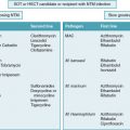
Species identification and susceptibility testing should be performed on all invasive Candida isolates. Resistance testing should be considered on isolates from superficial specimens in certain scenarios (e.g., the initial therapeutic choice is not effective). When a patient is known to have IC but susceptibility testing is not yet available, the choice of initial antifungal therapy should take into account the patient’s history (e.g., prior Candida infections, prior antifungal exposure), the hospital’s antibiogram for Candida spp., and published data on resistance profiles for a given species ( Table 26.1 ). Importantly, if a child is receiving antifungal prophylaxis and IC develops, a different antifungal class should be used until results of antifungal susceptibility testing are available.
Related posts:


Stay updated, free articles. Join our Telegram channel

Full access? Get Clinical Tree


