Cancer of the Bladder, Ureter, and Renal Pelvis


Cancer of the Bladder, Ureter, and Renal Pelvis


Adam S. Feldman

Jason A. Efstathiou

Richard J. Lee

Douglas M. Dahl

M. Dror Michaelson

Anthony L. Zietman


INTRODUCTION

This chapter details the incidence, epidemiology, pathology, and treatment of cancers of the bladder, ureter, and renal pelvis. Transitional cell carcinomas (TCC) constitute 90% to 95% of all the urothelial tumors diagnosed in North America and Europe. TCCs occur throughout the lining of the urinary tract from the renal calyceal system to the proximal two-thirds of the urethra, at which point squamous epithelium predominates. In this 10th edition, cancers of the renal pelvis and ureter are grouped with bladder cancer rather than with cancers of the kidney. This is a natural fit, because approximately 90% of the urothelial cancers of the renal pelvis, ureter, and bladder are transitional cell cancers, all of which share similarities in epidemiology, pathology, biology, patterns of spread, molecular tumor markers, and treatment. The chapter presents the common characteristics of urothelial cancers in an initial section and then deals in subsequent sections with the separate characteristics of these organs. The multidisciplinary treatment of this chapter reflects the current approach to patients with these diseases.


UROTHELIAL CANCERS


Epidemiology

Bladder cancer is almost three times more common in males than in females and more common in whites than in blacks. In 2013, there were approximately 72,570 new cases in the United States, over a 20% increase from 20 years ago. The incidence increases with age and peaks in the 6th, 7th, and 8th decades of life.1 Simultaneous or subsequent development of TCC of the urethra in patients with TCC of the bladder occurs with an incidence of 6% to 16% more commonly in women than men and in those with recurrent multifocal bladder cancers, and bladder neck or trigonal involvement with either invasive cancer or carcinoma in situ (CIS).2,3

The incidence of ureteral TCC is 0.7 per 100,000, whereas renal pelvic TCCs have an incidence of 1 per 100,000.4 Renal pelvic tumors constitute 5% of all renal tumors, and 90% of them are TCCs. Squamous cell carcinoma and adenocarcinoma constitute the majority of the remainder. Renal pelvic transitional cell cancers constitute 5% of all TCCs of the urinary tract. Patients who have primary TCCs of the renal pelvis or ureter have a 20% to 40% incidence of either synchronous or metachronous bladder cancer. Conversely, patients with bladder cancer have a 1% to 4% incidence of synchronous or metachronous upper tract urothelial tumors.5,6 However, if the bladder cancer is grade 3, there is associated CIS, or the patient has failed intravesical chemotherapy, some reports suggest a doubling of the incidence of upper tract tumors.7 Patients with Balkan nephropathy have an increased incidence of upper tract tumors; these tumors are usually low grade and multiple.8 Recently, aristolochic acid, a component of all Aristolochia plants, was identified as the etiologic agent causing Balkan nephropathy and the associated urothelial carcinoma.9,10,11 In the Balkan region, the exposure seems to occur via consumption of bread made from flour contaminated with Aristolochia clematitis seeds.12 There are also specific areas of Taiwan where TCC of the renal pelvis accounts for 40% of all renal tumors, whereas in other nonendemic areas, the upper tract tumors account for only 1% or 2% of renal tumors.13 Aristolochic acid has also recently been identified as the etiology in this population due to widespread use of Aristolochia herbal remedies.14,15

Risk factors for urothelial cancer may be classified into one of three categories: (1) gene abnormalities that result in perturbations in cell cycle regulatory processes, (2) chemical exposure, or (3) chronic irritation. Those risk factors that involve genetic abnormalities include chromosome deletions or duplications, proto-oncogene expression, tumor suppressor gene mutation, and abnormalities of specific cell cycle regulatory proteins. In non-muscularis propria-invasive transitional cell cancers, deletions of part or all of chromosome 9 and alterations in the gene encoding for fibroblast growth factor receptor 3 (FGFR3) are often encountered. Inactivation of the cohesion subunit stromal antigen 2 (Stage 2), which regulates sister chromatid cohesion and segregation, is frequently found in low-grade and low-stage bladder cancer.16,17,18 Other proto-oncogenes that have been implicated in bladder cancer include the RAS and p21 proteins.19 Genetic abnormalities associated with CIS include alterations in the retinoblastoma gene (Rb), p53, and phosphatase and tensin homolog (PTEN). In muscularis propria-invasive disease, the tumor suppressor genes that have been associated with an altered biology and more aggressive behavior include the p53 and the Rb gene.20 Abnormalities in specific cell-cycle regulatory proteins such as epidermal growth factor (EGF), Ki-67, cyclin D1, metalloproteinase (MMP), and tissue inhibition of metalloproteinase (TIMP) have also been implicated.20,21,22,23,24,25 At this time, there is no single molecular marker that is capable of predicting the tumor with a high degree of accuracy, which may result in muscularis propria invasion or distant metastases.

Chemical exposure has perhaps the most epidemiologic evidence to support it as an inciting agent. Aromatic amines, aniline dyes, and nitrites and nitrates have all been implicated. There are genetic polymorphisms that appear to increase the susceptibility of affected patients exposed to carcinogens. N-acetyltransferase, which detoxifies nitrosamines and glutathione-S transferase, which conjugates reactive chemicals, have been implicated in increasing the risk for the development of bladder cancer in patients so afflicted. Tobacco use carries with it, for those who continue to smoke, a threefold increased risk of developing bladder cancer, and even ex-smokers have a twofold increased risk.26 Numerous reports have shown strong associations between the development of both bladder and upper tract TCCs with industrial contact to chemicals, plastics, coal, tar, and asphalt, and aristolochic acid, as discussed previously. Cyclophosphamide administration over the
long term, particularly in patients who have upper tract or bladder outlet obstructions, results in an increased risk of bladder cancer. These cancers, when discovered, tend to be particularly aggressive. Coffee, tea, analgesics, alcohol, and artificial sweeteners have not been shown to act as independent risk factors.

Chronic irritants include catheters, recurrent urinary track infections, Schistosoma haematobium, and irradiation. Chronic irritation due to indwelling catheters associated with chronic infection increases the risk for the development of squamous cell carcinoma; a S. haematobium infestation results in an increased risk of squamous cell and TCCs; pelvic irradiation also carries with it an increased risk of developing a urothelial cancer.

There are many studies that suggest high water consumption, vitamin intake, and various diets as beneficial in preventing bladder cancer. However, none of these have shown any clear benefit with respect to prevention.


Screening and Early Detection

Screening has not been particularly useful in the detection of bladder cancer and the most recent statement from the U.S. Preventive Services Task Force concludes that the current data are insufficient to make a definitive recommendation on screening for bladder cancer in asymptomatic adults.27 The only test of proven usefulness is a urinalysis to detect microhematuria. If significant microhematuria is detected, then specific diagnostic studies are performed. When individuals are screened, 4% to 20% are found to have microhematuria. Of those with microhematuria, 0.5% to 8.1% have bladder tumors.28,29,30 In these particular studies, high-grade disease was identified in 2.4% to 3.5% of those presenting with dipstick microhematuria, and invasive disease was identified in 0.4% to 1%. Although one of these studies suggests that routine screening results in a reduced mortality from bladder cancer, the data are unconvincing due to a lack of randomization and likely significant selection bias.30 Others have suggested that screening in high-risk populations increases the early detection rate of high-grade cancers. Early treatment of these would be expected to be associated with an increased survival, although this hypothesis in this group of patients has not been substantiated. Screening does not generally improve the detection rate of low-grade tumors because the methods used for screening have a large number of false-negative findings for low-grade tumors. When urothelial cancer is suspected, noninvasive screening may be performed using cytology, nuclear matrix protein, telomerase, or fluorescence in situ hybridization analysis, but the definitive diagnosis is made only by cystoscopy and biopsy.

Cytology has been regarded as the gold standard for noninvasive screening of urine for bladder cancer. It has a sensitivity of 40% to 60% and specificity in excess of 90%. Nuclear matrix protein31 fibrin or fibrinogen degradation products,32 urinary bladder cancer antigen,33 and basic fetoprotein34 have all been compared with cytology in bladder cancer screening studies. Other methods used include fluorescence in situ hybridization,35 microsatellite analysis of free DNA,36 and telomerase reverse transcriptase determination.37 Unfortunately, all of these tests have a sensitivity that ranges from only 40% to 75% with a specificity of 50% to 90%, thus making it impossible to eliminate the need for cystoscopy by the use of these tests.38 These urinary biomarkers have not been studied yet for sensitivity and specificity in detecting upper tract TCCs.

Cytology remains the preferred bladder tumor marker for specificity39; however, many of the other bladder tumor markers have a better sensitivity.40


Pathology

More than 90% of the TCCs throughout the lining of the urinary tract occur in the urinary bladder and of the remaining 10%, most are in the renal pelvis and fewer than 2% are in the ureter and urethra. Squamous cell carcinomas, defined by the presence of keratinization, account for 5% of bladder tumors. Other, even less common bladder tumor types include adenocarcinoma and undifferentiated carcinoma variants such as small-cell carcinoma, giant-cell carcinoma, and lymphoepitheliomas.41,42,43 A TCC histology can also demonstrate areas of a variant subtype within a tumor, including micropapillary, squamous, glandular, or sarcomatoid differentiation. These are considered variants of TCC, and stage for stage, they do not portend a worse prognosis,44,45 likely with the exception of sarcomatoid carcinoma, which presents with a higher stage and more distant metastases than conventional TCC.46 Pure adenocarcinoma of the bladder may also arise in the embryonal remnant of the urachus on or above the bladder dome. Other adenocarcinomas may closely resemble intestinal adenocarcinoma and must be distinguished from direct spread to the bladder from an intestinal primary by careful clinical evaluation. Rarely, these demonstrate a signet ring cell or clear cell histology.


Primary Tumors of the Bladder

The differential diagnosis of TCC usually does not pose a diagnostic difficulty for experienced pathologists, but tumors that are grade 1 and invasive are occasionally difficult to distinguish from von Brunn nests.47 Also, rarely, an invasive TCC may be overdiagnosed when the glandular component of a nephrogenic adenoma is mistaken for TCC with glandular differentiation or for a pure adenocarcinoma. When invasion of the lamina propria has occurred, the pathologist must report whether muscularis propria is present in the submitted tissue and whether there is invasion of the muscularis propria. If muscularis propria is not present in the submitted tissue, this should be noted by the pathologist. Identification of invasion of the muscularis propria by the tumor may occasionally be difficult, because it may be confused with involvement of the muscularis mucosa, which is in the lamina propria.48 More than two-thirds of newly diagnosed cases of bladder tumors are exophytic papillary TCCs that are confined to the epithelium (stage Ta) or invade only into the lamina propria (stage T1). These tumors are generally managed endoscopically and, in some cases, with the addition of intravesical therapy (discussed in the following paragraphs). Approximately one-half to two-thirds of patients with such tumors have a recurrence or a new TCC in the bladder within 5 years.

Bladder tumors are also classified by their cytologic characteristics as low grade (G1) or high grade (G2, G3).43 Low-grade tumors may also be referred to as papillary tumors of low malignant potential (PNLMP). Tumor grade is clinically more significant for noninvasive tumors because nearly all of the invasive neoplasms are high grade at diagnosis. Papillary carcinomas of low grade are considered to be relatively benign tumors that histologically resemble the normal urothelium. They show only very slight pleomorphism or loss of polarity and rarely progress to a higher stage. On the contrary, CIS is cytologically synonymous with high-grade disease and carries a high risk of progression to invasive disease. Primary CIS (stage Tis) that presents without a concurrent exophytic tumor constitutes only 1% to 2% of newly detected cases of bladder cancer, but CIS is found accompanying more than half of bladders presenting with multiple papillary tumors. CIS, in this instance, is either adjacent to or involves mucosal sites remote from papillary lesions.49 CIS is believed to be an important precursor of invasive cancer and, if untreated, will develop into muscularis propria-invasive disease within 5 years from the initial diagnosis in more than 50% of patients.


Upper Tract Tumors

Like bladder tumors, 90% of upper tract tumors are TCCs with similar morphology.50 Squamous cell carcinomas account for most of the remaining carcinomas, with adenocarcinoma representing, at most, 1% of upper tract malignancies. The cytologic characteristics for the classification of TCC by grade are the same for upper tract TCCs as they are for those in the bladder.


Molecular Tumor Markers

Because the natural history of superficial urothelial tumors is that of recurrence, an area of controversy is if tumors that occur at separate sites or at separate times in the urothelial tract are derived from the same clone or are polyclonal in origin. A report by Sidransky et al.51 demonstrated the clonality of multiple bladder tumors from different sites. Miyao et al.52 showed concordant genetic alterations in asynchronous tumors from individual patients. These studies suggest that urothelial TCCs appearing at different times and sites can be derived from the same neoplastic clone. Moreover, many studies have reported an increasing frequency of specific genetic abnormalities in bladder tumors of more advanced stages.53,54,55,56 Many tumor suppressor gene modifications, including those of p53, pRB, p16, p21, thrombospondin-1, glutathione, and factors controlling the expression and function of the epidermal growth factor receptor (EGFR), have been shown in retrospective analyses to be adverse prognostic factors in patients with TCC after various treatments.58,59,60,61,62 However, even in the most intensively studied tumor suppressor gene in advanced TCC, the p53 gene, retrospective analyses give. There is conflicting retrospective data on the association of p53 mutation status and responsiveness to chemotherapy or radiation59,60 led to a phase III trial that randomized 114 postcystectomy patients with p53 alteration to three cycles of adjuvant methotrexate, vinblastine, doxorubicin, and cisplatin (MVAC) or observation. Neither p53 status nor MVAC adjuvant chemotherapy impacted the risk of recurrence.57

The enthusiasm engendered by the development of novel biologic agents targeted against tumor-specific growth factor pathways or against angiogenesis has been fortified by positive studies in a variety of solid tumors. Two classes of agents that have received great attention are inhibitors of EGFR, including EGFR1 and EGFR2 (or HER2/neu), and inhibitors of vascular endothelial growth factor (VEGF) or its receptors. Ample preclinical evidence has shown that (1) many, if not most, bladder tumors express products of the EGFR family, (2) overexpression correlates with an unfavorable outcome, and (3) inhibition of these pathways may have an antitumor effect.64,65,66,67,68,69

Evidence suggests that p53, p16, and pRB altered expression are of no prognostic significance in patients treated with chemoradiation, but that overexpression of HER2 correlated with a significantly inferior complete response rate. The recently closed Radiation Therapy Oncology Group (RTOG) 0524 protocol evaluated the addition of trastuzumab to chemoradiation for Her2 overexpressing tumors. EGFR overexpression, which occurred in only 19% of the patients, was associated with improved diseasespecific survival.70

Another potential therapeutic avenue is the inhibition of angiogenesis. Several studies have correlated elevated VEGF levels or cyclooxygenase-2 (COX-2) expression with disease recurrence or progression, often as an independent prognostic factor by multivariate analysis.69,71

Preclinical data support the concept that COX-2 inhibitors may inhibit the development of non-muscle-invasive bladder cancer. A randomized, double-blind, placebo-controlled trial sought to determine whether celecoxib, a COX-2 inhibitor, could reduce the time-to-recurrence of superficial tumors. No benefit was observed in patients receiving daily celecoxib.72

The major challenge for clinical and translational investigators is to design appropriate trials that will identify which molecular tumor markers will be prognostic of outcome and also be predictive of whether a patient will do better treated by surgery, radiation, chemotherapy, molecular targeted therapy, or a combination of these. An example of recent encouraging results include the identification of MRE11, a protein involved in DNA damage double-strand break repair, as a predictive marker of disease specific survival following radiation or chemoradiation for muscle invasive bladder cancer.73,74 Only when such molecular biomarkers are validated and incorporated into clinical decision making will physicians be able to make better treatment choices on behalf of their patients.


CANCER OF THE BLADDER

Cancers of the bladder can be grouped into three general categories by their stages at presentation: (1) those that do not invade the muscularis propria, (2) muscularis propria-invasive cancers, and (3) metastatic cancers. Each differs in clinical behavior, primary management, and outcome. When treating non-muscularis-invasive tumors, the aim is to prevent recurrences and progression to a stage that is life threatening. With muscularis propria-invasive disease, the main issue is to determine which tumors require cystectomy, which can be successfully managed by bladder preservation using combined modality therapy, and which tumors, by virtue of a high metastatic potential, require an integrated systemic chemotherapeutic approach from the outset. Combination chemotherapy is the standard for treating metastatic disease. Despite reports of complete responses (CR) in more than 40% of cases, the duration of response and overall cure rates remain low.


Clinical Presentations and Staging

Bladder cancer is rarely incidentally discovered at autopsy. Indeed, almost all cases show symptoms in the premortem period. The most common presentation is gross painless hematuria. Unexplained urinary frequency and irritative voiding symptoms should alert one to the possibility of CIS of the bladder or, less commonly, muscularis propria-invasive cancer.


Workup

The workup of suspected bladder cancer should include urine cytology, a cystoscopy, and an upper tract study. The preference for the upper tract study is a renal computed tomography (CT) urogram because both ureters and renal pelves as well as the relevant lymph nodes and the kidney parenchyma can be particularly well visualized.

Careful staging is important, because treatment depends on the initial stage of the disease. The clinical stage of the primary tumor is determined by transurethral resection of the bladder tumor (TURBT). This resection should include a sample of the muscularis propria for appropriate diagnosis, particularly if the tumor appears sessile or high grade. Once the specimen has been resected, the base of the resected area should be separately biopsied. Any suspicious areas in the remainder of the bladder should be biopsied, and many advocate additional selected biopsies of the bladder mucosa and a prostatic urethral biopsy as well. Urethral biopsies are clearly indicated in patients with risk factors for urethral involvement, as previously discussed, and in those who have persistent positive cytologies in the absence of a demonstrated bladder lesion. Patients who have T1, G3 tumors on biopsy without muscularis propria in the specimen require a second biopsy in order to obtain muscularis propria to reduce the risk of understaging. Indeed, the authors rebiopsy all patients with T1, G3 disease, because it has been shown that even if muscularis propria is in the initial specimen, a significant number of patients will be upstaged (T2) on the second biopsy.

5-Alpha amino levulinic acid installation into the bladder, resulting in porphyrin-induced fluorescence of vascular lesions when viewed with blue light, and narrow band imaging, which increases the contrast between vascular lesions and normal mucosa, have been recommended by some to increase the yield of positive biopsies. Several studies have shown a slight advantage to these techniques in reducing disease recurrence, but it remains difficult to differentiate inflammatory lesions from urothelial carcinomas with either technique, and not all well-designed clinical trials have shown a benefit.75,76,77,78


Staging

The primary bladder cancer is staged according to the depth of invasion into the bladder wall or beyond (Table 39.1).70,79 The urothelial basement membrane separates non-muscularis propria bladder cancers into Ta (noninvasive) and T1 (invasive) tumors. Stage T2 and higher T-stage tumors invade the muscularis propria, the true muscle of the bladder wall. If the tumor extends through the muscle to involve the full thickness of the bladder and into the serosa, it is classified as T3. If the tumor involves contiguous structures such as the prostate, the vagina, the uterus, or the pelvic sidewall, the tumor is classified as stage T4 (nonstromal invasive urothelial tumors of the prostate are not classified as T4, because the prognosis in this group is quite good). In a fragmented TURBT specimen, in contrast to a cystectomy specimen, it is relatively infrequent for the pathologist to be able to make an accurate assessment as to the depth of invasion of the tumor into the muscularis propria. Thus, the primary pathologic substages of the TNM (tumor, nodes, metastasis) staging system shown in Table 39.1, such as pT2a and pT2b, cannot be determined from TURBT specimens. Of note, significant rates of clinical-pathologic stage discrepancy and clinical (TURBT) understaging have been described.138 CT scans or magnetic resonance images (MRI), even those done prior to the TURBT, are not reliable for staging of the primary tumor. Neither scan can differentiate a Ta/T1 tumor from a T2/T3 tumor because neither can visualize the depth of invasion of the primary tumor into the bladder wall. These scans are helpful, however, when they show unequivocal tumor extension outside the bladder (stage T3) (Fig. 39.1). CT scans or MRIs following a TURBT also are not reliable for staging of the primary tumor because either surgically induced edema in the resected portion of the bladder wall or postsurgical extravesical inflammatory stranding may be confused with extravesical tumor extension. For this reason, it is preferable and recommended to perform a staging CT or MRI prior to TURBT.


TABLE 39.1 American Joint Committee on Cancer 2009 TNM Bladder Cancer Staging


Primary Tumor (T)


Tis


Carcinoma in situ


Ta


Noninvasive papillary tumor


T1


Tumor invades the lamina propria, but not beyond


T2


Tumor invades the muscularis propria


pT2a


Tumor invades superficial muscle (inner half)


pT2b


Tumor invades deep muscle (outer half)


T3


Tumor invades perivesical tissue


pT3a


Microscopically


pT3b


Macroscopically (extravesical mass)


T4


Tumor invades any of the following: prostatic stroma, uterus, vagina, pelvis, or abdominal wall


T4a


Tumor invades prostate, uterus, vagina


T4b


Tumor invades pelvic or abdominal wall


Regional Lymph Nodes (N)


NX


Regional lymph nodes cannot be assessed


N0


No regional lymph node metastasis


N1


Metastasis in a single lymph node in primary drainage region


N2


Metastasis in multiple lymph nodes in primary drainage region


N3


Common iliac lymph node involvement


Distant Metastasis (M)


MX


Distant metastasis cannot be assessed


M0


No distant metastasis


M1


Distant metastasis


Used with the permission of the American Joint Committee on Cancer (AJCC), Chicago, Illinois. The original source for this material is the AJCC Cancer Staging Manual, Seventh Edition (2010) published by Springer Science and Business Media LLC, www.springer.com, page 500.


Figure 39.1 A computed tomography scan of a patient with a muscularis propria-invasive bladder cancer performed before a transurethral tumor resection, unequivocally showing an extravesical extension of tumor (stage T3). The tumor projecting into the bladder lumen (black arrow); portion of the tumor extending into the ureter outside the bladder (white arrow).

Patients who have documented muscularis propria-invasive bladder cancer require an additional set of studies: a chest x-ray or CT scan, liver function studies, creatinine and electrolytes level studies, and a CT evaluation of the pelvic and retroperitoneal lymph nodes. A bimanual examination is also performed at the time the tumor is transurethrally resected to evaluate for possible extravesical extension of the tumor and to determine mobility of the pelvic contents. An MRI lymphangiography, using a lymphotropic iron nanoparticle administered intravenously, shows potential.80 Nodes that appear to be enlarged on a CT may be differentiated by this technique as to whether they are inflammatory or malignant. The sensitivity and specificity of the test are quite high.

If there is a history of functional bowel abnormality, a barium study of the segment of bowel to be used for the diversion should be performed. It is the authors’ practice when using colon in the reconstruction of the urinary tract to obtain a barium enema or colonoscopy so that there are no surprises at the time of surgery. Finally, patients with muscularis propria-invasive bladder cancer must have a prostatic urethra and bulbous urethra biopsy to determine whether an orthotopic bladder may be placed or whether the procedure should encompass the urethra—that is, a cystoprostatourethrectomy in males or a cystourethrectomy and anterior exenteration in females.


Treatment of Non-Muscularis Propria-Invasive Bladder Cancer (Ta, Tis, T1)

Of patients with bladder cancer, 70% have disease that does not involve the muscularis propria at presentation. Approximately 15% to 20% of these patients will progress to stage T2 disease or greater over time. Of those presenting with Ta or T1 disease, 50% to 70% will have a recurrence following initial therapy. Low-grade tumors
(G1 or G2) and low-stage (Ta) disease tend to have a lower recurrence rate at about 50% and a 5% progression rate, whereas high-risk disease (G3, T1 associated with CIS, and multifocal disease) has a 70% recurrence rate and a 30% to 50% progression rate to stage T2 disease or greater. Less than 5% of patients with non-muscularis propria-invasive bladder cancer will develop metastatic disease without developing evidence of muscularis propria invasion (stage T2 disease or greater) of the primary lesion.

Patients who are at significant risk for developing progressive or recurrent disease following TURBT are generally considered candidates for adjuvant intravesical drug therapy. This includes those with multifocal CIS, CIS associated with Ta or T1 tumors, any G3 tumor, multifocal tumors, and those whose tumors rapidly recur following TURBT of the initial bladder tumor. A number of drugs have been used intravesically, including bacillus Calmette-Guérin (BCG), interferon (IFN) and BCG, thioTEPA, mitomycin C, doxorubicin, and gemcitabine. Complications generally include frequency, dysuria, and irritative voiding symptoms. Over the long term, bladder contracture may occur with these agents. Other complications, which are specific for each drug, are as follows: BCG administration may result in fever, joint pain, granulomatous prostatitis, sinus formation, disseminated tuberculosis, and death; thioTEPA may cause myelosuppression; mitomycin C may cause skin desquamation and rash; and doxorubicin may cause gastrointestinal upset and allergic reactions. The proposed benefit of intravesical chemotherapy is to lessen the rate of recurrences and reduce the incidence of progression. Unfortunately, it cannot be clearly stated that any of these drugs accomplish these goals over the long term.

The use of electromotive installation as an adjunct to intravesical therapy remains controversial. A randomized trial sought to clarify the benefit of electromotive installation of mitomycin. Patients were randomized to TURBT alone (n = 124), immediate post-TURBT mitomycin (n = 126), or pre-TURBT electromotive mitomycin (n = 124). Trial results demonstrated that intravesical electromotive installation of mitomycin before TURBT reduced recurrence and improved the disease-free interval compared with intravesical mitomycin after TURBT and TURBT alone.81

Intravesical BCG therapy is typically initiated with an induction course of 6 weekly instillations, followed by a cystoscopic evaluation 1 month after induction. In cases in which CIS is present or suspected, only a biopsy can differentiate this from inflammatory change secondary to treatment. For those who respond to induction, maintenance BCG therapy for up to 3 years is a standard of care, although patients frequently discontinue therapy early due to bladder toxicity.82

A European Organisation for Research and Treatment of Cancer (EORTC) phase III trial in over 1,300 patients sought to evaluate whether a third of a dose versus a full dose and a 1-year treatment versus a 3-year treatment could suffice.83 The trial thus had four different doses and schedules of BCG maintenance therapy. No meaningful differences in toxicity, progression, or survival were observed across dose and schedules. However, the recurrence rate was lowest in high-risk patients treated with the full dose therapy for 3 years, supporting current treatment recommendations.

A number of studies have compared one intravesical chemotherapeutic agent with another. For the most part, BCG in these comparisons has a slight advantage in reducing recurrences.84 However, when the follow-up is more than 5 years, it appears that there is minimal overall effect at reducing the recurrence rate when compared with no treatment. BCG and epirubicin are the most commonly used agents in this setting and both are considered effective for the treatment of superficial bladder cancer. However, superiority of one over the other is unknown. A meta-analysis of over 1,100 patients treated with either drug reported that intravesical BCG was more efficacious, although also more toxic.85

BCG failure is a clinical concern and a treatment dilemma with limited truly effective nonsurgical options. The precise definitions of BCG failure are well outlined by the 2005 International Consensus Panel on T1 bladder cancer and include four subtypes of BCG failure.86 BCG refractory T1 disease should be of paramount concern and raises the concern for understaged diseases. Options for further intravesical treatment after BCG failure include BCG plus IFNα-2B, gemcitabine, valrubicin, docetaxel, and other novel agents.85,86,87,88,89,90,91,92 Unfortunately, however, no single agent has yet proven to be more reliably or durably effective than another, and a true consensus on continued intravesical treatment in this setting remains to be determined.

Approximately 70% of patients with high-grade disease will experience recurrence whether or not they are treated with intravesical therapy. Moreover, there is no well-documented evidence that the use of these agents prevents disease progression, for example, from stage Ta/T1 disease to stage T2 or greater disease. One-third of patients who are at high risk for disease progression (those with G3, T1 disease) will progress to stage T2 or greater disease whether or not they are treated with BCG.93 One-third of patients at 5 years who have disease progression and undergo a cystectomy die of metastatic disease. Thus, approximately 15% of patients with superficial disease at high risk for disease progression (CIS with associated Ta or T1 disease, rapidly recurrent disease, or G3 disease), irrespective of treatment modality, will die of their disease.94 If definitive therapy (cystectomy) is performed when the disease is found to progress into the muscularis propria (T2 or greater), there is no difference in cure rate when these patients are compared with those who present primarily with T2 or greater disease. These statistics have encouraged some to perform a preemptive cystectomy in those patients at high risk for progression before muscularis propria invasion is documented. Ten-year cancer-specific survivals of 80% are given as justification for this approach, as compared with 50% in patients in whom the cystectomy is performed when the disease progresses to involve the muscularis propria.95 Unfortunately, this approach subjects approximately two-thirds of these patients who are included in the 80% cancer-specific survival figure to a needless cystectomy, making it questionable as to whether there is in fact any survival advantage whatsoever. Although cystectomy remains the gold standard for recurrent BCG refractory T1 disease, there is an open protocol RTOG 0926 evaluating chemoradiation for such patients who opt for an attempt at bladder preservation or are otherwise not good cystectomy candidates.96


Treatment of Muscularis Propria-Invasive Disease


Surgical Approaches

The standard of care for squamous cell carcinoma, adenocarcinoma, TCC, and sarcomatoid or spindle cell carcinoma that invade the muscularis propria of the bladder is a bilateral pelvic lymph node dissection and a cystoprostatectomy, with or without a urethrectomy in the male. In the female, an anterior exenteration is performed, which includes the bladder and urethra (the urethra may be spared if uninvolved and an orthotopic bladder reconstruction is performed), the ventral vaginal wall, and the uterus. A radical cystectomy may be indicated in non-muscularis propria-invasive bladder cancers when G3 disease is multifocal or associated with CIS or when bladder tumors rapidly recur, particularly in multifocal areas following intravesical drug therapy. When the prostate stroma is involved with TCC or when there is concomitant CIS of the urethra, a cystoprostatourethrectomy is the treatment of choice.97 If the urethra needs to be removed, the type of urinary reconstruction is limited to an abdominal urinary diversion. In selected circumstances in the male, the neurovascular bundles coursing along the lateral side of the prostate caudally and adjacent to the rectum more cephalad may be preserved, sometimes preserving potency. Partial cystectomies may rarely be performed in selected patients, thus preserving bladder function and affording in the properly selected patient the same cure rate
as a radical cystectomy.98 Patients who are candidates for such procedures must have focal disease located far enough away from the ureteral orifices and bladder neck to achieve at least a 2-cm margin around the tumor and a margin sufficient around the ureteral orifices and bladder neck to reconstruct the bladder. Practically, this limits partial cystectomies to those patients who have small tumors located in the dome of the bladder and in whom random bladder biopsies show no evidence of CIS or other bladder tumors.


Survival

The probability of survival from bladder cancer following a cystectomy is determined by the pathologic stage of the disease. Survival is markedly influenced by the presence or absence of positive lymph nodes. Some have argued that the number of positive nodes impacts survival in that, when resected, there is a potential for cure provided there are less than four to eight positive nodes.99,100 Positive perivesical nodes have a less ominous prognosis when compared with involvement of iliac or para-aortic nodes. Pathologic type may also impact outcome, but in most series, survival is more dependent on pathologic stage than on the cell type of the cancer. Most large series of survival statistics following treatment include all patients regardless of cell type. These series are generally constituted as to histologic type as follows: TCC, 85% to 90%; combination of TCC and either squamous cell or adenocarcinoma, 6%; pure squamous cell carcinoma, 3%; pure adenocarcinoma, 3%; small-cell and sarcomatoid or spindle cell carcinoma, 2% (Table 39.2).


Types of Urinary Diversion

Urinary diversions may be divided into continent and incontinent. Incontinent urinary diversions or conduits involve the use of a segment of ileum or colon and, less commonly, a segment of jejunum. The distal end is brought to the skin, and the ureters are implanted into the proximal end. The patient wears a urinary collection appliance. The advantages of a conduit (ileal or colonic) are its simplicity and the reduced number of immediate and long-term postoperative complications. In most series, 13% of patients who undergo a cystectomy and urinary diversion of this type will have a significant complication that impacts on hospital stay or recovery. Generally, the distal ileum is used for the urinary conduit or reservoir; however, if it has been irradiated or is otherwise involved, one may select the right colon or a short segment of jejunum. The latter is the least desirable choice because electrolyte problems may be significant. On occasion, during exenterative surgery when an end colostomy is created, a segment of distal bowel is used, thus obviating the need for an intestinal anastomosis.


TABLE 39.2 Survival After Radical Cystectomy According to Pathologic Stage at 10 Years


Pathologic Stage


Disease-Specific Survival (%)


Overall Survival (%)


pTa, Tis, T1 with high risk of progression


82


Organ confined, negative nodes (pT2, pN0)


73


49


Non-organ confined (pT3-4a or pN1-2)


33


23


Lymph node positive (any T, pN1-2)


28, 34


21


From Gschwend JE, Dahm P, Fair WR. Disease specific survival as endpoint of outcome for bladder cancer patients following radical cystectomy. Eur Urol 2002;41:440-448; Stein JP, Cai J, Groshen S, et al. Risk factors for patients with pelvic lymph node metastases following radical cystectomy with en bloc pelvic lymphadenectomy: concept of lymph node density. J Urol 2003;170:35-41; Stein JP, Lieskovsky G, Cote R, et al. Radical cystectomy in the treatment of invasive bladder cancer: long-term results in 1,054 patients. J Clin Oncol 2001;19:666-675; Dalbagni G, Genega E, Hashibe M, et al. Cystectomy for bladder cancer: a contemporary series. J Urol 2001;165:1111-1116; Grossman HB, Natale RB, Tangen CM, et al. Neoadjuvant chemotherapy plus cystectomy compared with cystectomy alone for locally advanced bladder cancer. N Engl J Med 2003;349:859-866; and Zehnder P, Studer UE, Skinner EC, et al. Super extended versus extended pelvic lymph node dissection in patients undergoing radical cystectomy for bladder cancer: a comparative study. J Urol 2011;186:1261-1268.


Figure 39.2 An intravenous urogram of a patient with an orthotopic bladder after a radical cystoprostatectomy. The orthotopic bladder was constructed of the right colon and distal ileum.

Continent diversions may be divided into two types: abdominal and orthotopic. Abdominal diversions require a continence valve, whereas an orthotopic neobladder depends on the urethral sphincter for continence. The reservoir is made of bowel that is fashioned into a globular configuration. In the abdominal type of continent diversion, the stoma is brought through the abdominal wall to the skin. The patient catheterizes the pouch every 4 hours. Orthotopic urinary diversions entail the use of bowel brought to the urethra, thus allowing the patient to void by Valsalva (Fig. 39.2). Patients must have the facility to catheterize themselves, because it is mandatory in the abdominal continent diversion and occasionally necessary in the orthotopic reconstruction. The advantage of continent diversions is the avoidance of a collection device. The advantage of an orthotopic bladder over all other types of continent diversions is that it rehabilitates the patient to normal voiding through the urethra, often without the need for intermittent catheterization or the need to wear a collection device. Postoperative and long-term complications of continent diversion are increased over the conduit types of diversions. Indeed, in some series, postoperative complications range from 13% to 30%. Long-term metabolic complications are also increased.


Complications of Cystectomy and Urinary Diversion

The complications of all types of urinary diversion may be divided into three groups: metabolic, neuromechanical, and surgical.


Metabolic Complications of Urinary Intestinal Diversion. When the intestine is interposed in the urinary tract, there is the potential for a number of metabolic complications.104 These may involve electrolyte abnormalities and altered drug metabolism, which may result in altered sensorium, infection, osteomalacia, growth retardation, calculi both within the reservoir as well as in the kidney, short bowel syndrome, cancer, and altered bile metabolism.

Depending on the segment used, different specific electrolyte abnormalities may occur. When the ileum and colon are used, hyperchloremic metabolic acidosis may result; when jejunum is used, hypochloremic or hyperkalemic metabolic acidosis may follow.

Hypokalemia is more common when the colon is used, whereas hypocalcemia is more common when the ileum and colon are used, and hypomagnesemia is more common when the ileum and the colon are used.

The most pervasive detrimental effect created by all urinary intestinal diversions is due to acidosis. Acidosis may result in electrolyte abnormalities, osteomalacia, growth retardation, altered sensorium, altered hepatic metabolism, renal calculi, and abnormal drug metabolism. In general, patients with normal renal function as well as normal hepatic function are less prone to acidosis and its complications.

Treatment for the metabolic acidosis is straightforward and can be accomplished with bicarbonate or with Bicitra solution, which is sodium citrate and citric acid. Polycitra, which is a combination of potassium citrate, sodium citrate, and citric acid, may also be employed. It has the advantage of supplying potassium, which, on occasion, is deficient. Chlorpromazine and nicotinic acid have been used to block the chloride bicarbonate exchanger, and thus lessen the potential for the acidosis.

Decreased renal function is seen in a majority of patients in the decade following a radical cystectomy, and choice of diversion does not predict the decline. Postoperative hydronephrosis, pyelonephritis, and uretero-enteric strictures represent factors that, if addressed, may mitigate the loss of function.105

Patients with conduits may have a 3% to 4% incidence of renal calculi over the long term. Those with reservoirs have up to a 20% incidence of calculi within the reservoir. The pathogenesis may be a metabolic alteration or infection, whereas reservoir stones are most commonly due to a surgical foreign body or mucus serving as a nidus.

There is a high incidence of bacteriuria in patients with either conduits or pouches, and the incidence of sepsis is 13%. There appears to be diminished antibacterial activity of the intestinal mucosa, with the immunoglobulins, which are normally secreted by the mucosa, being altered. In addition to this, when the bowel is distended, there can be a translocation of bacteria from the lumen into the bloodstream.

Because the intestine is interposed in the urinary tract, drugs that are eliminated unchanged from the body through the kidney and have the potential to be reabsorbed by the gut can in fact result in significant alterations in metabolism of that drug. Patients with a urinary diversion, when given systemic chemotherapy, have a higher incidence of complications and are more likely to have their chemotherapy limited when compared with patients without diversion who receive the same drugs and dose.106

The loss of the distal ileum may result in vitamin B12 malabsorption, which then manifests itself as anemia and neurologic abnormalities. Bile salt malabsorption may occur and result in diarrhea. Loss of the ileocecal valve may result in diarrhea with bacterial overgrowth of the ileum and malabsorption of vitamin B12 and fat-soluble vitamins A, D, E, and K. Loss of the colon may result in diarrhea and bicarbonate loss.

Neuromechanical Complications. Neuromechanical complications may be of two types: atonic, resulting in an atonic segment with urinary retention, and hyperperistaltic contractions. The latter is relevant in continent diversions, as this may result in incontinence and a low-capacity reservoir.

Surgical Complications. There are a number of complications that occur following any major surgical procedure, which include thrombophlebitis, pulmonary embolus, wound dehiscence, pneumonia, atelectasis, myocardial infarction, and death. Complications specific to cystectomy and urinary diversion are divided into short term and late. The short-term complications include acute acidosis (16%), urine leak (3% to 16%), bowel obstruction or fecal leak (10%), and pyelonephritis (5% to 15%). The longer term complications include ureteral or intestinal obstruction (15%), renal deterioration (15%), renal failure (5%), stoma problems (15%), and intestinal stricture (10% to 15%).107,108

The morbidity of salvage cystectomy for a recurrence following bladder sparing chemoradiation has also been described and appears acceptable when compared to primary cystectomy series.109


Selective Bladder-Preserving Approaches

The treatment options for muscularis propria-invasive bladder tumors can broadly be divided into those that remove the bladder and those that spare it. In the United States, a radical cystectomy with pelvic lymph node dissection remains the standard method used to treat patients with this tumor. Several reports from North America and Europe have described long-term results using multimodality treatment of muscularis propria-invading bladder cancer, with appropriate safeguards for early cystectomy should this treatment fail. For bladder-conserving therapy to be more widely accepted, this treatment approach must have a high likelihood of eradicating the primary tumor, must preserve good organ function, and must not result in compromised patient survival. It does appear that, for selected patients, bladder sparing therapy with salvage cystectomy reserved for tumor recurrence represents a safe and effective alternative to immediate radical cystectomy.110

Successful bladder-preserving approaches have evolved during the past 3 decades. They began with the use of radiation therapy but expanded when the National Bladder Cancer Group first demonstrated the safety and efficacy of cisplatin as a radiation sensitizer in patients with muscle-invasive bladder cancer that was unsuitable for cystectomy.
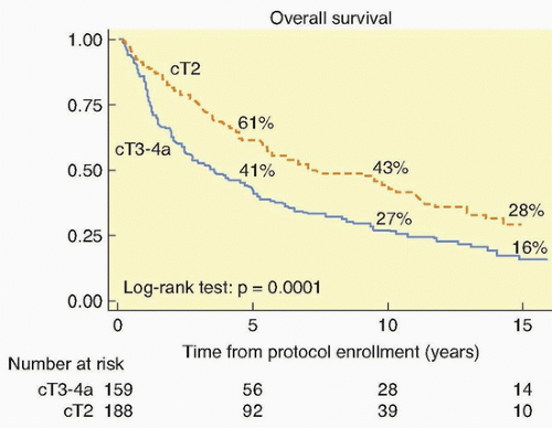
111 The long-term survival with stage T2 tumors (64%) and stage T3 to T4 tumors (22%) was encouraging. This was validated by the National Cancer Institute-Canada randomized trial of radiation (either definitive or precystectomy) with or without concurrent cisplatin for patients with T3 bladder cancer, which showed a significant improvement in long-term survival with pelvic tumor control (67% versus 47%) in the patients who were assigned cisplatin.112 Additional single-institution studies showed that the combination of a visibly complete TURBT followed by radiation therapy or radiation therapy concurrent with chemotherapy safely improved local control.
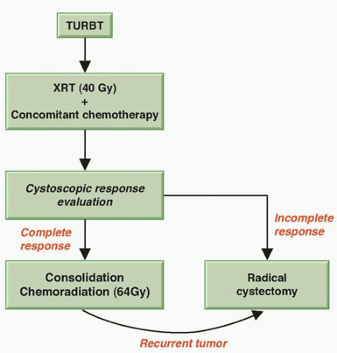
113,114 These findings led the RTOG to develop protocols for bladder preservation beginning with a TURBT of as much of the tumor as is safely possible, followed by the combination of radiation with concurrent radiosensitizing chemotherapy. One key to the success of such a program is the selection of patients for bladder preservation on the basis of the initial response of each individual patient’s tumor to therapy. Thus, bladder conservation is reserved for those patients who have a clinical CR to concurrent chemotherapy and radiation. A prompt cystectomy is recommended for those patients whose tumors respond only incompletely or who subsequently develop an invasive tumor (Fig. 39.3). Up to 30% of the patients entering a potential bladder-preserving protocol with trimodality therapy (initial TURBT followed by concurrent chemoradiation) will ultimately require a salvage radical cystectomy.

For over 2 decades, the Massachusetts General Hospital (MGH), the RTOG, and several centers in Europe have evaluated in phase II and III protocols concurrent chemoradiation plus neoadjuvant or adjuvant chemotherapy (Table 39.3). Radiosensitizing drugs studied in these series, either singly or in various combinations, include cisplatin, carboplatin, paclitaxel, 5-fluorouracil (5-FU), mitomycin C,
and gemcitabine.113 The first RTOG study of patients treated with once-daily radiation treatment and concurrent cisplatin yielded a 5-year survival of 52% (42% with intact bladder).115 RTOG studies 8802 and 8903 used methotrexate, cisplatin, and vinblastine (MCV) chemotherapy as neoadjuvant treatment.116 In the latter study, the neoadjuvant therapy was tested in a randomized fashion.117 No improvement was seen in survival or in local tumor eradication as a result of neoadjuvant therapy, although the trial was closed early and underpowered to give a definitive answer. The toxicity of the MCV arm was considerable, with only 67% of patients able to complete the planned treatment. The use of contemporary neoadjuvant chemotherapy (dose-dense methotrexate, vinblastine, adriamycin, cisplatin [ddMVAC] or gemcitabine and cisplatin [GC]) regimens with appropriate supportive therapy in well-selected bladder-sparing patients may warrant further investigation.


Figure 39.3 Schema for trimodality treatment of muscularis propria-invasive bladder cancer with selective bladder preservation. XRT, radiation therapy.


TABLE 39.3 Results of Multimodality Treatment for Muscle-Invading Bladder Cancer


Series (Ref.)


Multimodality Therapy Used


Number of Patients


5-Year Overall Survival (%)


5-Year Survival with Intact Bladder (%)


RTOG 8512, 1993115


External-beam radiation with cisplatin


42


52


42


RTOG 8802, 1996116


TURBT, MCV, external-beam radiation with cisplatin


91


51


44 (4 y)


RTOG 8903, 1998117


TURBT with or without MCV, external-beam radiation with cisplatin


123


49


38


University of Paris, 1998118


TURBT, 5-FU, external-beam radiation with cisplatin


120


63


N/A


Erlangen, 2002119


TURBT, external-beam radiation, cisplatin, carboplatin, or cisplatin and 5-FU


415 (cisplatin, 82; carboplatin, 61; 5-FU/cisplatin, 87)


51


42


RTOG 9906, 2009122


TURBT, TAX plus CP plus XRT; adjuvant CP plus GEM


80


56


47


MGH, 2012130


TURBT, external-beam radiation and cisplatin with or without 5-FU or TAX; neoadjuvant or adjuvant chemotherapy


348


52


42


MCV, methotrexate, cisplatin, vinblastine; 5-FU, 5-fluorouracil; N/A, not available; TAX, paclitaxel; CP, cisplatin; GEM, gemcitabine.


Other studies from Paris and Germany have reported their large experience with bladder sparing.118,119 The CR rate in the German study was 72%, and local control of the bladder tumor after the CR without a muscle-invasive relapse was maintained in 64% of the patients at 10 years. The 10-year disease-specific survival was 42%, and more than 80% of these survivors preserved their bladder. This series reported the sequential use of radiation with no chemotherapy (126 patients), followed by concurrent cisplatin (145 patients), then concurrent carboplatin (95 patients), and finally concurrent cisplatin with 5-FU (49 patients). The CR rates in these four protocols were 51%, 81%, 64%, and 87%, respectively.120,121 The 5-year actuarial survival with an intact bladder in these studies was 38%, 47%, 41%, and 54%, respectively. These results strongly suggest that radiochemotherapy, when given concurrently, is superior to radiation therapy alone; that carboplatin is less radiosensitizing than cisplatin; and that cisplatin plus 5-FU may be superior to cisplatin alone.

The RTOG protocols have subsequently explored both twicedaily radiation therapy and novel radiosensitization using cisplatin with or without 5-FU or paclitaxel.62,122,123,124 Complete response and bladder preservation rates are consistently high, with no one regimen clearly superior.62

Gemcitabine has been also tested in bladder-treatment protocols. In a phase I trial from the University of Michigan, 23 patients, mostly T2, were treated with gemcitabine and concurrent daily radiation. At a median follow-up of 5.6 years, an impressive 91% CR rate was observed, and the 5-year actuarial estimates of survival include a bladder-intact survival of 62%, an overall survival of 76%, and a disease-specific survival of 82%.125 A phase II study from the United Kingdom of 50 patients treated with concurrent weekly gemcitabine and hypofractionated radiation reported an 88% complete endoscopic response rate, a 3-year overall survival of 75%, and cancer-specific survival of 82%.126 Twice weekly low-dose gemcitabine was recently evaluated as a radiosensitizer with daily radiation in protocol RTOG 0712.

Cisplatin is not always an ideal drug for bladder cancer patients, because it may cause impaired renal function in many. A British group observed high response rates using the combination of 5-FU
and mitomycin C with pelvic radiotherapy.127 These results led to the phase III Bladder Cancer 2001 (BC2001) trial, in which 360 patients with muscle-invasive bladder cancer were randomized to either radiotherapy alone or to radiotherapy with concomitant 5-FU and mitomycin C chemotherapy. Local-regional disease-free survival was superior for those patients receiving chemotherapy (67% versus 54% at 2 years; hazard ratio [HR] 0.68, p = 0.03 with median follow-up of 70 months). Survival at 5 years was higher with chemoradiotherapy (48% versus 35%), but did not reach statistical significance (HR 0.82; p = 0.16).128


Aug 27, 2016 | Posted by in ONCOLOGY | Comments Off on Cancer of the Bladder, Ureter, and Renal Pelvis

Full access? Get Clinical Tree

Get Clinical Tree app for offline access