Blood Management in the Cardiovascular Surgical Patient


Blood Management in the Cardiovascular Surgical Patient


Lawrence Tim Goodnough

L. Henry Edmunds Jr.


Bleeding is a potential complication of any surgery and a major concern of all surgeons. All surgeons are trained to control visibly bleeding vessels, but additional skills and information are necessary for successful management of complex operations, trauma victims, and patients who require mechanical circulatory assistance. Surgical hemostasis now requires knowledge of perioperative coagulation disorders, blood conservation techniques, and the risks and benefits of blood component replacement and transfusion therapy. This chapter outlines a practical approach to the principles of blood management related to all surgery, but particularly cardiac surgery.

Blood management is defined as the appropriate use of blood and blood products, with the goal of minimizing their use.1 The conservation of blood is historically based on principles related to blood risks, blood inventory, and blood costs. Cardiothoracic surgery programs led the development of intraoperative and postoperative salvage techniques because of concerns regarding blood availalbity.2 Subsequently, recognition of blood-transmitted hepatitis and HIV infections fueled further efforts to limit blood transfusions3, 4 (Table 129.1). Accurate accounting of blood costs has identified that alternatives to allogeneic blood (e.g., autologous blood salvage and reinfusion) may be cost equivalent.5 Blood risks and known and unknown emerging pathogens will continue to drive blood conservation and blood transfusion practices.6, 7

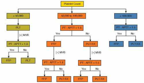
Considerable variation in transfusion practices has been identified in several studies over the last 20 years,8, 9, 10 despite guidelines emphasizing more prudent transfusion practices, with suggested transfusion triggers of 6 to 8 g/dL in the perioperative setting.11, 12, 13 A randomized trial in patients undergoing cardiac surgery demonstrated that a restrictive transfusion strategy (Hgb ≤ 8 g/dL) resulted in equivalent patient outcomes, compared to patients treated with a liberal (Hgb ≤ 10 g/dL) transfusion strategy.14 This trial supported a previous randomized trial in critical care patients that demonstrated similar clinical outcomes in patients transfused for Hgb ≤ 7 g/dL compared to Hgb levels <10 g/dL.15 Point-of-care testing (FIGURE 129.1) linked with a transfusion algorithm for plasma and platelet transfusions has improved patient care and reduced transfusions of blood and blood components16 along with substantial cost savings.17


PREOPERATIVE ASSESSMENT

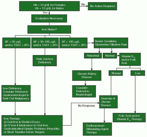
Preoperative planning is essential to reducing or avoiding perioperative allogeneic transfusion.1, 18 Preadmission testing should take place as far in advance as possible (e.g., 30 days) of elective surgery to allow time for adequate identification, evaluation, and management of anemia,19 as illustrated in FIGURE 129.2. The estimated equivalent to number of units “saved” by various blood conservation strategies available in blood management of the surgical patient in the preoperative, intraoperative, and postoperative intervals is listed in Table 129.2.20

The preoperative evaluation of a patient who requires surgery varies with the type and urgency of the operation. For all patients, a thorough history is the best method to discover clinically significant bleeding disorders. A good history reveals bleeding related to previous surgical and dental procedures; epistaxis; menorrhagia; excessive bleeding with major trauma; and easy bruising or joint or muscle swelling after minor trauma. Positive answers raise the possibility of hereditary or acquired coagulation deficiencies, antibodies to specific coagulation proteins, von Willebrand disease (vWD), or platelet disorders. The patient’s medication profile may reveal ingestion of inhibitory drugs, such as aspirin or aspirin-containing compounds, nonsteroidal anti-inflammatory drugs (NSAIDs), warfarin, chronic steroid use, platelet inhibitors, or injections of standard or low molecular weight heparin. The history may also reveal acquired hemorrhagic conditions associated with parenchymal liver disease, renal failure, or myeloproliferative syndromes.

The physical examination includes assessment of a possible bleeding tendency. One may find purpura, which is particularly important if not associated with a known history of trauma. Other findings include petechiae, splenomegaly, hepatomegaly, lymphadenopathy, joint deformities, lack of mobility, palpable collections of blood arising as deep hematomas, and evidence of gastrointestinal (GI) bleeding on digital rectal examination. Discovery of blood in stool is the most effective way to detect occult sources of GI bleeding from esophageal varices, duodenal ulcer, gastritis, diverticulitis, polyps, cancer, ingestion of NSAIDs, and other disorders. Comorbid diseases, such as uremia, cirrhosis, aortic stenosis, polycythemia vera, or continuous flow ventricular assist devices, may enhance bleeding from local lesions in the GI tract.

The results of the history and physical examination and the proposed operation determine the selection of preoperative laboratory tests for coagulation disorders and the need for special preoperative or intraoperative therapy.

Blood must be typed, and a sample sent to the blood bank for antibody screening (type and screen/crossmatch) for all patients prior to scheduled surgery. A complete blood count is obtained in all patients, and traditionally both a prothrombin time (PT) and activated partial thromboplastin time (aPTT) are measured. A prolonged PT detects deficiencies of the extrinsic coagulation pathway, which may be due to vitamin K deficiency or liver disease; a prolonged aPTT detects deficiencies of proteins in the intrinsic coagulation pathway. Results of these measurements
are generally laboratory specific, and the predictive value for bleeding is low, but values exceeding the normal range should be investigated. For example, familial factor XI deficiency may not be detected by a bleeding history, but does prolong the aPTT. A history of bleeding episodes or physical signs of bleeding mandate a full bleeding workup and consultation with a hematologist to assess platelet numbers and function and the presence of inherited or acquired coagulation factor deficiences.21


Table 129.1 Risks of major infectious and noninfectious complications of transfusion in the United States


Complications


Risk


Transfusion-transmitted Disease Agents


Human immunodeficiency virus


1:1,800,000


Hepatitis C virus


1:1,600,000


Hepatitis B virus


1:220,000


Bacteria (platelets, apheresis)


1:75,000


Noninfectious Complications


Acute hemolytic reaction


1:25,000


Mistransfusiona


1:19,000


Transfusion-related acute lung injury


1:5,000


Transfusion-associated circulatory overloada


1:400


Fever/allergy


1:100


a Can occur in association with transfusion of autologous blood as well as allogeneic blood.


Modified from Uhl L. Patient blood management: a 68 year old woman contemplating autologous blood donation before elective surgery. JAMA 2011;306:1902-1910.


FIGURE 129.1 Transfusion algorithm based on preoperative platelet count. An initial platelet count of <50,000 (left arm: severe platelet deficiency) warrants platelet transfusion. If the initial platelet count is >50,000 (middle and right arms), the PT:aPTT ratio (patient vs. normal reference) would delineate whether platelet therapy or plasma was to be administered. Platelets, Platelet transfusion (6 random donor units); PLT RX, platelet therapy (6 random donor units or 0.3 mg/kg DDAVP at physician’s discretion); FFP, 2 units of thawed plasma; [+] MVB, persistent microvascular bleeding. (Reprinted from Despotis GJ, Santoro SA, Spitznagel E, et al. Prospective evaluation and clinical utility of on-site monitoring of coagulation in patients undergoing cardiac operation. J Thorac Cardiovasc Surg 1994;107:271-279, with permission.)

The role of erythropoiesis-stimulating agents (ESAs) in management of preoperative anemia patients undergoing cardiovascular surgery and its safety in this setting are unresolved issues. In a European trial,22 the authors found no differences in mortality, thrombotic events, or serious adverse events among their 76 patients, whether treated with ESA or placebo. A US study also concluded that no differences in adverse events between ESA- and placebo-treated patients were observed and that ESA therapy was well tolerated.23 The safety of ESA in surgical settings has been under re-review since the completion of the SPINE study, in which patients scheduled for elective spine surgery had higher rates of thrombovascular events with an ESA versus placebo in subjects who did not receive prophylactic ESA therapy during spinal surgery.24 Nevertheless, there was an uneven distribution of adverse events between the two treatment groups. Use of ESA remains a valuable tool for patients with special requirements, such as Jehovah’s Witness patients for whom blood transfusion is not an option.18 Until additional
safety data are forthcoming, however, the off-label use of ESA in patients undergoing cardiac or vascular surgery in the United States cannot be recommended.25


FIGURE 129.2 Algorithm for the detection, evaluation, and management of preoperative anaemia. SF, serum ferritin; TSAT, transferrin saturation. (From Goodnough LT, Maniatis A, Earnshaw P, et al. Detection, evaluation, and management of preoperative anaemia in the elective orthopaedic surgical patient: NATA guidelines. Br J Anaesth 2011:106:13-22.)


CONDITIONS THAT INCREASE PERIOPERATIVE BLEEDING


Genetic and Acquired Clotting Abnormalities

Hereditary coagulation deficiencies are usually identified by history or screening test and are confirmed by specific laboratory evaluation. The most common inherited coagulation protein deficiencies are hemophilia A and B and vWD. Patients with hemophilia A or B may develop IgG antibodies to the deficient coagulation proteins, FVIII or FIX, from chronic replacement therapy. Deficiencies in coagulation proteins are usually treated by replacement therapy, and many of these are now available as recombinant human proteins. Inherited deficiencies in fibrinolytic inhibitors, α2-antiplasmin or plasminogen activator inhibitor-1, may also cause substantial bleeding due to excessive fibrinolysis, and these are effectively treated by lysine analogs, epsilon aminocaproic acid, or tranexamic acid.26 A variety of genetic defects may affect platelets and disturb platelet numbers and function.27 Some may respond to desmopressin or require platelet transfusions, but consultation from an expert hematologist is needed, particularly since patients may develop antibodies against the diminished receptors from prior transfusions. Osler-Weber-Rendu and Ehlers-Danlos syndromes may cause troublesome bleeding problems during surgery even though circulating coagulation factors and platelets are normal. Marfan syndrome is not associated with increased risk of bleeding.27

Acquired bleeding disorders of coagulation proteins, fibrinolysis, and platelets occur more often than hereditary bleeding diseases. Drug-induced antibodies (quinidine, quinine, some antibiotics) against specific coagulation proteins are uncommon. Surgeons, however, may encounter patients with antibodies to factor V due to prior use of topical bovine fibrin glue for local hemostasis.28, 29 Advanced liver disease may diminish nearly all
coagulation and regulatory proteins of the coagulation system to levels that affect hemostasis and/or promote severe fibrinolysis.30 However, drugs prescribed to inhibit platelet function cause the most common acquired bleeding disorders that concern surgeons.


Table 129.2 Approximate contributions of selected modalities to blood management in the surgical patient


Options


Number of Blood Units


Tolerance of anemia (reduce transfusion trigger)


1-2


Increase preoperative red blood cell mass


2


Preoperative autologous donation


1-2


Intraoperative options


Meticulous hemostasis and operative technique


1 or more


Acute normovolemic hemodilution


1-2


Blood salvage


1 or more


Postoperative options


Restricted phlebotomy


1


Blood salvage


1


Modified from Goodenough LT, Spence R, Shander A. Bloodless medicine: clinical care without allogenic blood transfusion. Transfusion 2003;43:668-676.


Surgeons must also be aware of a large menu of genetic procoagulant clotting disorders that increase the likelihood of thrombotic and embolic complications. Factor VLeiden mutation, which impairs activation of the natural anticoagulant protein C, is prevalent (2% to 5%) in Northern European whites and their descendants and the prothrombin 20,210 mutation is prevalent (1% to 3%) in Southern Europeans.31 Both mutations increase the risk of venous thrombosis.31 Lupus anticoagulant and anticardiolipin antibodies may produce thrombotic complications during and after major surgery.32

Platelets. Thrombocytopenia or platelet dysfunction that are not drug related occur in patients with myeloproliferative diseases, uremia, acquired vWD, antiplatelet antibodies (e.g., immune thrombocytopenia), posttransfusion purpura, and cirrhosis.33 Aspirin and some NSAIDs irreversibly inactivate platelet cyclooxygenase for the life of the platelet (˜10 days). Cyclooxygenase blockade partially inhibits platelet function by preventing endoperoxide and thromboxane A2 synthesis.34 Aspirin prolongs the bleeding time35 and blocks platelet aggregation,36 but may increase the bleeding time to a greater extent if other, mild hemostatic deficiencies (e.g., vWD) are present. Aspirin resistance caused by genetic or acquired mechanisms occurs in approximately 10% of patients with acute coronary syndromes.37

Major surgery, including cardiac surgery, in patients with aspirin-inhibited platelets may increase perioperative bleeding.36, 38, 39, 40 Nearly all patients presenting with acute coronary syndromes receive aspirin as part of their initial therapy, but usually the impact on surgical hemostasis is minor.38 Aspirin-related bleeding is treated by platelet transfusions, since aspirin is cleared from blood in <90 minutes.41

Clopidogrel and more recently prasugrel are thienopyridines that are irreversible platelet inhibitors that prevent adenosine diphosphate binding to platelet P2Y12 receptors. Both drugs have rapid platelet inhibition, but the kinetics of prasugrel are faster and more consistent and the drug is preferred for management of acute coronary syndromes.42, 43 Median plasma half-life is 7 to 8 hours, but platelet inhibitory effects vary and last 3 to 10 days after the drug is discontinued.33 Both drugs are routinely used in combination with aspirin to reduce adverse cardiac ischemic events.44 Major bleeding complications from the combination of aspirin and prasugrel are slightly, but significantly higher (2.4%) than with aspirin and clopidogrel (1.8%).42 For this reason, the Society of Thoracic Surgeons’ guidelines recommend stopping thienopyridines at least 3 days and preferably 5 days before elective coronary arterial bypass surgery.39 However, reversal of thienopyridines is highly variable between patients. A point-of-care device, which assesses platelet responsiveness to ATP, showed reduced platelet aggregation in some patients who had stopped thienopyridines within 7 days of operation.45

Platelet GPIIIa/IIb (αIIbβ3) receptor antagonists (abciximab, eptifibatide, and tirofiban) inhibit most platelet functions and prolong bleeding times considerably longer than aspirin.46 Abciximab (Reopro) rapidly binds to the platelet receptors and remains bound for the life of the platelet.47 Eptifibatide and tirofiban are reversible inhibitors and relatively short acting. Inhibitory effects of abciximab last approximately 48 hours, whereas that of eptifibatide lasts <1 hour.47 Certain prostaglandins inhibit platelets by activating platelet adenylate cyclase or inhibiting cyclic nucleotide phosphodiesterases (e.g., prostacyclin and dipyridamole).33 All of these platelet inhibitors increase the bleeding time.33

Warfarin. For minor or superficial operations, it is usually not necessary to reverse warfarin, but for major operations, warfarin should be stopped for 3 to 5 days before surgery, if the likelihood of a clotting event within that interval is low.13 The PT should be checked before operation to be sure that most, if not all, of the warfarin effect has been reversed. In patients that cannot safely tolerate interruption of anticoagulation (e.g., mechanical heart valves), intravenous heparin may be used to maintain anticoagulation until 2 to 3 hours before incision.

Oral vitamin K (2 to 4 mg) administration usually corrects the PT to normal within 24 hours.48 In an emergency, warfarin anticoagulation can be reversed by intravenous vitamin K (10 mg IV, infused slowly over 30 minutes) and plasma infusion (15 to 30 mL/kg, or 4 to 8 units in an adult) over 4 to 6 hours, with careful attention to volume status of the patient.49, 50) Transfusions of prothrombin complex concentrates (PCCs), some of which contain high concentrations of vitamin K-dependent coagulation factors II, VII, IX, and X, have also been utilized. PCC acts rapidly but may have a risk of thrombosis.51, 52 If replacement therapy is not effective, administration of recombinant factor VIIa (rFVIIa) (2 mg IV, or 20 to 40 µg/kg for 100 to 50 kg patients) is another alternative in patients with emergency or life-threatening bleeding39, 53; however, this agent also has defined risks of arterial thrombotic complications, particularly in the elderly or at higher doses.54

Dabigatran is a recently approved, oral inhibitor of thrombin that does not require blood monitoring.55, 56, 57 The drug is taken twice daily and is effective prophylaxis in patients with paroxysmal or chronic atrial fibrillation (AF),58 in treatment of acute venous thromboembolism,57 and in acute coronary
syndromes.59 Plasma half-life is 12 to 17 hours,56 and the drug is cleared by the kidney.

Rivaroxaban is a new, oral factor Xa inhibitor that is rapidly absorbed and has a half-life of 9 to 12 hours and can be given as a fixed dose.59, 60. Most of the drug is eliminated in urine and the remainder in feces. Rivaroxaban has been approved for preventing stroke in patients with AF (ROCKET-AF trial),61 as it reduced the incidence of stroke and intracranial bleeding compared with warfarin treatment (0.49 vs. 0.74%). If emergency surgery is required before either dabigatran or rivaroxaban is eliminated, rFVIIa may be attempted to control bleeding, but its value is not certain as an antidote for anticoagulants.62 Fibrinolytic drugs. If operation occurs within 12 hours of the last dose of streptokinase or tissue-plasminogen activator (t-PA), the risk of surgical bleeding is increased.63 If excessive bleeding occurs in such patients, plasma fibrinogen can be replaced with cryoprecipitate, fresh frozen plasma, or fibrinogen concentrate (available in Europe). Platelet transfusion may also be needed, especially if a long-acting platelet inhibitor such as a thienopyridine or abciximab is circulating. Antifibrinolytic drugs are not generally utilized, as the thrombolytic agent will have been cleared from the circulation in most cases. Fibrinolytic drugs are cautiously recommended for treating thrombotic strokes. Potential benefits appear to outweigh the risk of intracranial hemorrhage or surgical site bleeding.64, 65, 66


ANTICOAGULANTS USED DURING CARDIAC AND VASCULAR SURGERY

Standard, unfractionated heparin (UFH) is universally used to maintain the fluidity of blood during mechanical circulation outside the body.67 Heparin greatly accelerates inhibition of thrombin and to a lesser extent activated coagulation factors IXa and Xa by the binding action of antithrombin, which is an abundant plasma protein. The drug does not inhibit thrombin bound in fibrin clots.68 UFH acts rapidly, can be given intravenously, and is reversed by protamine sulfate (PS), but disadvantages include incomplete suppression of thrombin formation; heparin resistance (low antithrombin concentrations); interaction with platelets, complement, and various plasma proteins that variously affect clearance; and stimulation of IgG antiplatelet factor 4 antibodies in some patients. “Heparin resistance” occurs when plasma antithrombin concentrations are deficient from either malnutrition in cyanotic infants or cachectic patients or from sustained heparin administration just prior to operation.69


Low Molecular Weight Heparin

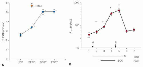
Low molecular weight heparins (LMWHs) catalyze antithrombin, but primarily inhibit factor Xa. Theoretically, these heparins should prevent thrombin formation, but they fail to do so during extracorporeal circulation, as monitored by protein fragment, F1.2, in recirculated human blood70 or in baboons.71 LMWHs can be given intravenously, act rapidly, are long acting (plasma half-life 4 to 8 hours), are monitored by anti-factor Xa activity, are not completely reversed by PS, may generate antiplatelet IgG antibodies (10% as frequently as with UFH), do not reliably prevent clot formation in the circuit, and are associated with excessive postoperative bleeding.72, 73 LMWH anticoagulants are not suitable for cardiopulmonary bypass (CPB).

Bivalirudin (Hirulog) is a synthetic, 20-amino acid, reversible, direct thrombin inhibitor,74 which acts rapidly, has a half-life in plasma of 25 to 36 minutes,75, 76 is cleared by proteolysis and the kidneys,77 but has no antidote.76 Dosage is preferably monitored by ecarin clotting time,78, 79 but it has also been successfully monitored with the activated clotting time (ACT)80, 81 and aPTT. Bivalirudin is essentially nonantigenic and has no major side effects except bleeding. The drug has been evaluated in patients with acute coronary artery syndromes,82 during off-pump coronary arterial surgery,83 in cardiac surgery with CPB,84, 85 and during extracorporeal membrane oxygenation.86 The pharmokinetics of the drug are attractive for use during CPB, but very careful and rigorously monitored protocols must be followed to prevent clotting within the perfusion circuit. Drug dosage remains under investigation: initial dosing schedules combine 1.5 mg/kg iv and 50 mg in the pump prime and a continuous infusion at 2.5 to 3.5 mg/kg/h.84, 87

Lepirudin (recombinant hirudin) directly binds and inactivates thrombin, including thrombin within fibrin clots. The drug has a plasma half-life of approximately 80 minutes and is primarily excreted by the kidney.88 The duration of anticoagulant activity of lepirudin is inversely proportional to creatinine clearance and is prolonged up to 3 to 5 hours by anesthesia.88 Plasma concentrations are monitored by ecarin clotting time or by aPTT.89 There is no antidote, the major side effect is bleeding, and allergic reactions are minimal. Lepirudin is approved by the US Food and Drug Administration for use during CPB in patients with heparin-induced thrombocytopenia (HIT) or heparin-induced thrombocytopenia with thrombosis (HITT) and is the most widely used alternative to standard heparin.88, 90

Argatroban is a synthetic, low molecular weight, reversible, direct thrombin inhibitor that avidly binds thrombin even in fibrin clots.91 Given intravenously, the drug acts rapidly, has a half-life in plasma of approximately 45 minutes,92 and is metabolized by the liver. Anticoagulant activity is monitored by aPTT, but when high doses are used, ACTs are recommended.91, 92 There is no antidote, bleeding is the major side effect, and no toxic or allergic reactions are known. Argatroban has been used for CPB in patients for whom heparin was contraindicated,93 but difficulties with excessive bleeding or clotting within the circuit are reported.94, 95


EXCEPTIONAL SURGICAL BLEEDING PROBLEMS


Heparin-Induced Thrombocytopenia

An unexpected low platelet count in patients who received heparin 5 to 15 days earlier raises the possibility of HIT and the possibility of complicating thrombosis (HITT).96 HIT is mediated by IgG antibodies to a complex of heparin and platelet factor 4 (PF4).87, 97 The disease affects approximately 2% of all cardiac surgical patients.98, 99, 100 HIT is strongly suggested when the platelet count precipitously decreases by 30% to 50% or by 100,000/µL after exposure to heparin.87 A serotonin release test or enzyme immunoassay to detect the presence of anti-heparin/PF4 IgG antibodies101, 102 is mandatory before more heparin is given. Thrombocytopenia and thrombosis due to massive platelet consumption may produce platelet counts below 30,000/µL, but platelet transfusions are not recommended.103


FIGURE 129.3 A: F1.2 concentrations taken from the perfusion circuit during cardiac surgery. Thirty to forty-five minutes after beginning CPB, simultaneous samples were drawn from the pericardial well surrounding the heart (PERC) and the circuit (PERF). HEP, heparin injection; post, immediately after CPB stopped; PROT, at the time of protamine administration. (Reproduced from Chung JH, Gikakis N, Drake TA, et al. Pericardial blood activates the extrinsic coagulation pathway during clinical cardiopulmonary bypass. Circulation 1996;93:2014, with permission.) B: Serial measurements of prothrombin fragment, F1.2, during CPB. S, duration of operation; ECC, duration of CPB; H, heparin injection; P, protamine administration. (Reproduced from Boisclair MD, Lane DA, Philippou H, et al. Thrombin production, inactivation and expression during open heart surgery measured by assays for activation fragments including a new ELISA for prothrombin fragment F1+2. Thromb Haemost 1993;70:253, with permission.)

Disseminated intravascular coagulation (DIC) is a consumptive coagulopathy that may complicate severe trauma, particularly if brain is injured, or complicate postoperative septicemia. The underlying disease (e.g., trauma or infection) initiates thrombin generation primarily by tissue factor exposed in injured tissues105, 106 and on monocytes and endothelial cells.107, 108, 109, 110, 111, 112 DIC may coexist with a fibrinolytic state.105, 113

Only gold members can continue reading. Log In or Register to continue

Stay updated, free articles. Join our Telegram channel

Jun 21, 2016 | Posted by in HEMATOLOGY | Comments Off on Blood Management in the Cardiovascular Surgical Patient

Full access? Get Clinical Tree

Get Clinical Tree app for offline access