and Christian Waldherr1
(1)
Bern, Switzerland
5.1 Apocrine Metaplasia
General Information/Clinical Issues
The patient was a 56-year-old female. Previous bilateral breast reduction. She came for a routine screening examination. Synthetic 2D mammograghy and 3D tomosynhesis were obtained on (17.6.2013) followed by tomosynthesis guided vacuum-assisted biopsy (T-VAB) on (27.06.2013).
Synthetic 2D and 3D Mammographic Analysis (17.06.2013)
The breasts are heterogeneously dense, which may obscure small masses; American College of Radiology (ACR) III. Synthetic 2D (Figs. 5.1a and 5.2a) in cc/mlo, and 3D (Figs. 5.1b and 5.2b) in cc/mlo of the left breast show, in the middle third of the upper outer quadrant, a microlobulated low-density mass with clustered punctate calcifications. Posterior to the lesion, another clustered punctate microcalcifications without an associated mass.
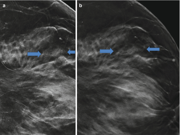
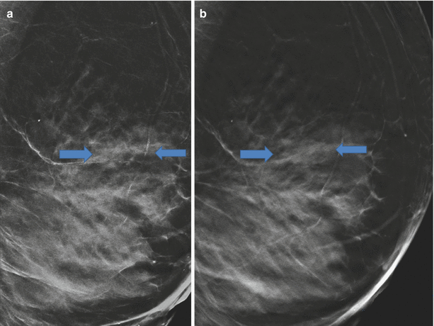

Fig. 5.1
(a) 2D and (b) 3D in Lcc. Synthetic 2D (Figs. 5.1a and 5.2a) in cc/ mlo and 3D (Figs. 5.1b and 5.2b) in cc/mlo of the left breast show, in the middle third of the upper outer quadrant, a microlobulated low-density mass with clustered punctate calcifications. Posterior of the lesion shows clustered punctate microcalcifications without a mass. Tomosynthesis did not expose any signs of an obscured malignancy and showed the same benign morphological findings as 2D. Due to DBT’s capability of an improved conspicuity and characterization of lesions and distortions, the existance of an masked mass is low. The reader confidence in a benign diagnosis is increased. However, Tomosynthesis guided vacuum assisted biopsy of the grouped calcifications is need to exclude DCIS. (BI-RADS 4)

Fig. 5.2
(a) 2D and (b) 3D in Lmlo. Synthetic 2D (Figs. 5.1a and 5.2a) in cc/ mlo and 3D (Figs. 5.1b and 5.2b) in cc/mlo of the left breast show, in the middle third of the upper outer quadrant, a microlobulated low-density mass with clustered punctate calcifications. Posterior of the lesion shows clustered punctate microcalcifications without a mass
Differential Diagnosis (DD) on Synthetic 2D
DD, fibrocystic change, usual ductal hyperplasia, ductal carcinoma in situ, or apocrine metaplasia within benign lesions such as fibroadenoma, sclerosing adenosis, papillomas, and complex sclerosing lesion (Breast Imaging Reporting and Data System; BIRADS 0/4). Ultrasound is recommended to exclude obscured masses. Tomosynthesis guided vacuum assisted biopsy of the grouped calcifications is need to exclude DCIS. (BIRADS 0/4).
Conclusion/Diagnosis with 3D
Tomosynthesis did not expose any signs of an obscured malignancy and showed the same benign morphological findings as 2D. Due to DBT’s capability of an improved conspicuity and characterization of lesions and distortions, the existance of an masked malignant mass is low. The reader confidence in a benign diagnosis is increased. DD, fibrocystic change, usual ductal hyperplasia, ductal carcinoma in situ, or apocrine metaplasia within benign lesions such as fibroadenoma, sclerosing adenosis, papillomas, and complex sclerosing lesion (BI-RADS 0). Tomosynthesis guided vacuum assisted biopsy of the grouped calcifications is needed to exclude DCIS. (BI-RADS 4).
Ultrasound (US)
Mild fibrocystic changes. Tomosynthesis guided vacuum-assisted biopsy (T-VAB) was recommended to exclude ductal carcinoma in situ (DCIS).
Tomosynthesis Guided Vacuum-Assisted Biopsy
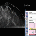
Stereo image suggests correct prefire needle position close to the clustered microcalcifications. Postfire tomosynthesis reveals correct sampling of the calcifications and the adjacent mass (Fig. 5.3a, b).
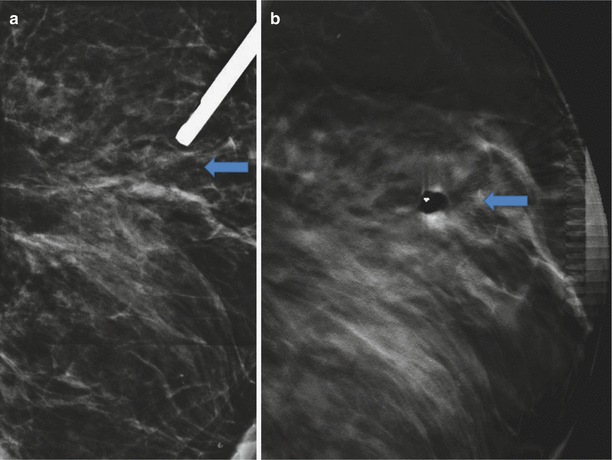

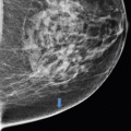
Fig. 5.3
(a) T-VAB 2D Lobl; (b) T-VAB 3D Lml. Postfire tomosynthesis reveals correct sampling of the calcifications and the adjacent mass
Histology
Apocrine metaplasia.
Comment
Apocrine metaplasia is characterized by dilated acini lined by columnar secretory epithelium with granular, eosinophilic cytoplasm. The best diagnostic clue is the finding of microcysts on US. Mammographic features are a microlobulated mass with or without punctate or amorphous calcifications. Also, clustered microcalcifications without a mass are possible. Apocrine metaplasia cannot be distinguished from DCIS or the other histologies named above. In this case, tomosynthesis just highlights the benign character of the mass and facilitates correct biopsy. Underlying invasive breast cancer is unlikely, because 3D did not expose an suspicious mass or distortion.
Image Interpretation Pearls
The best diagnostic clues for apocrine metaplasia are clustered microcysts on US. Unfortunately these lesions are frequently overlooked or are occult in dense fibrocystic breast tissue. In cases where clustered microcalcifications cannot be correlated with benign microcystic changes, DCIS has to be ruled out. Tomosynthesis facilitates correct localization and sampling of masses and calcifications.
5.2 Botox-Injections, Foreign Body Granulomatous Mastitis, Simple Cysts
General Information/Clinical Issues
The patient was a 50-year-old female. History of Botox injections in both breasts, done in a foreign country. Routine screening mammography (synthetic 2D) and tomosynthesis (24.06.2014), no previous mammography.
Synthetic 2D and 3D Mammographic Analysis (24.06.2014)
The breast tissue is heterogeneously dense, ACR III. Synthetic 2D (Figs. 5.4a, 5.5, 5.6, and 5.7a) and 3D (Figs. 5.4b, 5.5, 5.6, and 5.7b) show very small bilateral diffuse fluid-dense nodules with circumscribed margins, particularly in the left breast, and diffuse skin thickening. Overall the breast parenchyma appears to be more dense than normal due to trabecular thickening. A morphologically simple cyst is located in the right breast at the 11–12 o’clock position anterior; it is 1 cm in diameter, round, circumscribed, and with fluid-like density. On synthetic 2D Rcc, the cyst has obscured margins (Fig. 5.4a), in 2Dmlo the halo is as nicely shown as in 3D (Fig. 5.5a), as in 3D (Figs. 5.4b and 5.5b). No distortions and no suspicious calcifications are seen.
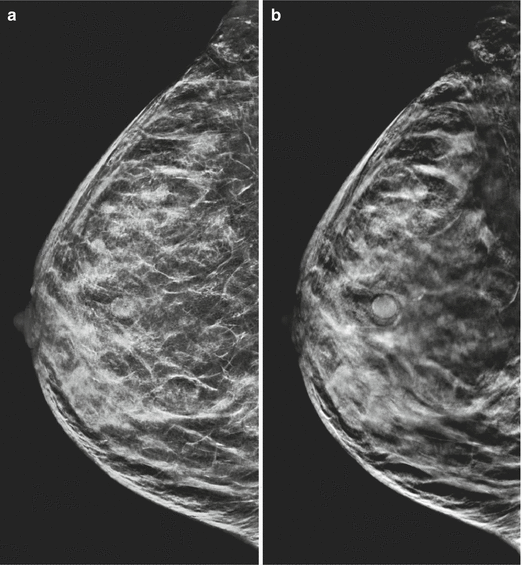
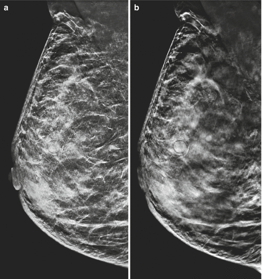
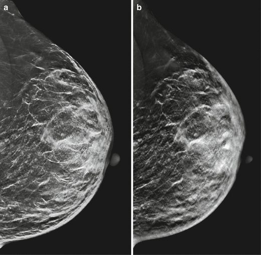
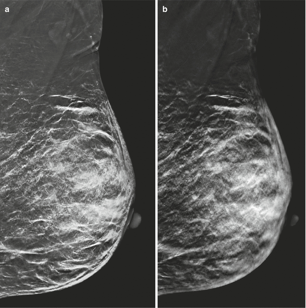

Fig. 5.4
(a) Synthetic 2D Rcc. (b) 3D Rcc

Fig. 5.5
(a) Synthetic 2D Rmlo. (b) 3D Rmlo

Fig. 5.6
(a) Synthetic 2D Lcc. (b) 3D Lcc

Fig. 5.7
(a) Synthetic 2D Lmlo. (b) 3D Lmlo
Differential Diagnosis on Synthetic 2D
Diffuse bilateral skin thickening, a diffuse micronodular alteration of the breast parenchyma, and a diffuse bilateral trabecular thickening without suspicious masses, distortions, or microcalcifications are presented. Knowing the history of bilateral Botox injections (probably in combination with hyaluronic acid), a chronic granulomatous mastitis is most likely on 2D. Further imaging or follow up is needed to exclude underlying malignancy. (BIRADS 3/0).
Conclusion/Diagnosis with 3D
Tomosynthesis with its ability to highlight the symmetric benign character of masses and scar tissue as well as to exclude suspicious radial distortions, makes the existence of malignancy unlikely (BI-RADS 2/0). Knowing the history of the patient, no further examination is need to classify the study BIRADS 2 (BIRADS 2).
Recommendation
US correlation to exclude suspicious masses and abnormal vascularity. In cases of suspicious clinical signs or symptoms, magnetic resonance imaging (MRI) is recommended.
Comment
Any type of cosmetic injection may result in a foreign body granulomatous reaction. The best diagnostic clue was the bilateral appearance of skin thickening, granulomas, and trabecular thickening. Tomosynthesis improves sensitivity and specificity, allowing us to rule out malignany with a very high likelyhood, due to DBT’s ability of unmasking obscured margins and distortions. However, mammography and US are usually not useful for cancer detection in patients with cosmetic augmentations; thus, MRI has to be recommended in such patients with clinical signs and symptoms.
Image Interpretation Pearls
Rule out asymmetric underlying suspicious masses and distortions by DBT. History is of greatest value to diagnose foreign body granulomatous mastitis.
5.3 Chondroid Lipoma
General Information/Clinical Issues
The patient was a 61-year-old female. A routine breast US detected an oval, circumscribed mass in the right breast with a mixture of sonolucent fat and echogenic glandular elements, as well as posterior shadowing. Synthetic 2D full-field digital mammography (FFDM), 3D-tomosynthesis (5.1.2015), and US-guided biopsy were carried out (5.1.2015).
2D-FFDM and 3D Mammographic Analysis (1.3.2012)
There are scattered areas of fibroglandular density, ACR II. In the upper inner quadrant of the right breast, in the anterior third, there is an oval, circumscribed, dense mass with dystrophic macrocalcifications inside, 1.7 cm in diameter.
Despite the obscured margins in 2D, the mammographic morphology is pathognomonic for benign masses as fibroadenoma (same appearance), hamartoma/fibroadenolipoma, galactocele (radiolucent, fat fluid level), fat necrosis (history of trauma), chondrolipoma (BI-RADS 2).
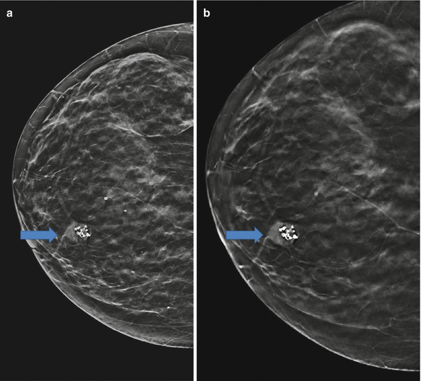
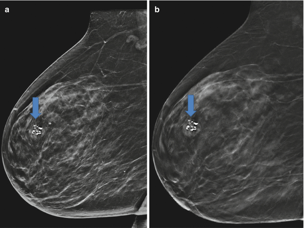

Fig. 5.8
(a) 2D and (b) 3D in Rcc. In the upper inner quadrant of the right breast, in the anterior third, there is an oval, circumscribed, dense mass with dystrophic macrocalcifications inside, 1.7 cm in diameter

Fig. 5.9
(a) 2D and (b) 3D in Rmlo. 3D highlights a halo of compressed fat, the sign of circumscribed margins
In contrast to 2D imaging, 3D highlighted/ exposed. 3D highlighted a halo of compressed fat, the sign of benign circumscribed margins. Due to the pathognomonic morphology no further examination needes to be conducted. (BIRADS 2).
US and US Guided Core Biopsy (Fig. 5.10a–d)
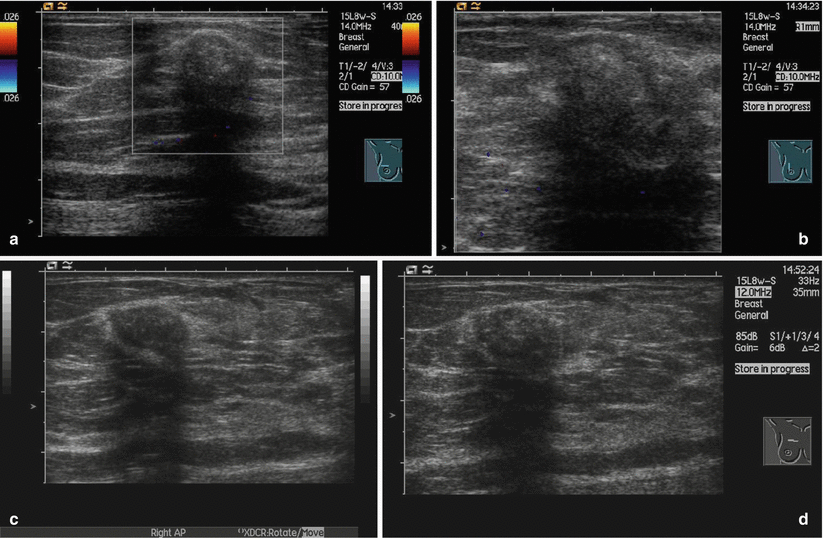
Fig. 5.10
(a–d) Oval, circumscribed, slightly hypoechoic mass without vascularity. Hyperechogenic calcifications with shadowing
Oval, circumscribed, easy hypoechoic mass without vascularity. Hyperechogenic calcifications with shadowing.
Histology
Chondroid lipoma.
Comment
Chondroid lipomas are rare benign soft-tissue tumours that contain a varied ratio of both fat and cartilage. These lesions can be diagnostically confusing as they may mimic or be confused with other fat-containing neoplasms.
Image Interpretation Pearls
Chondroid lipomas have the same image features as fibroadenomas. 3D highlights the benign circumscribed shape of the mass and excludes adjacent distortions.
5.4 Epidermal Inclusion Cyst
General Information/Clinical Issues
The patient was a 63-year-old female. Palpable mass in the upper inner quadrant of the right breast. Diagnostic mammography (synthetic 2D) and tomosynthesis were conducted (08.08.2014).
Synthetic 2D and 3D Mammographic Analysis (08.08.2014)
The breast is almost entirely fat: <25 % fibroglandular tissue, ACR I. Synthetic 2D (Fig. 5.11a, b) in cc/mlo and 3D (Fig. 5.12a, b) in cc/mlo of the right breast show a subcutaneous, circumscribed, dense mass. The subcutaneous location is pathognomonic of an epidermal inclusion cyst or simple cyst.
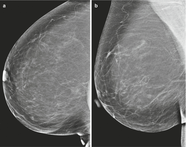
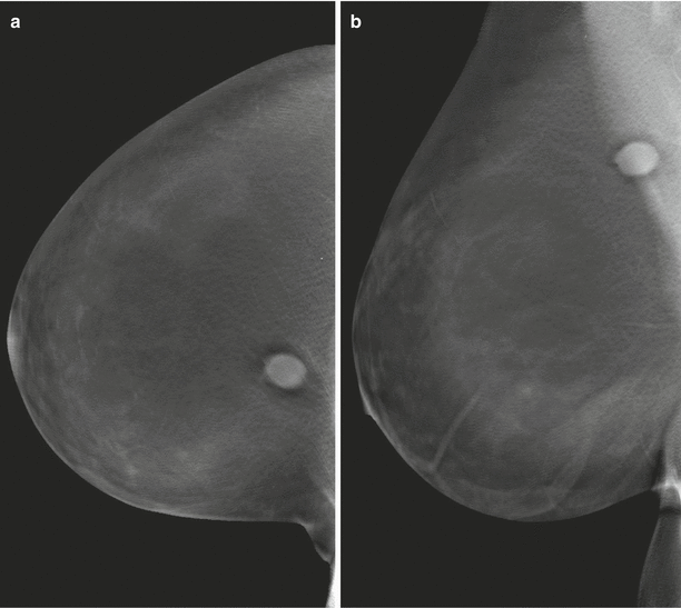

Fig. 5.11
(a) 2D Rcc. (b) 3D Rmlo. Synthetic 2D (a, b) in cc/mlo and 3D (Fig. 5.12a, b) in cc/mlo of the right breast show a subcutaneous, circumscribed, dense mass

Fig. 5.12
(a) 3D Rcc. (b) 3D Rmlo. Tomosynthesis demonstrates the subcutaneous location clearly. In this location it is very likely an epidermal inclusion cyst, a breast sebaceous cyst, or a simple cyst
Differential Diagnosis on Synthetic 2D
The subcutaneous location is not clear with this modality. On 2D the mass could be in the posterior third of the right breast. A dense, circumscribed mass in no man’s land could be a tubular or medullary carcinoma. Further imaging is need to clearify the location of the mass. (BI-RADS 0).
Conclusion/Diagnosis with 3D
Tomosynthesis demonstrates the subcutaneous location clearly. A cutaneous/ subcutaneous mass with benign criteria as demonstared in 3D is most likely an epidermal inclusion cyst, a breast sebaceous cyst, or a simple cyst. Skin metastases of a carcinoma of unknown origin are very unlikely (BI-RADS 2/0).
US
Superficial circumscribed, oval mass with variable echogenicity involving the skin. A hypoechoic line from the mass to the skin confirms the diagnosis.
Histology
Epidermal inclusion cyst.
Comment
Epidermal inclusion cysts or simple epidermoid cysts, sometimes referred to as breast sebaceous cysts, despite not having the same histology, are benign breast lesions. They arise from an obstructed hair follicle. These lesions are typically located in the skin or subcutaneous tissue.
Image Interpretation Pearls
The best diagnostic clue for epidermal inclusion cysts or breast sebaceous cysts is the superficial location involving the skin that can be easily proven by 3D.
5.5 Fibroadenolipoma
General Information/Clinical Issues
The patient was a 36-year-old female. Family risk of breast cancer. Bilateral mastodynia. A diagnostic 2D-FFDM and a 3D-Tomosynthesis were performed (26.10.2010).
2D and 3D Mammographic Analysis (26.10.10)
The breast tissue is extremely dense, >75 % fibroglandular tissue ; breast density ACR IV. 2D Lmlo (Fig. 5.13a) and 3D Lmlo (Fig. 5.13b) of the left breast show, in the anterior upper half, a lobulated, circumscribed mass with a mixture of fibroglandular and fat elements. 3D highlights a pseudocapsule of compressed parenchyma. No calcifications were detected with 2D and 3D.
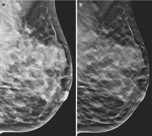

Fig. 5.13
(a) 2D and (b) 3D in Lmlo. 2D Lmlo (a), and 3D Lmlo (b) of the left breast show, in the anterior upper half, a lobulated, circumscribed mass with a mixture of fibroglandular and fat elements
Differential Diagnosis on 2D
On 2D there is a lobulated, incompletely circumscribed mass with a mixture of fibroglandular and fat elements. 2D misses the demonstration of a pseudocapsule. The mixture of fat and fibroglandular septae may originate from a distortion. An exclusion of an underlying malignant mass is neccessary, thus additional imaging is requested. (BIRADS 0).
Conclusion/Diagnosis with 3D
3D clearly shows the pseudocapsule of the fibroadenolipoma and the fat inside the mass. The 3D imaging findings are pathognomonic of a fibroadenolipoma (BI-RADS 2).
US
On US there is a mixture of hypoechoic fat and echogenic fibroglandular tissue. The pseudocapsule shows variable echogenicity.
Histology
Fibroadenolipoma.
Comment
Fibroadenolipoma results from a benign proliferation of fibrous, glandular, and fatty tissue surrounded by a thin capsule of compressed parenchyma. The main differential diagnoses are all benign, such as fibroadenoma, galactocele, fat necrosis, lipoma, hibernoma, chondrolipoma, and hamartoma.
Image Interpretation Pearls
3D imaging may clearly demonstrate the pseudocapsule and exclude a suspicious distortion as a reason for the mixture of tissue elements. 3D imaging localizes the fat inside the mass, a sure sign of benignity
5.6 Fibroadenoma
General Information/Clinical Issues
The patient was a 57-year-old female. Known fibroadenomas. Screening mammography and mammography and tomosynthesis (3D) (27.03.2014), previous mammography (16.08.2012).
2D-FFDM and 3D Mammographic Analysis (27.03.2014)
The breast tissue is heterogeneously dense: 50–75 % fibroglandular tissue, ACR III. Two lobulated, circumscribed dense masses are found in the mid and posterior thirds of the outer upper quadrant of the right breast. The mass in the mid third shows a single dense macrocalcification.
Stay updated, free articles. Join our Telegram channel

Full access? Get Clinical Tree


