Figure 126.1 Laguna Paron is the largest lake in the Cordillera Blanca of the Peruvian Andes. It is 30 km from Caraz, in the Ancash region: one of the most endemic areas for Bartonella bacilliformis. Author: Nuria Sanchez Clemente.
Young children under 10 are the most affected age group in endemic communities, partly because of a predominantly younger population but also due to the presumed protective immunity that develops with repeated infection.
Clinical features
There are two well-described phases of the illness.
The initial acute phase, known as Oroya fever, occurs typically around 2 to 6 weeks after inoculation of the microorganism by the bite of an infected sandfly. It is characterized by fever, pallor, malaise, joint pain, headache, and anorexia. In severe cases, with high parasitemia, this progresses to severe hemolytic anemia.
High mortality rates of 44% to 88% have been reported in untreated individuals in endemic areas. Most deaths are associated with secondary bacterial infection, most commonly with Salmonella species but also with Toxoplasma, Histoplasma, mycobacteria and fungi. Other complications include pericardial effusion, acute respiratory distress syndrome, hepatitis, convulsions, and coma. In pregnancy, infection can lead to miscarriage, premature labor, and maternal death.
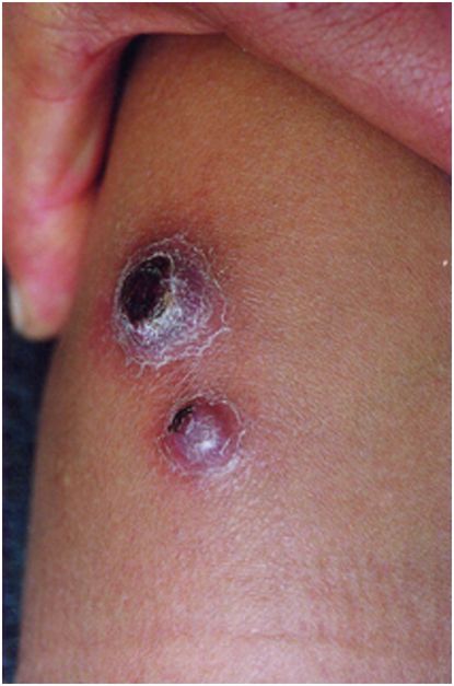
The subsequent chronic eruptive phase, which may occur weeks to months after the acute illness, is characterized by the eruption of crops of miliary, mular, or nodular verrugas or warts, containing sero-sanguinous fluid. These occur mainly over the extremities but can also affect the face and trunk. Miliary lesions (Figure 126.2, lower lesion) are the most common; occurring in the upper dermal layers they can crop in large numbers and can be pruritic. There may also be mucosal involvement. Mular lesions (Figure 126.2, upper lesion) are >5 mm in diameter and have an eroded center. Nodules are larger diffuse subdermal swellings. The latter two can be painful especially if occurring over joints. Secondary bacterial infection is a recognized complication.

Figure 126.2. Mular (above) and miliary (below) lesions in verruga peruana. Author: Ciro Maguiña.
The eponym Carrion’s disease recognizes the contribution of Daniel Alcides Carrion, a Peruvian medical student who in 1885 asked a fellow student to inoculate him with blood from a cutaneous lesion from a diseased patient, in order to test his hypothesis that the two clinical entities were actually manifestations of the same disease. His hypothesis was proven to be tragically correct as he developed, and soon after succumbed to, the acute febrile form of the illness hitherto known as Carrion’s disease, becoming a martyr of Peruvian medicine.
Diagnosis
In endemic areas, cases are treated empirically during outbreaks. However, the most frequently used diagnostic test is peripheral blood smear (Figure 126.3). When stained with Giemsa, the blue intra- and extra-erythrocytic coccobacilli of B. bacilliformis
Stay updated, free articles. Join our Telegram channel

Full access? Get Clinical Tree


