B-Lymphocyte Receptors, Signaling Mechanisms, and Activation


B-Lymphocyte Receptors, Signaling Mechanisms, and Activation


Akanksha Chaturvedi

Angel Davey

Wanli Liu

Hae Won Sohn

Susan K. Pierce


INTRODUCTION

The primary function of mature B lymphocytes in response to foreign antigens is to proliferate and differentiate into antibody-producing plasma cells that secrete large quantities of antibodies. Effective antibody responses are highly specific and of high affinity for the inducing antigen and of the appropriate isotype to allow the antibodies to carry out the effector functions, as described in Chapter 5, that best serve to eliminate an antigen-containing pathogen. For B cells to produce the most effective antibodies, they must have mechanisms that allow them to discern their specificity and affinity for antigens and to produce plasma cells that secrete antibodies of the appropriate isotype. In this chapter, we explore what we know and are learning about these mechanisms. Understanding how B cells recognize and respond to antigens will aid in our efforts to develop effective vaccines and to target therapies to block hyper-B-cell responses as in systemic autoimmune diseases and in some B-cell tumors.

B cells are activated by antigen by a process of clonal selection, a fundamental feature of the adaptive immune response. Each B cell expresses a membrane form of a single heavy (H) chain (VHCH) and a single light (L) chain (VLCL) that are assembled together with a noncovalently bound immunoglobulin (Ig)α-Igβ heterodimer into an antigen receptor, the B-cell receptor (BCR), which is expressed on the B-cell surface (Fig. 9.1). It is conservatively estimated that during a lifetime, an adult human expresses more than 1013 unique clonally distributed BCRs. This estimate is based, in part, on the diversity of H and L chain variable (V) regions that can be generated given the germ line encoded V gene segments and the molecular combinational and mutational mechanisms that create the V genes described in Chapter 6. When an antigen enters the immune system, it selects from among this extraordinarily large array those B cells whose receptors fit it best and signal these to proliferate and differentiate into antibody-secreting plasma cells. Even very simple antigens are predicted to find hundreds of B cells that express BCRs with sufficient specificity and affinity to be activated. This process of antigen selecting best fits from among the enormous array of preexisting B cells to proliferate and differentiate into antibody-secreting cells is a complicated process that takes place in highly specialized microenvironments in the secondary lymphoid organs and involves the functions of both T cells and innate immune system cells, as will be described in Chapter 10. Here, we focus on the events that follow antigen binding to the BCR that initiate signaling, an essential, critical first step in B-cell activation.

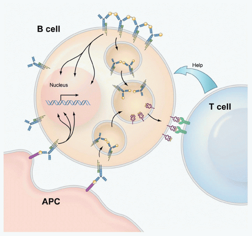
Antigen binding to the BCR triggers several signaling cascades that lead to the transcriptional activation of a variety of genes associated with B-cell activation (see Fig. 9.1). The BCR is also an internalizing receptor; under the influence of the signaling cascades, the BCR and bound antigen are actively endocytosed into the cell and trafficked to specialized intracellular compartments in which the antigen is processed and presented on major histocompatibility complex (MHC) class II molecules, as described in Chapter 22. The MHC-peptide complexes then move to the B-cell surface where they activate antigen-specific helper T cells to provide T-cell help, a complex process involving both direct interactions between B cells and T cells as well as the release of soluble cytokines by the T cells.

A considerable amount of what we know about the biochemical nature of the signaling pathways triggered by antigen binding to the BCR comes from studies in which multivalent antigens are provided to the B cell in solution (see Fig. 9.1). Although B cells can respond to antigens in solution, a variety of recent studies suggest that antigens are presented to B cells on the surface of antigen-presenting cells (APCs) including macrophages and dendritic cells present in the local microenvironments of the lymph nodes and spleen in which B cells are activated in vivo (see Fig. 9.1). Antigens may be presented on macrophages and dendritic cells as part of complement-fixed complexes bound to the complement receptors or immune complexes bound to FcRs expressed by these cells. We do not yet know if the molecular mechanisms by which antigens in solution and antigens on cell surfaces initiate signaling are identical, even though it appears in both cases that the engagement of antigen by the BCR results in the triggering of similar signaling cascades.

In this chapter, we describe what we are learning about the events that occur following the binding of the BCR to antigen that trigger signaling cascades that lead to B-cell activation. The question that will be addressed is how is the BCR’s specificity and affinity for an antigen read by the BCR and transduced across the B cell’s membrane to trigger intracellular signaling cascades that induce the B cell’s response. The signaling cascades that are set off upon antigen binding have been described in considerable biochemical detail and will be reviewed in this chapter. However, biochemical analyses do not capture the spatial and temporal dynamics of the events that are triggered by antigen binding to the BCR. The recent applications of live cell imaging technologies are providing the first views of B cells as they encounter antigen with the resolution to follow individual BCRs. These images are showing us
that the activation of B cells by antigen is far more dynamic than originally thought. It has been possible to infer from the behavior of the B cells and BCRs in these images the mechanisms underlying antigen-driven events that lead to B-cell activation. Such images are changing our view of BCR-mediated B-cell activation and are providing the tools to gain an understanding of the mechanisms underlying diseases that result from inappropriate B-cell activation including systemic autoimmune diseases and certain B-cell tumors.


FIG. 9.1. Overview of the Activation of B Cells by Antigen. B-cell activation is initiated by the binding of antigen to the B-cell receptor (BCR). B cells encounter antigen either in solution or on the surface of an antigen-presenting cell (APC), likely as complement coupled antigens or as immune complexes bound to APC complement receptors or FcRs. Antigen binding triggers signaling cascades that lead to the activation of a variety of genes associated with B-cell activation. The BCR is also a trafficking receptor; under the influence of BCR signals, it transports antigens into intracellular compartments where the antigens are processed and presented on major histocompatibility complex class II molecules for recognition by helper T cells.

As we consider the mechanisms by which antigens trigger B cells through BCRs, we need to be aware that B cells encounter antigen in both developmental and environmental contexts, and that these contexts greatly influence the outcome of antigen engagement. Consider, for example, that B cells express BCRs throughout their development from immature B cells to memory B cells. However, engagement of the BCR by antigen at different developmental stages has different outcomes. The binding of self-antigens to the BCRs expressed on immature B cells signals these cells, but the signals do not result in activation. This means that the developmental state of the B cell dictates the outcome of the BCR’s engagement of antigen, either driving proliferation and differentiation in mature B cells or triggering mechanisms that lead to elimination or silencing of self-reactive immature B cells, as described in Chapter 8. The basic structure of the BCR does not change from the immature to the mature B-cell stage, but the BCR signals differently in different developmental cellular contexts. The outcome of BCR signaling is also influenced by the environmental context in which the antigen is encountered. Antigens enter the immune system in various contexts as relatively simple vaccines to complex microorganisms including viruses, bacteria, and parasites. Both vaccines and pathogens bring with them materials that can activate B cells through coreceptors other than the BCR as well as activate T cells and cells of the innate immune system to secrete products that bind to B cell coreceptors that influence how the BCR signals in response to the antigen. The molecular form of the antigen itself can influence the outcome of BCR antigen binding, as described in Chapters 10 and 23. Bacteria and some viruses display rigid arrays of antigens on their surface that induce antibody responses in the absence of helper T cells (coined T-independent antigens), as do polysaccharides on bacteria in which the carbohydrate moieties are arrayed as multimers. Thus, in predicting the outcome of BCR-antigen binding, we need to consider not only the antigen’s interaction with the BCR but also the developmental state of the B cells, the environment in which the B cell is activated, and the nature of the antigen itself. In other words, BCR signaling always occurs in a context; to predict the outcome of antigen binding to the BCR, we need to be aware of that context.

The BCR also signals during development to provide survival signals to the B cell, often referred to as tonic signaling, and to keep B cells in nonresponsive or tolerant states, termed anergic signaling; however, we do not yet know how these signals relate to those that activate mature B cells.
Concerning the impact of the environmental context in which B cells are activated, this chapter will focus on the effect of B-cell coreceptors that directly affect BCR signaling. We will provide a comprehensive list of the coreceptors and what they respond to and describe how two of the best studied coreceptors, the enhancing cluster of differentiation (CD)19/CD21 complex and the inhibitory FcγRIIB receptor, function to influence B-cell responses. We will also comment on the interactions between the innate immune system’s toll-like receptors (TLRs) and the BCR that appear to serve to regulate each other’s signaling.
We hope that this chapter leaves the reader with a clear view of the early events that follow the engagement of antigen by the BCR and the signaling cascades that are triggered that ultimately activate B cells to proliferate and differentiate into antibody-secreting cells. We also hope that the reader gains an appreciation that B-cell activation occurs in both a developmental and environmental context that dictates the outcome of antigen binding to the BCR. Lastly, we consider the repercussions of uncontrolled BCR signaling that may result in B-cell tumors and in systemic autoimmune diseases.


THE STRUCTURE OF THE B-CELL RECEPTOR

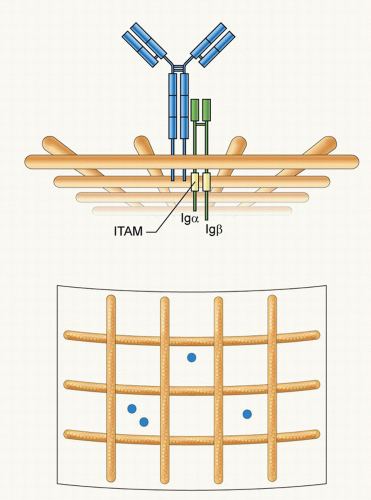
The BCR belongs to the multichain immune recognition receptor (MIRR) family that includes the T-cell receptor for antigen and the high-affinity receptor for IgE. MIRR family members contain ligand-binding chains, which for the BCR is a membrane form of Ig (mIg) (Fig. 9.2). B cells express BCRs composed of Igs of all isotypes that are expressed in a developmentally controlled fashion beginning with IgM-BCRs in immature B cells, IgM- and IgD-BCRs in mature B cells, and then isotype switched BCRs, containing IgGs, IgAs, and IgEs in memory B cells. The mIgs have short cytoplasmic tails of 3 to 28 amino acids that, with the exception of IgG- and IgE-BCRs, do not connect directly with the cell’s signaling apparatus.1,2,3 Rather, the MIRRs ligand binding chains noncovalently associate with membrane proteins that contain immunoreceptor tyrosine-based activation motifs (ITAMs) in their cytoplasmic tails. For the BCR, the mIg associates with a disulfide linked heterodimer, Igα and Igβ, each chain of which has a single ITAM in its cytoplasmic tail (see Fig. 9.2).4 The stoichiometry of the BCR complex is 1mIg:1Igα-Igβ determined by biochemical analysis as well as by live cell imaging (see Fig. 9.2).5,6

In considering how the BCR’s engagement of antigen triggers signaling, it is helpful to consider what we know about the structure of the BCR. From x-ray crystallographic studies, we know that the antibody Fab does not undergo large conformational changes between the free and antigen-bound states that could transduce the information that the BCR has bound antigen to the BCR’s cytoplasmic domains to initiate signaling.7 However, the BCR is a complicated multichain complex and to date, there is no structure of a complete BCR containing mIg, Igα, and Igβ. Thus, a full understanding of the molecular basis of the antigen-induced initiation of BCR signaling will await the determination of the structure of the BCR. Recently, as an effort toward this goal, the structure of the disulphide-linked homodimer of Igβ was solved, which allowed the modeling of an Igα-Igβ heterodimer with the existing structure of the Cα4 domain.8 These results predicted an unexpectedly extensive contact surface between the ectodomains of mIg and both Igα and Igβ through multiple charged residues that could potentially be sensitive to antigen-induced changes in the BCR’s mIg.


FIG. 9.2. The Structure and Organization of the B-Cell Receptor (BCR). The immunoglobulin (Ig)M BCR is composed of a membrane form of IgM associated with a covalently linked heterodimer of Igα and Igβ that contain in their cytoplasmic domains immunoreceptor tyrosine-based activation motifs. The BCR is depicted compartmentalized on the plasma membrane by actin cytoskeleton “fence”9 as proposed by Batista et al.21 Two different views are provided: a side view showing the BCR short cytoplasmic tail and the Igα-Igβ tails in the cytosol and a view from above showing the BCRs compartmentalized by actin “fences.”

In considering how the BCR’s engagement of antigen triggers signaling, it is also useful to consider the organization of the plasma membrane in which the BCR resides (see Fig. 9.2). Our current understanding of the plasma membrane is that it is not simply a fluid mosaic structure of freely diffusing proteins in a phospholipid bilayer, but rather, the plasma membrane appears to be partitioned into highly dynamic compartments formed by actin cytoskeleton “fences” and actin-anchored protein “pickets.”9,10 These compartments serve to organize the plasma membrane to control the diffusion of membrane proteins and concentrate proteins in one compartment or conversely segregate proteins into separate compartments. The fences and pickets are also
dynamic structures that can disassemble and reassemble during B-cell activation.


THE ANTIGEN-INDUCED CLUSTERING OF THE B-CELL RECEPTOR

Like other MIRRs, the BCR has no intrinsic receptor kinase activity, but upon antigen binding, the ITAM tyrosines in the Igα and Igβ chains are phosphorylated by the membrane-tethered kinase Lyn.11,12 The phosphorylation of the BCR ITAMs leads to the recruitment of the Src homology 2 (SH2) domain-containing kinase Syk and the initiation of a variety of downstream signaling pathways described later in this chapter. The focus of this section is on the early events following antigen binding that lead to the phosphorylation of the BCR.

Until recently, most of what we learned about the responses of B cells to antigens came from biochemical studies of B cells responding to antigens in solution. Such studies showed that BCR signaling can only be initiated by the binding of multivalent antigens. As an example, only the bivalent F(ab′)2 fragments, but not the monovalent Fab fragments, of anti-IgM antibodies trigger BCR signaling.12 By immunofluorescence imaging, following multivalent antigen binding, BCRs were observed to form microscopic clusters or patches on the cell surface that then move to one pole of the B cell to form a cap. Based on these biochemical studies, a widely accepted concept emerged that the physical aggregation of the BCR by multivalent antigens promoted the patching and capping of BCR that initiated signaling.

However, there is growing evidence both from studies in vitro as well as from intravital imaging in live animals that B cells are activated by membrane-bound antigens and not by antigens in solution. B cells were shown to be efficiently activated by antigens expressed on the surfaces of APCs in vitro resulting in the formation of a polarized bull’s eye-like structure in which the BCRs are concentrated in the center, surrounded by the adhesion molecule lymphocyte function-associated antigen, which engages intercellular adhesion molecule on the APC surface.13 Recent intravital imaging studies showed that B cells interacted with antigens on the surfaces of APCs in lymph nodes in vivo.14,15,16,17,18 Antigens in lymphatic fluid enter lymph nodes through efferent vessels and gain access to B cells through various mechanisms.19 Small soluble antigens move through follicular conduit networks and are presented to B cells within the follicles. Particle-like antigens, including viruses and immune complexes, are captured by macrophages lining the subcapsular sinuses and are then transported into the cortex of the lymph node where they are presented to B cells. In addition, B cells are also able to engage antigens on dendritic cell surfaces in the lymph nodes.20 These findings provided a new view of the initiation of antigen-driven BCR signaling in which BCR signaling is initiated at the contact interface between B cells and APCs. Live cell imaging technologies are providing the tools to observe B cells as they first engage antigen on membrane surfaces. To facilitate these studies and gain high-resolution images, B cells are often activated by antigens incorporated into fluid planar lipid bilayers as surrogate APCs. These studies are revealing the B cells’ engagement of membrane-bound antigens to be a remarkably dynamic event21,22 that contrasts with the view of the patching and capping of BCRs that resulted from the simple physical cross-linking of BCRs by multivalent antigens in solution.

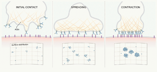
B cells first touch the antigen-containing bilayer through finger-like protrusions of their plasma membrane from which the BCRs engage antigen and form microclusters (Fig. 9.3).
These BCR microclusters are enriched in tyrosine phosphorylated proteins and are the site of Lyn and Syk recruitment and thus appear to be the elemental signaling units. B cells subsequently spread over the bilayer allowing the engagement of additional antigens (see Fig. 9.3). Following maximal spreading, the BCR-antigen microclusters are actively moved toward the center of the contact area, and the B cell contracts to form an immunologic synapse. This dynamic process of touching, spreading, and contraction occurs within minutes of the BCR’s first contact with the bilayer.23 These observations bring us back to the fundamental questions: How are BCR microclusters formed, and how do BCR microclusters trigger BCR signaling? Our understanding of these processes is still incomplete. However, based on current data, several models have been proposed that address these questions.


FIG. 9.3. The Activation of B Cells on Antigen-Presenting Cell (APC) Surfaces. The initial contact of the B cell with an antigen-containing APC surface is through finger-like protrusions of the membrane. Antigen binding induces B-cell receptor (BCR) clustering and signaling that triggers the B cell to spread over the APC surface, forming additional BCR clusters as antigens are encountered. After maximal spreading, the B cell contracts, actively moving BCR clusters to the center of the contact area ultimately forming an immune synapse.

One model, the conformation-induced oligomerization model, is based on evidence from live cell imaging at the level of individual BCRs. Single BCR tracking provided evidence that BCRs are dispersed and freely diffusing in the plasma membrane of resting B cells. Upon binding antigens presented on lipid bilayers, BCRs formed immobile oligomers that grew into microclusters by trapping additional antigen-bound BCRs.24 Remarkably, contrary to soluble antigens, monovalent antigens presented on fluid lipid bilayers, which could not physically cross-link BCRs, activated B cells equivalently to multivalent antigens. This observation led to the conclusion that the physical cross-linking of BCRs by multivalent antigens was not a requirement for BCR oligomerization or clustering. Early oligomerization and microcluster formation that occurred within 60 seconds of antigen binding were shown to be BCR-intrinsic events that did not require the signaling apparatus of the BCR.25 Mutagenesis studies showed that the membrane-proximal Cµ4 portion of the ectodomain of mIgM (or the Cγ3 membrane-proximal domain of mIgG) was both necessary and sufficient for BCR oligomerization and signaling.25 Thus, the membrane proximal domain of the mIg appeared to contain an oligomerization domain that was not exposed in the absence of antigen binding. It was hypothesized that the B cell’s binding antigen presented on an APC surface exerted a force on the BCR that revealed the oligomerization domain, allowing oligomerization of antigen-bound BCRs as they randomly bumped on the membrane. This model provides a mechanism by which the universe of foreign, structurally distinct antigens can bring BCRs into a precise signaling-active oligomer by the monovalent binding of BCRs to epitopes on APC-presented antigens. This model also suggests how BCRs are able to so exquisitely discriminate their affinity for antigen by monovalent binding, avoiding the affinity obscuring effects of the avidity contributed by multivalent antigen binding to bivalent BCRs.

Another model for the initiation of BCR signaling based on live cell single molecule imaging focuses on the role of the actin cytoskeleton in signaling.26,27,28,29 In this model, the membrane cytoskeleton fences and pickets restrict BCR mobility and interactions with signaling molecules and coreceptors. BCR signaling results in the disruption of the cytoskeleton barriers and increases the likelihood that the antigen-engaged BCR will encounter activated kinases and coreceptors.29 The actin cytoskeleton may also segregate BCRs from kinases and phosphatases at steady state. Current evidence suggests that the cytoskeleton fences and pickets confine the BCR and inhibitory phosphatases to the same areas, and that BCR signaling serves to disrupt the cytoskeleton and allow the BCRs and phosphatases to diffuse away, promoting B-cell activation.

A third model, the dissociation activation model, is based on biochemical studies.6,30 In this model, in resting B cells in the absence of antigen most BCRs exist on the B-cell surface as signaling-inactive, autoinhibited oligomers in equilibrium with a small number of signaling-active BCR monomers.30 The binding of multivalent antigen disrupts the oligomers shifting the equilibrium toward BCR active monomers, which are then clustered by the multivalent antigen in a manner that prevents the formation of inactive oligomers within the cluster. This model also accounts for the ability of structurally diverse antigens to activate B cells by proposing that antigens keep BCRs apart rather than bringing them together into well-ordered oligomers.


THE ROLE OF B-CELL RECEPTOR AFFINITY AND ISOTYPE IN SIGNALING

Affinity maturation and class switching are two hallmark features of humoral immune responses.31 In a typical T-cell-dependent antibody response, antigen-specific antibodies become increasingly higher in affinity and predominantly of the IgG isotype through the linked molecular processes of somatic hypermutation and class switching. Because the B-cell immune response functions through clonal selection, it is presumed that affinity maturation and class switching reflect an advantage of B cells expressing high-affinity, classswitched BCRs in the selection process. Indeed, adoptive transfer studies provided clear evidence that high-affinity B-cell outcompete low-affinity B-cell clones for survival in vivo.32,33,34,35,36,37,38 Similarly, when comparing IgM and class-switched BCRs, several seminal studies showed that class-switched B cells outcompete IgM B cells for survival in vivo, and that this survival advantage can be attributed to the 28 amino acid cytoplasmic tail of mIgG,1,2 missing in mIgM and mIgD. The question is at what point in the antigen-selection process does a high-affinity, class-switched BCR gain an advantage?

Biochemical experiments suggested that the signals that are triggered through high-affinity BCRs are qualitatively different from signals through low-affinity BCRs.39 At an earlier point in B-cell activation, the affinity of the BCR for antigen was shown to determine the degree to which B cells spread over antigen-containing fluid lipid bilayers, allowing increased accumulation of antigen into the immune synapse and subsequent enhanced responses.23 The IgG-BCR cytoplasmic tail is responsible for the enhanced B-cell proliferative responses to antigen in vitro and signaling cascades that have been shown to be qualitatively different from those triggered by IgM-BCRs.3,40,41,42 Enhanced signaling through IgG-BCRs is dependent on the phosphorylation of a tyrosine in the IgG tail that serves to recruit growth factor-receptor-bound protein 2 (Grb2), resulting in sustained kinase activation and enhanced B-cell proliferation.3


Recent studies using high-resolution imaging to follow single BCRs showed that the earliest events that occur following antigen binding to the BCR are also highly sensitive to both the affinity of the BCR for antigen and the isotype of the BCR. Comparing BCRs that differed 50-fold in their affinity for antigen, it was observed that high-affinity BCRs more readily formed BCR microclusters that grow more rapidly, thereby resulting in larger microclusters that recruit more Syk and signal for more robust calcium responses.24 These imaging experiments also demonstrate that IgG-BCRs are dramatically enhanced in their ability to oligomerize and to grow microclusters, ultimately leading to increased recruitment of Syk and more robust calcium responses as compared to IgM-BCRs of the same affinity.43 The enhanced function of IgG-BCRs was mapped to a novel membrane proximal 15 amino acid region of the cytoplasmic tail. These studies place the effects of both affinity maturation and class switching at the earliest steps in the initiation of BCR signaling.


HOW B-CELL RECEPTOR CLUSTERING TRIGGERS SIGNALING

Another fundamental yet still open question related to the initiation of BCR signaling is how BCR clustering in response to antigen recruits Lyn to phosphorylate the antigen-bound BCRs and triggers signaling. One simple explanation is that Lyn is constitutively associated with the cytoplasmic domains of some monomeric BCRs, but only phosphorylates BCRs in trans when the BCRs are clustered. However, recent studies suggest that the mechanism for recruitment of Lyn may be more complicated. Lyn is lipidated and tethered to the inner leaflet of the plasma membrane. BCR clustering has been shown to perturb the local lipid environment leading to the transient coalescence of lipid rafts around the BCR oligomers followed by a more stable association of the BCR microcluster with lipid tethered Lyn.44 Thus, the BCR’s perturbation of the membrane may serve to recruit Lyn to the BCR cluster. BCR clustering has also been shown to alter the way the Igα and Igβ chains’ cytoplasmic domains associate. In the absence of antigen, the domains are in close proximity in a “closed” conformation and upon antigen binding, the domains “open”; the opening is simultaneous with Lyn’s phosphorylation of the ITAMs.5 Thus, BCR clustering may serve to reveal the ITAMs for phosphorylation by Lyn.


B-CELL RECEPTOR-TRIGGERED SIGNALING CASCADES

BCR signaling is a multistep process that involves the initiation of signaling by the activation of protein tyrosine kinases (PTKs), serine-threonine kinases, and lipid kinases; amplification of signaling by recruiting adaptors; generation of second messengers; and finally activation of the transcription of genes involved in B-cell responses.


Initiation of B-Cell Receptor Signaling—Protein Tyrosine Kinase Activation

Following BCR clustering, three different families of PTKs, Src, Syk, and Tec, are activated sequentially. This sequential activation of the members of the three different PTK families is essential to trigger and regulate downstream signaling. The importance of these PTKs in the B-cell signaling is underscored by the fact that deficiencies in any one of the three families result in aberrant B cell development and function (Table 9.1). The first kinases that are activated following BCR cross-linking are the Src family PTKs, primarily Lyn, but also Blk and Fyn followed by the activation of Syk and the Tec family kinase, Btk45 (Fig. 9.4). ITAM phosphorylation of Igα-Igβ by Lyn generates phosphotyrosine motifs that allow the binding of the SH2 domains (Box 9.1) of the second kinase, Syk, resulting in rapid Syk activation. Upon binding to phosphorylated ITAMs, Syk undergoes autophosphorylation at multiple tyrosines within its linker regions that not only prolongs Syk’s activation but also creates SH2 binding sites on Syk for the recruitment of downstream signaling molecules, including PLC-γ2,46 leading to a positive feedback of BCR signaling and the concomitant influx of calcium.12 Btk is the third PTK that is activated upon BCR cross-linking.47 The importance of Btk in BCR signaling for the development, activation, and differentiation of B cells is underscored by the fact that the loss-of-function mutations of the gene encoding Btk lack circulating B lymphocytes, are unable to generate Igs, and cannot mount humoral immune responses.48 This primary immunodeficiency is named X-linked agammaglobulinemia.49,50 Similarly, a spontaneous mutation in the mouse Btk gene leads to X-linked immunodeficiency.51 Btk consists of multiple protein domains including PH, SH2, SH3, and kinase domains (see Box 9.1), which define its subcellular location and regulate its activity. For Btk activation, plasma membrane localization is important, which is governed by the interactions between the PH domain of Btk with phosphatidylinositol (PI)(3,4,5)P3, the product of PI3K activity and between the SH2 domain of Btk with phosphorylated BLNK, an adaptor protein. Mutation in the PH domain of Btk (R28C) leads to classical X-linked agammaglobulinemia,52 substantiating the importance of plasma membrane localization for Btk activation. Following BCR cross-linking, Btk translocates from the cytosol to the plasma membrane where it is activated by phosphorylation at Y551 in its catalytic domain by Lyn53,54,55 followed by an autophosphorylation of its SH3 domain.54,56 Once activated, Btk triggers a cascade of signaling events that culminate in calcium mobilization through phosphorylation of PLC-γ2, cytoskeletal rearrangements, Vav activation, and transcriptional activation involving NF-κB.


Amplification of B-Cell Receptor Signaling— Recruitment of Adaptors

BCR-mediated signaling is a complex process in which each phosphorylation event is linked with another in a regulated manner, thereby generating a large number of protein-protein interaction networks ultimately resulting in the formation of a large, well-ordered structure often referred to as a signalosome. Interactions between signaling networks are regulated by a number of scaffolding or adaptor proteins, which regulate BCR-mediated signaling cascades not only by recruiting multiple signaling intermediates to the proper location but also by controlling interactions between signaling components. Recent studies have identified the roles of
adaptors in B-cell activation, leading to important insights into how they integrate BCR signaling. Although adaptors generally lack any enzymatic activity, they consist of multiple protein-protein or protein-lipid interaction domains, including SH2, SH3, PH and PX homology domains (see Box 9.1). Each of these domains has the potential to interact with a number of proteins that are critical to amplify BCR signaling by facilitating the coupling of multiple downstream signaling pathways. Essentially, the adaptors define where and when macromolecular complexes are assembled, allowing both spatial and temporal regulation of signaling cascades. To illustrate the importance of adaptors, we describe the functions of three: B-cell linker protein (BLNK); B-cell adaptor for PI3K (BCAP), and B-cell adaptor molecule of 32 kDa (Bam32).


TABLE 9.1 The Phenotype of Mice Deficient in Key B-Cell Signaling Components


Target Protein


Major Phenotype


Reference


Igα cytoplasmic domain


Normal pre-B-cell development; completely impaired mature B-cell development


184


Igβ


Complete block at the pro-B-cell stage


185


Igµ


Deletion of mature B cells; increased Fas expression


186


Lyn


Normal B-cell development in the bone marrow; reduced number of peripheral B cells; increased proportion of immature B cells; enhanced BCR induced ERK activation and hyperproliferative responses


187


Syk


Block in transition of the pro-B-to the pre-B-cell stage; intact Igα-Igβ ITAM phosphorylation in remaining B cells; abolished BCR-induced calcium influx; failure to transmit downstream signals


188,189


Btk


Reduced number of peripheral B cells; increased immature B cells; complete loss of B1 B cells; fail to respond to TI-II antigens


190,191,192,193


BLNK


Block in transition of the pro-B-to the pre-B-cell stage; incomplete block in B-cell development; fail to respond to both TD and TI antigens


194,195,196


PLC-γ2


Decreased mature B cells; block in pro-B-cell differentiation; B1 B-cell deficiency; block in BCR-induced calcium influx and proliferation


197,198


BCAP


Reduced number of B cells; B1 B-cell deficiency; reduced serum IgM and IgG3 levels; abolished TI antibody response; reduced BCR induced calcium influx and proliferation


82


PI3K, p110 subunit


Reduced numbers of B1 and marginal zone B cells; reduced serum Ig levels; defective primary and secondary response to TD antigens; diminished response to TI-II antigens; reduced BCR, CD40, and LPS induced proliferation


199,200


PI3K p85 subunit


Reduced number of peripheral B cells and B1 cell; reduced serum Ig levels; reduced BCR- and CD40-induced proliferative response; abolished TI antibody response


201,202


PKC-β


Failure to activate IKK and degrade IκB; failure to upregulate NF-κ-dependent survival signals; impaired humoral immune responses and reduced cellular responses


203,204


Bam32


Normal B-cell development; impaired TI-II antibody responses; reduced responses to BCR cross-linking


89


Vav1/2


Reduced number of B cells; defects in formation of germinal centers and class switching; defective immune responses against TD and TI antigens; impaired BCR-induced calcium influx and proliferation


205,206


Rac


Reduced number of mature and marginal zone B cells and B1-a cells and IgM secreting plasma cells; increased number of peripheral B cells in blood; reduced serum IgM and IgA concentration


207


CD19


Defective response to TD antigens; failure to form germinal centers and undergo affinity maturation; lack of B-1, marginal zone B cells; reduced BCR- and CD40-induced proliferative responses


191,208,209


CD22


Decreased surface IgM levels; augmented BCR-induced calcium; compromised marginal zone B-cell compartment


210,211


PIRB


Increased number of peritoneal B1 cells; constitutive activation of follicular B cells; increased BCR-induced proliferation


212


Calcineurin


Reduced number of B1 cells; reduced plasma cell differentiation; decreased TD antibody response; BCR-induced proliferation defects


102


IKKα


Reduced mature B-cell population; impaired basal and Ag-specific Ig production; disrupted splenic architecture including germinal center formation; decreased expression of NF-κB target genes


213,214


IKKβ


Disappearance of mature B cells 215 NEMO Disappearance of mature B cells


215


BCR, B-cell receptor; CD, cluster of differentiation; Ig, immunoglobulin; ITAM, immunoreceptor tyrosine-based activation motif; LPS, lipopolysaccharide; TD, T-dependent; TI, T-independent.


BLNK—Integrating Protein Tyrosine Kinases and PLC-γ2

BCR clustering activates Lyn and Syk that regulate a variety of effectors including PLC-γ2, an essential phospholipase for
calcium responses. Although Syk can directly phosphorylate PLC-γ2 in vitro,57 expression of a functional BCR, Lyn, and Syk in nonlymphoid cells did not induce PLC-γ2 phosphorylation or calcium mobilization,58 leading to the discovery of the B-cell-specific adaptor protein, BLNK. BLNK (also known as SLP-65 or BASH) is a cytoplasmic protein consisting of an N-terminal region containing a short leucine zipper motif, a proline-rich region within the middle third of the molecule, and a C-terminal SH2 domain. BLNK contains 13 tyrosine residues, of which 6 are in putative SH2 binding motifs and are phosphorylated upon BCR clustering. BLNK is recruited to the clustered BCR by binding through its SH2 domain to the phosphorylated non-ITAM tyrosine 204 in Igα. Mice with a Igα Y204 mutation exhibit reduced BLNK phosphorylation and calcium fluxes.59 Once translocated to the plasma membrane, BLNK is phosphorylated by Syk, which creates docking sites for SH2 domains of multiple effector molecules facilitating interactions among them and allowing them to phosphorylate and activate their respective signaling pathways. One of the best studied examples of how BLNK bridges multiple signaling components is provided by the activation of PLC-γ2. PLC-γ2 activation is completely abolished in Syk-deficient B cells.60 In addition, BCR-induced PLC-γ2 activation is diminished in Btk-deficient B cells, suggesting that both Syk and Btk are required for PLC-γ2 activation.61 However, the mechanisms underlying how these two families of PTKs regulate PLC-γ2 activation became clear only after the discovery of BLNK. After phosphorylation by Syk, BLNK creates binding sites for the SH2 domains of both Btk and PLC-γ2, bringing them into close proximity with each other, and hence facilitating PLC-γ2 phosphorylation at Tyr753 and Tyr759 by Btk, which is required for PLC-γ2 activation.62,63,64,65,66,67,68 Cells expressing a mutant BLNK lacking either Btk or PLC-γ2 binding sites
show significant reduction in calcium activation, further suggesting that BLNK acts as a scaffold for bridging Btk to PLC-γ2.69 A single BLNK molecule can bind three PLC-γ2 molecules, thus BLNK also functions as an amplifier of BCR signaling.62 Following BCR clustering, both BLNK and Btk are also translocated to the plasma membrane bringing along PLC-γ2 and allowing it to gain access to its substrate PI(4,5)P2 in the inner leaflet of the plasma membrane.66,67,68 In addition to phosphorylating PLC-γ2 and recruiting it to the plasma membrane, Btk also recruits PIP5 kinase to the plasma membrane, which catalyzes PI(4,5)P2 synthesis from PIP(4)P to ensure that activated PLC-γ2 does not run out of its substrate.70 Once activated, PLC-γ2 hydrolyzes PI(4,5) P2 to generate two important second messengers: inositol trisphosphate (IP3) and diacyl glycerol (DAG). BLNK not only couples Btk and PLC-γ2, but it also binds to the SH2 domains of Vav and Nck, resulting in activating the mitogen-activated protein (MAP) kinase pathway, cytoskeletal rearrangements, and BCR internalization.71

Only gold members can continue reading. Log In or Register to continue

Stay updated, free articles. Join our Telegram channel

Aug 29, 2016 | Posted by in IMMUNOLOGY | Comments Off on B-Lymphocyte Receptors, Signaling Mechanisms, and Activation

Full access? Get Clinical Tree
Get Clinical Tree app for offline access