B-Lymphocyte Development and Biology


B-Lymphocyte Development and Biology


Richard R. Hardy


INTRODUCTION

B lymphocytes constitute one of the major arms of the immune system, being responsible for humoral immunity. B cells in humans and mice are produced throughout life, primarily in the fetal liver before birth and in the bone marrow after birth. Their development from hematopoietic stem cells (HSCs) has been extensively characterized in mice, and the generation of numerous gene-targeted and transgenic lines in many cases has provided crucial information on the role of transcription factors, cellular receptors, and interactions that are critical in their generation. Recently, the role of microribonucleic acids (miRNAs) in regulating hematopoietic development has also emerged. The complexity of this process is now apparent, and B-cell progenitor differentiation into multiple peripheral subsets with distinctive functions is also widely appreciated. This chapter will focus on B-cell development and function in the mouse, touching more briefly on aspects of human B cells that are similar or distinctive, with a focus on immunodeficiency and B-cell neoplasias. It will conclude with a brief description of novel aspects of B-lymphocyte development in other species, highlighting differences from development in mouse and human.


B-CELL DEVELOPMENT IN MICE

In mice, B cells are produced from HSCs through a complex process of differentiation that has been uncovered over the past 30 years or so. One of the goals of classical hematology has been the delineation of differentiation pathways for different lineages of blood cells. There has been considerable progress in utilizing the ordered expression of a diverse set of cell surface and internal proteins, some with known functions, others whose roles are only suspected, to construct a description of the intermediate stages that cells transit as they develop into B lymphocytes. A simplified example of such a description is presented in Figure 8.1. Thus, HSCs with the capacity to generate all the cell types in blood generate progeny with a more restricted capacity, recognizable in this example by expression of the receptor for interleukin (IL)-7. These in turn produce yet more restricted progenitor cells identified by expression of cluster of differentiation (CD)45R/B220 (and, importantly, by absence of CD19). This kind of pathway can be constructed by isolation and short-term culture of intermediate stages, allowing progression to occur, which helps to define the order. This framework for development serves as a starting point for analysis of the effects of transcription factors, microenvironmental interactions, cytokines, and natural or engineered mutations. It can also be extended by analysis of gene or protein expression at distinct intermediate stages. Critical processes, such as D-J rearrangement and immunoglobulin (Ig) heavy chain expression, can also be mapped onto this framework. Progress in this work facilitates experiments that address additional issues, such as identification of key regulatory interactions, developmental checkpoints, and the mechanism of B-lineage commitment.

The following sections will cover the sites of B-lineage development at different stages of ontogeny, then focus on what is known about their development in the bone marrow of adult mice, highlighting the function of the pre-B-cell receptor and the crucial role of Ig heavy and light chains in guiding development. Later sections will consider their differentiation into various specialized peripheral populations and emphasize insights into B-cell selection gained from various transgenic models of tolerance.


Early Development


Sites of B Lymphopoiesis during Ontogeny

In the mouse, hematopoiesis occurs predominantly in the fetal liver prior to birth, in the spleen just prior to and shortly after birth, and in the bone marrow thereafter. Prior to liver hematopoiesis, the blood islands of the yolk sac (YS) contain the first identifiable hematopoietic cells, nucleated erythrocytes with embryonic forms of hemoglobin.1 However, these early YS precursors appear incapable of generating other blood cell lineages, and generation of all blood cell types, including lymphocytes,2,3 initiates at around 9 to 10 dpc in an embryonic region referred to as the splanchnopleura/AGM (or simply Sp/AGM). Cells from this site are capable of longterm repopulation of lethally irradiated adult recipients with all blood lineages.4,5 These cells colonize the fetal liver at about 11 dpc, initiating hematopoiesis there. Thus, there are two sites of very early hematopoietic precursors, with one in the YS largely limited to erythropoiesis and the other in the Sp/AGM capable of complete (referred to as “definitive”) hematopoiesis. However, it may be that precursors in the YS have a broader lineage potential in the fetal microenvironment, as when they are injected directly into the newborn liver.6 HSCs capable of developing into all the blood cell types are produced in the Sp/AGM and migrate to the fetal liver at about d10. Thereafter, B-lineage cells develop largely in a wave, with earlier stages present at earlier times and later stage predominating at later times, close to (and shortly after) birth.7,8 This progression with gestation day is easily visualized by staining with antibodies that delineate B-cell
development, as shown in Figure 8.2. Early precursors can also be found in the fetal omentum.9 In contrast with the bone marrow, cells at most differentiation stages in the fetal liver appear to be rapidly proliferating so that larger and larger numbers of B-lineage cells are detected at progressive days of gestation. Another distinction of fetal liver from bone marrow development in the adult is the absence of terminal deoxynucleotidyl transferase (TdT),10,11 an enzyme that mediates nontemplated addition of nucleotides at the D-J and V-D junctions of Ig heavy chain. 12,13,14 Therefore, heavy chains produced during fetal development have little or no N-region addition, and CDR3 diversity is constrained even further by favoring of short stretches of homology at the V-D and D-J junctions.15,16 Rearrangement of certain V or D elements may also differ between fetal and adult development, as for example, the reported high utilization of the DFL16.1 segment in fetal liver.17 Differential expression of genes other than TdT also distinguish B-cell development during fetal life from that in the adult including precursor lymphocyte regulated myosin light chain like PLRLC transcripts11,18 and major histocompatibility complex (MHC) class II.19,20 Interestingly, although absence of the cytokine IL-7 completely eliminates bone marrow B-lineage development,21 it nevertheless spares some fetal development,22 suggesting a difference in growth requirements. The B-cell progeny of this early fetal wave may largely consist of B cells quite distinct from adult-derived cells, populating the B-1 subset.23


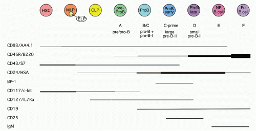
FIG. 8.1. Differentiation Diagram for Development of B Cells from Hematopoietic Stem Cells. Expression of the each surface protein is indicated by a line. Changes in level of expression is indicated by line thickness. CLP, common lymphoid progenitor; ELP, early lymphoid progenitor; Fo B, follicular B cell; HSC, hematopoietic stem cell; MLP, multilineage progenitor; NF B, newly formed B cell.

At birth, B-cell development can also be detected in spleen, but development at this site gradually decreases to very low levels by 2 to 4 weeks of age. Over this same period, B-cell development shifts to the bone marrow and thereafter it continues for the life of the animal. B lymphopoiesis decreases in aged mice. This may be due to diminished responsiveness of precursors to IL-7.24,25


Stem Cells, Commitment, and Early B-Cell Progenitors in Bone Marrow

B cells are continually generated from HSCs in the bone marrow of adult mice. Considerable effort has focused on evaluating the functional capacity of fractions of bone marrow cells to repopulate different lineages of blood cells; this work has progressed to the stage of defining a phenotype for such cells, with expression of c-KIT constituting an important marker in the so-called lineage negative subset.26,27 This is the small fraction of bone marrow cells (< 5%) that lacks expression of a panel of “differentiation markers,” cell surface molecules that are expressed on later stages of various hematopoietic cell lineages. Careful analysis of this HSC fraction using additional markers has shown that it represent perhaps 1/30,000 of nucleated bone marrow cells with as few as 10 mediating multilineage repopulation in cell transfer assays.28,29,30 An important capacity of “true” or “long-term repopulating” HSCs is their ability to give rise to cells in a recipient mouse that can also repopulate all the blood cell lineages upon retransfer into a second host, indicating a capacity for extensive self-renewal without differentiation into more restricted progenitors.

A major focus in research on hematopoiesis has been defining and characterizing lineage-restricted progenitors, such as the common myeloid and common lymphoid

progenitors (CLPs).31,32,33 The CLP cell fraction was identified by lack of a panel of “lineage markers,” expression of the IL-7Rα chain, and distinctive, intermediate levels of c-KIT, compared to higher levels on HSCs. Initial characterization of these cells in various functional assays suggested that these cells could generate B, T, natural killer (NK), and a subset of dendritic cells but no other blood cell lineages. The reason for this restriction has been intensively studied, and downregulation of the receptor for granulocyte-myeloid colony stimulating factor has been suggested to be a key event in this process.34 Cells with the phenotype of CLP constitute about 1/3000 of bone marrow cells. Prior to the CLP stage, multipotent progenitors exhibit low-level expression of genes characteristic of diverse cell lineages, leading to the idea that such promiscuous expression indicates chromatin accessibility that facilitates flexibility in cell fate decisions.35


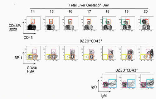
FIG. 8.2. Phenotypic Progression of Developing B-Lineage Cells in Mouse Fetal Liver Analyzed at Different Days of Gestation. Note that B220+cluster of differentiation (CD)43+ cells precede B220+CD43- cells and that within the B220+CD43+ fraction, heat stable antigen (HSA)- cells precede HSA+ cells, and BP-1- cells precede BP-1+ cells. Within the B220+CD43- fraction, the immunoglobulin M+ percentage increases until birth (at about day 20).

CLP cells can give rise in short-term cultures to cells of the B lineage, naturally raising the issue of when cells become restricted to the B lineage. Most cells growing in stromal cultures give rise only to B cells upon transfer into mice, and the phenotype of these cells has been well characterized. 36 Most have at least some heavy chain rearrangement and bear the B-lineage marker CD19.37,38 There is less certainty concerning the cells isolated directly from primary lymphoid tissues, as such cells are quite rare similar to CLPs and HSCs. Most of the CD45R/B220+ cells in bone marrow are also CD19+; such cells are committed to the production of B cells.39 However, a subset of B220+ cells lacks detectable CD19 expression; cells within this fraction can generate CD19+ cells in short-term stromal culture with IL-7. Such cells are included in the CD43+CD24low fraction (Fr. A, 1% of bone marrow) of B220+ cells in bone marrow, but this fraction also contains other cell types including NK-lineage precursors.38,40 Thus, it is necessary to exclude cells lacking AA4.1 (about half38) and expressing Ly6c.41 Many of these Ly6c+ cells also express CD4; recent work suggests that these are plasmacytoid dendritic cells.42,43 A phenotypic approach for enriching and fractionating very early B-lineage subsets is shown in Figure 8.3A.

Careful analysis of the LIN− (including CD19) IL7Rα+cKIT+ CD45/B220− (CLP) and CD45/B220+ (Fr. A), as delineated in Figure 8.3, modified our understanding of the earliest stages of B-cell development in bone marrow.44 First, while CLP stage cells fail to efficiently generate myeloid cells upon transfer into irradiated hosts, they nevertheless retain significant capacity to produce such cells in short-term cultures, likely due to continued (albeit reduced) expression of receptors for myeloid growth factors. In contrast, this myeloid capacity is greatly reduced as cells begin to express CD45R/B220 (ie, become “Fr. A”), concomitant with reduced expression of receptors for myeloid growth factor receptors. Yet these Fr. A cells, while poorly reconstituting T cells in cell transfer assays, nevertheless retain the capacity to generate T-lineage cells in culture, mediated by engagement of Notch by its ligand DL1.45 Thus, the potential for alternate hematopoietic lineages appears to be lost somewhat later in progression down the B-lineage pathway in mouse bone marrow than previously thought. On the other hand, it appears that initiation of Ig rearrangement is initiated earlier than some studies have indicated. Determination of the extent of germline deoxyribonucleic acid (DNA) segments lost upon D-J rearrangement, and the formation of such D-J segments in individual cells isolated by electronic cell sorting showed that 30% to 50% of cells in CLPs and more than 80% of cells in Fr. A contained a D-J rearrangement on at least one chromosome.44 This is consistent with high-level expression of genes important in Ig rearrangement, including TdT, Rag-1, and Rag-2, in CLP46 and Fr. A stage cells.

An emerging view of CLPs (and possibly even the earlier multilineage progenitor [MLP]) stage cells considers them to be early B-lineage precursors rather than branch points in the production of other hematopoietic cell lineages. Thus, analysis of CD4/CD8/CD3 “triple-negative” cells in thymus failed to identify cells with a surface phenotype comparable to CLPs, and mutant mice lacking CLPs in bone marrow nevertheless have relatively intact thymic development, leading these authors to suggest a distinct “early T progenitor” different from CLPs.47 Furthermore, cells with T/myeloid potential, but lacking B-lineage capacity, have been described.48 It seems reasonable to hypothesize that MLP, CLP, and Fr. A stage cells occupy a distinctive microenvironmental niche in bone marrow where they receive signals that guide them along the early stages of B-lineage development culminating in CD19+ pro-B cells that are irreversibly committed to becoming B cells due to expression of PAX539 (see following section). Interestingly, cells considered to be progressing down a lymphoid/B-lineage path can be redirected to become dendritic cells by signals through toll-like receptors (TLRs), suggesting that infection can profoundly alter early stages in hematopoietic development.49

Additional issues remain regarding the lineage restriction of cells at these early stages in B-cell development. For example, there is evidence that cells restricted to generating B and myeloid/macrophage (but not T) lineage may exist in the fetal liver50 and even in bone marrow.51 There is also apparently a different dependence of fetal liver B lymphopoiesis on expression of the transcription factor BSAP compared to bone marrow, as determined by analysis of PAX5 null mice.52 Further comparison of B-cell development in fetal liver with that in bone marrow is needed to clarify this point. Finally, the precise delineation and characterization of B-cell precursors earlier than CLPs, prior to IL-7 expression, remains imprecise. It seems likely that at least some of the MLP stage cells mentioned previously are initiating a B-lineage program based on their expression of E2A, Rag-1, Rag-2, and TdT.44 However, potential heterogeneity in this fraction needs to be assessed. Determination of Rag-1 transcriptional activity at the single cell level by a green fluorescent protein reporter, used for identification of the early lymphoid progenitor fraction,53 may provide a key approach for such studies.


Transcription Factors Regulating B-Lineage Development

The GATA-2 and Runx1/AML1 transcription factors are required for the development of HSCs that are the precursors of all the blood cell lineages, including B cells54,55,56,57,58 (Fig. 8.4). Experiments with the core-binding factor-associated leukemia fusion protein CBFbeta-SMMHC, whose expression

inhibits RUNX function, have revealed that its expression negatively impacts pre-pro-B-cell through pre-B-cell populations in bone marrow.59 Although the frequency of CLP stage cells was unaffected, the expression of B-lineage associated genes (such as CD79a and λ5) was decreased, demonstrating the key importance of early RUNX activity.


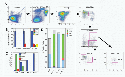
FIG. 8.3. A: An approach for purifying the earliest stage of B-lineage cells in mouse bone marrow. Bone marrow cells expressing cell surface proteins characteristic of differentiated stages of T, myeloid, erythroid, and B lineages are depleted sequentially by electronic gating in the first three panels. Cells with low-level expression of cluster of differentiation (CD)24/heat stable antigen (HSA) and intermediate levels of CD43(S7) are selected in the fourth and the distribution of CD45R/B220 versus CD93/AA4 is shown in the fifth. AA4+B220− cells contain multilineage progenitors (MLPs) and common lymphoid progenitors (CLPs), resolved by analysis for c-Kit versus interleukin (IL)-7Rα in panel six. AA4+B220+ cells, shown in the final panel, are enriched for c-Kit+IL-7Rα+ cells, termed Fr. A. CLP stage cells resemble Fr. A, but lack detectable expression of CD45R/B220. In contrast with CLP and Fr. A, MLP stage cells have higher levels of c-Kit and lack IL-7Rα expression. B: Functional analysis of early B-lineage cells by in vivo competition assay, showing absence of myeloid or T-lineage generation, but production of B-lineage cells from Fr.A. In contrast, CLP stage cells generate B and T cells, whereas MLPs repopulate B, T, and myeloid lineage cells. MLP, CLP, and Fr. A as identified in A. Fr. B stage cells are DJ/DJ rearranged pro-B cells, identified as CD19+CD43+CD24(HSA)+. C: Functional analysis of early B-lineage cells by in vitro S17 stromal cell assay, showing predominant B-lineage colony formation from Fr. A, but some myeloid generation from CLP stage cells. D: Functional analysis of early B-lineage cells by in vitro DL1-OP9 stromal cell culture, revealing significant T-lineage potential in Fr. A stage cells.


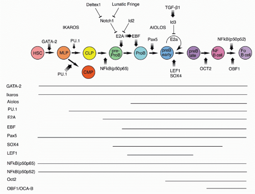
FIG. 8.4. Transcription Factors Important at Different Stages in B-Cell Development in Mouse Bone Marrow. Some regulatory networks are also shown. Positive/activating activity is indicated by arrows, whereas negative or blocking activity is indicated by bars. The rapidly cycling stage, early pre-B cell, is also indicated. Predominant stages of expression are indicated below the diagram.

Somewhat later acting, but still very early in development, is the Ikaros transcription factor.60,61,62 Ikaros and the related transcription factor Aiolos63 play important roles in lymphocyte development. Ikaros is expressed early in hematopoietic precursors. Ikaros null mice lack B-lineage cells62; a different Ikaros mutant that acts as a dominant negative completely blocks lymphoid development.60 Ikaros activates numerous early B-lineage genes, including TdT, Rag-1, λ5, and VpreB. Expression of EBF1 in Ikaros−/− hematopoietic progenitor cells restored generation of CD19+ cells, but these cells were not committed to the B-cell fate and failed to rearrange IgH genes.64 Thus, Ikaros acts in a transcription factor pathway, inducing EBF1 expression, and acting in concert with PAX5 to maintain B-lineage commitment, but also altering chromatin compaction around the IgH locus, which together with Rag expression results in heavy chain rearrangement. Aiolos is detected somewhat later in development at about the stage of B-lineage commitment, and its expression increases further at later stages. It is induced by pre-B-cell receptor (BCR) signaling (see section on the role of Ig heavy chain and the pre-BCR), and it acts to down-regulate transcription of λ5 (initially induced by action of Ikaros), a part of the pre-BCR complex. In this way, Ikaros family members play key roles both initiating and terminating pre-BCR signaling, a critical checkpoint in B-cell development.

The PU.1, an Ets family transcription factor, is critical for progression to the earliest stage of lymphoid development, as demonstrated by the inability of PU.1 null precursors to generate lymphocytes.65,66 An important target of PU.1 for B-lineage development is the gene for Igβ, known as MB-1. The level of PU.1 appears to be critical for development
along the B lineage, as, while low-level expression induced in PU.1 null mice allowed B-lineage development, high-level expression blocked this and fostered myeloid lineage development,67 likely due to differential induction of the IL-7Rα and macrophage colony-stimulating factor receptor chains.68 In fact, retroviral mediated expression of the IL-7Rα chain complements defective B lymphopoiesis in PU.1 null bone marrow HPCs.69 Surprisingly, recent work from several groups indicates that some B-cell development can occur in the absence of PU.1 expression.70 Furthermore, analysis of conditional PU.1 knockout mice showed that expression of this transcription factor was not required after the pre-B-cell stage.71

E2A codes for two proteins, E12 and E47, members of the basic helix-loop-helix family of transcription factors; its induction is crucial from the earliest stages of B-lineage development, as all stages after CD19 expression are absent from E2A null mice.72,73 These mice lack detectable D-J rearrangements, and, interestingly, such rearrangements can be induced in nonlymphoid cells by introduction of the Rag genes and ectopic expression of E2A,74 implicating this transcription factor in the process of chromatin remodeling of the Ig heavy chain locus that permits accessibility by the recombinase machinery.75 The regulation of E2A is crucial for B-lineage development, as negative regulators such as Notch1 and ID2 have been show to block this lineage and induce alternate cell fates, the T and NK lineages.76,77,78,79 Consistent with this picture, ectopic expression of genes that negatively regulate Notch1, lunatic fringe, and Deltex-1 induce the B-cell fate.80,81,82

Expression of the early B-cell factor, EBF1, a member of the O/E protein transcription factor family, is requisite for progression of early B-lineage progenitors to the D-J rearranged pro-B stage (Fr. B), as shown in EBF1 null mice.83 The expression of EBF1 is induced by action of the epigenetic histone H2A deubiquitinse MYSM1, as revealed by targeting this gene.84 EBF1 and E2A act at a similar stage in early in B-lineage development; these two transcription factors can act together to upregulate a family of early B-lineage-specific genes, including Ig-α/β, VpreB/λ5, and Rag-1/2.85,86 There is evidence that E2A upregulates expression of EBF1, found by transfection of E2A in a macrophage cell line,87 suggesting an ordering of these two in development. Furthermore, recent studies showed that there are two distinct promoters for EBF1 that are regulated differently.88 A distal promoter is activated by IL-7 signaling, E2A and EBF1, whereas a proximal promoter is regulated by PAX5, Ets1, and PU.1. Such complex regulation indicates that B-cell development occurs through the action of several feedback loops in a regulatory network that is becoming understood.89,90

BSAP, the product of the PAX5 gene, is expressed throughout B-cell development until the plasma cell stage.91 PAX5/BSAP transcriptional targets include CD19 and BLNK; expression of this transcription factor acts to upregulate V to D-J heavy chain rearrangement.52 Analysis of chromatin structure around the Ig heavy chain locus revealed that PAX5 induces V to D-J locus contraction, thereby promoting rearrangement.92 PAX5 null mutant mice show an arrest in bone marrow development at the pro-B stage, likely due to the lack of complete heavy chain rearrangements and also due to the absence of the critical B-cell adaptor protein BLNK that serves to link the pre-BCR to the intracellular signaling pathway via the tyrosine kinase Syk.93 BSAP/PAX5 also acts to repress alternate cell fates, as pro-B phenotype cells isolated from PAX5 null bone marrow can generate diverse hematopoietic cell lineages, in contrast with such cells from wild-type mice that are B-lineage restricted.39,94 This occurs by repression of the myeloid growth factor receptor gene c-fms95 and by repression of the Notch1 signaling pathway96 critical for T-cell fate specification.45,97 Conditional targeting of PAX5 in more mature B-cell stages shows that its continued expression is necessary for maintenance and function of mature B cells.98 Finally, as mentioned previously, in contrast with bone marrow, the absence of BSAP/PAX5 arrests B-cell development prior to the B220+ stage in fetal liver, suggesting a crucial difference in the early dependence on this transcription factor.52

The Forkhead family transcription factor FoxO1 plays important roles at several stages of B-cell development.99 Early in development, it acts to induce expression of the receptor for IL-7 at the CLP stage. It also functions to regulate Rag-1 and Rag-2 expression during heavy and light chain rearranging stages of development.100 Finally, in mature B cells, it is needed for normal expression of L-selectin, a homing receptor important for normal recirculation of peripheral B cells through the lymphatics. GA binding protein, a ubiquitously expressed Ets family transcription factor, is another player regulating expression of the IL-7R.101 Importantly, through interaction with PAX5, it acts in concert to induce expression of critical PAX5 target genes such as CD79a. There is recent evidence that FoxO1 regulates Ikaros activity by altering splicing of its messenger ribonucleic acid (mRNA), rather than altering Ikaros transcription.102 FoxO1 activation of Ikaros was sufficient for induction of rearrangement of proximal VH genes, but expression of PAX5 was required for rearrangement of distal VH genes. Thus, FoxO1, Ikaros, and PAX5 appear to function in a network to coordinate the ordered rearrangement of Ig genes during B-cell development.

Lymphoid enhancer binding factor (LEF-1) shows a pattern of expression restricted to the pro-B and pre-B stages of B-cell development.103 Targeted inactivation of the LEF-1 gene allows B-cell development but with reduced numbers.104 This is because LEF-1 regulates transcription of the Wnt/β-catenin signaling pathway whose activation increases proliferation and decreases apoptosis of early B-lineage cells. In fact, exposure of normal pro-B cells to Wnt protein induces their proliferation.104 Interestingly, there is a counterproliferative signal that can act at the pre-B proliferative stage, mediated by transforming growth factor-β1.105 It appears that this occurs due to induction of the ID3 inhibitor that negatively regulates the activity of E2A.106 Another transcription factor whose expression is similar to LEF-1 is SOX-4; its inactivation also results in the inability of normal early B-lineage cell expansion and a block at the pro-B stage.107


Several forms of NF-κb subunits are expressed throughout B-cell development; this transcription factor can regulate kappa light chain expression and also growth factor signaling.108 Mice lacking the p65 subunit die before birth, so development must be analyzed by transfer of fetal liver precursors into wild-type recipients. Such experiments showed diminished B-lineage cell numbers, but the major defect was in mature B-cell mitogenic responses.109 Mice lacking the p50 subunit showed relatively normal B-cell development but again poor response to mitogen by mature B cells.109 However, mice lacking both the p50 and p65 subunits failed to generate any B220+ B-lineage cells. Curiously, when mixed with wild-type fetal liver cells, normal numbers of mature B cells could be generated from the double-defective precursors, suggesting that the defect could be overcome by secreted or membrane-bound signals provided by the wild-type precursors. Another double mutant, p50p52, showed a late stage defect in B-cell development, with a failure to generate mature B cells in spleen.110

Inactivation of the Oct-2 transcription factor results in neonatal lethality, but transfers of fetal precursors can reconstitute lymphoid cells in wild-type recipients, allowing assessment of effects on the B lineage. Such studies have shown that fewer mature follicular B cells are generated in these mice and B-1 (CD5+) B cells are completely eliminated.111,112,113 Similarly, the Oct binding factor, OBF-1, also known as OCA-B and BOB-1, appears to function in the maturation of newly formed B cells in the bone marrow to become follicular B cells in the periphery, as inactivation of this gene resulted in a significant deficit in mature B cells.114,115,116 Both of these transcription factors have been shown to regulate the follicular B-cell chemokine receptor CXCR5, which may explain at least part of the defect.117 Curiously, unlike Oct-2 null mice, there was reportedly no deficit in B-1 B cells in OBF-1 null mice. Interestingly, when the OBF-1 mutant mouse is crossed with btk-deficient mice, there is a complete lack of peripheral B-cell generation,118 suggesting that this transcription factor may function in the BCR-mediated selection of mature B cells.


Impact of Microribonucleic Acids on B-Cell Development

The miRNAs are small noncoding ribonucleic acids that facilitate the degradation of mRNAs and thereby act at a posttranscriptional level to regulate gene expression. The generation of mature functional miRNAs requires action of Dicer, a protein that cleaves pre-miRNAs, so the targeting of Dicer allows assessment of the global effect of miRNA on B-cell development. Ablation of Dicer in early B-lineage progenitors results in a block at the pro- to pre-B cell stage, likely due to upregulation of the proapoptotic molecule Bim.119 Counteraction of Bim function by a Bcl-2 transgene reveal further dysregulation of normal development, including nontemplated nucleotide addition (N-regions) at the Ig light chain V-J junction, due to aberrant expression of the terminal deoxynucleotidyl transferase gene that is normally extinguished at the pre-B-cell stage.

Another approach for assessing the importance of specific miRNAs is direct overexpression or knockdown of expression; such a study with miR-150 reveal its role in regulating c-Myb, a transcription factor that regulates pro-B to pre-B progression and also the survival of mature B cells.120 Transgenic overexpression of miR-17-92, a miRNA often found to be amplified in lymphoma, resulted in a lympho-proliferative syndrome and autoimmune disease, resulting in premature death.121 One target of miR-17-92 is Bim; its decrease in the transgenic animals may have resulted in excessive cell survival and a loss of normal tolerance to self-antigens. The potential relevance to normal growth regulation is quite interesting, considering the amplification of this miRNA in some lymphomas. Another miRNA with a cancer association, miR-21, has been studied as a transgene in mice, where it results in tumors with a pre-B malignant lymphoid-like phenotype.122 The miRNAs that are amplified in cancers and likely contribute to the neoplastic process are now termed “oncomirs.”

miRNAs can also influence lineage choice or act at key checkpoints during hematopoietic development. Retroviral provision of miR-34a in bone marrow hematopoietic progenitors blocked B-cell development at the pro-B to pre-B checkpoint, resulting in reduced numbers of mature B cells in mice repopulated with such precursors. A possible explanation for the developmental block is action of miR-34a on Foxp1, a transcription factor that appears to be important at this stage, as cotransfection of FoxO1 lacking its normal 3′ UTR target of miR-34a restored B-cell development. A novel regulator of lineage choice appears to be Let-7, a family of miRNAs regulated by the highly conserved ribonucleic acid-binding protein Lin28.123 While Lin28 has been studied for its role in pluripotency, developmental timing, and oncogenesis, a recent study indicates that it may regulate a developmental switch in both B and T cells, such that its expression in adult hematopoietic progenitors results in reprograming development toward a more “innate-like” pattern, normally only seen during fetal/neonatal timing.124


Bone Marrow Developmental Stages


Functional Definition

Distinct stages of developing B-lineage cells can be delineated based on their capacity for growth under different culture conditions. That is, the earliest precursors require cell contact with the stromal microenvironment in addition to specific cytokines, notably IL-7.105,125 This stromal cell/precursor adhesive interaction is mediated, at least in part, by binding of VLA-4 to intercellular adhesion molecule-1.126,127 Later stage cells do not require cell contact but maintain a need for cytokines.128,129 Both cell types can undergo considerable cell proliferation in culture. Interestingly, the difference between cell contact requirement and independence is linked to the expression of heavy chain protein.128,130 A population of cytoplasmic heavy chain-expressing B-lineage cells later than either of these, the so-called late or small pre-B cells, does not proliferate in culture. These cells likely require different culture conditions for survival, as they usually do not persist for extended periods, but rather die with a half-life of less than 24 hours unless protected from apoptosis by a Bcl-2 transgene.131


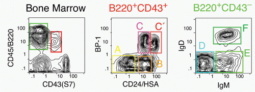
FIG. 8.5. Flow Cytometry Approach for Analyzing Different Stages of Cluster of Differentiation (CD)19+ B-Cell Development in Mouse Bone Marrow. Note that the antibody used for CD24/heat stable antigen (HSA) staining, 30F1, is important, as other monoclonal antibodies that recognize HSA do not resolve high from low-level expression as well. The cells expressing low levels of CD24/HSA, labeled “A”, are CD19- and is enriched for very early B-lineage precursors but also is contaminated by other cell types that can be detected by staining for AA4, NK1.1, DX5, and Ly6c; early B-lineage precursors are AA4+NK1.1-DX5-Ly6c-.


Phenotypic Definition

Further clarification of the heterogeneity in bone marrow can be achieved by analysis using fluorescent staining reagents and either microscopic or flow cytometric analysis. For example, the earliest determination that there were both heavy chain surface-positive B cell and cytoplasmic-positive pre-B cells was through microscopic examination using anti-Ig staining.132,133 Later studies in mouse showed that there were specific surface proteins or “markers” that could be useful in identifying these populations, notably a restricted isoform (CD45Ra) of the common leukocyte antigen, CD45.134 This largely B-lineage-restricted 200 kDa molecular mass isoform is often referred to as “B220.” Some highly B-lineage-restricted monoclonal antibodies, such as RA3-6B2, recognize a specific glycosylation of the CD45Ra isoform.135 However, as described previously, even highly specific antibodies such as 6B2 may also recognize other cell types, such as particular differentiation stages or subsets of NK or dendritic cells.

The application of multiparameter/multicolor flow cytometry and additional monoclonal antibodies specific for other cell surface proteins differentially expressed during B-lineage development has facilitated delineation of multiple additional intermediate stages in this pathway.129 For example, the B220+ population in bone marrow can be further fractionated into an earlier subset expressing CD43 (about 3% to 5% of marrow cells) and a later fraction with much lower CD43 expression (20% to 30% of marrow). The precursor/progeny relationship of cells in these two fractions can be readily demonstrated by short-term culture, with CD43+ cells giving rise to CD43− cells. These two populations can be further subfractionated based on additional developmentally regulated surface proteins, such as CD24/heat stable antigen (HSA), BP-1 (a zinc-dependent cell surface metallopeptidase also known as aminopeptidase A136), and the surface Ig molecules IgM and IgD.129 This is shown in Figure 8.5. Again, these cell populations can be isolated and short-term culture used to determine their order in the pathway. Alternative approaches based on other developmental markers can be correlated with this framework of cell stages, notably the system developed by Melchers’ group137 using expression of CD45R/B220, CD19, c-KIT, and the IL-2Rα chain. A diagram summarizing this type of phenotypic subdivision and relating different nomenclatures is shown in Figure 8.6.


Culture Systems and Critical Microenvironmental Interactions

The combination of phenotypic characterization coupled with analysis of growth and differentiation in culture has provided a powerful approach for the further understanding of B-cell development, as employed by many different investigators. Bone marrow cultures developed by Whitlock and Witte,138,139,140 and fetal liver cultures developed by Melchers’ group141,142 have allowed determination of the critical cytokines and some of the cell adhesion molecules important in the in vivo development of these cells. Many of these are summarized in Table 8.1. A typical B-lineage colony proliferating on S17 stromal cells in the presence of IL-7 is shown in Figure 8.7.

Survival and growth of the earliest stages of developing B-lineage cells require cell contact with nonlymphoid adherent cells that can be isolated from bone marrow, cells referred to generically as “stromal cells.” A number of lines have been derived from primary cultures of bone marrow adherent cells and characterized in terms of their capacity to support B lymphopoiesis in vitro.143 This work has led to the discovery of adhesion molecules that play important roles in mediating the organization of clusters of developing B-lineage cells on stromal layers, including CD44 interacting with hyaluronate and VLA-4 interacting with vascular cell adhesion molecule 1.126,127,144,145 Both of these interactions could be disrupted by addition of blocking antibodies
to CD44 and VLA-4 on B-cell precursors, resulting in a disruption of normal pre-B proliferation in vitro.146 Such adhesion interactions may serve to transmit signals directly to the stromal cells or B precursors, or both. There is some evidence that stromal cells are induced to elaborate specific growth mediators after interaction with B-cell precursors or soluble regulators.147


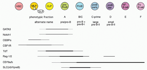
FIG. 8.6. Diagram of Distinct Phenotypic Stages and Characterization of Terminal Deoxynucleotidyl Transferase, Biphasic Rag Expression, Immunoglobulin-α/β, and Surrogate Light Chain Expression. Genes characteristic of myeloid and T-cell lineage are also shown. The cell type descriptions are cross-referenced to the alphabetic phenotypic fraction nomenclature and also to the Basel nomenclature. The early lymphoid progenitor population is identified by activation of the Rag-1 locus in a green fluorescent protein reporter mouse.

Another function of the stromal cells is to produce growth factors critical to B-lineage survival, proliferation, and differentiation; the most important of these for mouse B-cell development is IL-7.105,125,148,149 IL-7 promotes the survival and proliferation of pro-B and pre-B stage cells, both in vivo and in vitro.150,151 Neutralizing antibody to IL-7 can block B-cell development in vitro,129 and IL-7 expressed as a transgene can deregulate normal B-cell development, leading to B-cell lymphadenopathy.152 The IL-7 receptor consists of a unique IL-7Rα chain153 paired with the common gamma chain (λc) that is also found in the receptors for
IL-2, IL-4, IL-9, IL-15, and IL-21.154 IL-7Rα null mice have a severe deficit of both B and T cells in the periphery and lack most B-lineage cells in bone marrow.155 Mice with targeted inactivation of the γc or IL-7 do have some B-lineage development, suggesting an alternate cytokine; this appears to be thymic stromal lymphopoietin (TSLP). This protein was first identified as a pre-B-cell growth factor produced by a thymic stromal line156 and shows some of the same effects in culture as IL-7, although possibly inducing less proliferation and more differentiation.157 Its receptor has been cloned; it consists of two chains, the TSLP receptor and IL-7Rα.158 The TSLP receptor shares both sequence homology and genomic exon organization with the common gamma chain.159 Signaling through the IL-7 receptor requires JAK3 and activates the transcription factor STAT5, whereas signaling through TSLP is JAK3 independent but also activates STAT5.157,160 Unexpectedly, the growth response to TSLP requires synergy with the pre-BCR in bone marrow but not in fetal liver,161 leading some to propose that this might be a marker for distinctive B1 B-cell development in bone marrow.162 When TSLP is overexpressed from an inducible transgene, B1 B cells expand, apparently at the expense of marginal zone (MZ) B cells.163


TABLE 8.1 Regulators of Growth of Early B-Lineage Cells


Mediator


Effect


Reference


L-7


Stimulates CLP and B-precursor proliferation


105,148,149,590


TSLP


Alternate IL-7-like cytokine


156,157


IGF-1


Stimulates accumulation of Cµ+cells in culture


185


FLT-3/FLK2-L


Critical for earliest stages of B-lineage development


168,172,591,592


c-KIT-L


Synergizes in IL-7-induced proliferation


26,166


IL-3


Substitute for IL-7 in proliferation of pre-B clones


176


CXCL12/CXCR4


Crucial chemokine interaction for early B-lineage precursors


194,196,198,199


Hemokinin


Novel regulator of B lymphopoiesis


187


VLA-4/VCAM-1


Adhesive interaction; antibodies to either block B lymphopoiesis


126,127,593


CD44/hyaluronate


Adhesive interaction; mediates association of B-lineage/stromal cells


144,146


TGF-β


Inhibits proliferation stimulated by IL-7


105


Sex steroids


Decrease B-lineage precursors in bone marrow


594,595


Growth hormones


Required for normal B lymphopoiesis


188,189,596


TLRs


Innate immune system regulation


190


CD, cluster of differentiation; CLP, common lymphoid progenitor; IL, interleukin; TGF, transforming growth factor; TLR, toll-like receptor; TSLP, thymic stromal lymphopoietin; VCAM, vascular cell adhesion molecule.


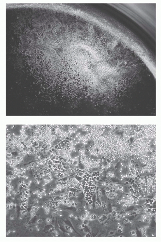
FIG. 8.7. Photomicrographs of B-Lineage Colony Proliferating on S17 Stromal Layer (in the Well of a 96-Well Microplate) in the Presence of Interleukin-7. Day 10 colony derived from a single Fr. A phenotype (see Fig. 8.5) cell. Low power and high power views. All of these cells now express CD19 and many have progressed to BP-1+.

The earliest precursors in the B-lineage pathway, probably including cells that are not B-lineage committed but that can efficiently give rise to B cells in a short time in vitro, have receptors for SCF/c-KIT-ligand26,27,164,165,166,167 and FLK2/FLT3-ligand.168,169,170,171,172 Thus, the most permissive cultures for expanding precursors of B-lineage cells will include these cytokines, in addition to IL-7 and a stromal adherent cell layer, such as S17.173,174,175 IL-3 has also occasionally been suggested as playing a support role for pre-B cells in vitro,176 although its role in vivo may be at a much earlier stage. While culture conditions have been reported that can support B-lineage development in the absence of stromal cells,177,178 the clear-cut alteration of contact-dependence prior and post heavy chain expression128,129,130,179,180 argues that the most physiologic model for early B-lineage growth will include stromal cells. Besides providing important cell-cell contacts that may signal survival, proliferation, and differentiation, it is also likely that stromal cells bind at least some cytokines to their surface, providing higher local concentrations to the clusters of B-lineage precursors that adhere.181,182 B-lineage development may be modified by exposure to hormones; considerable interest has focused on sex steroids released during pregnancy that serve to depress B lymphopoiesis, particularly the pre-B-cell pool.183 This may be important to avoid autoimmune responses by the mother but could have negative consequences due to possible transient immunodeficiency. Interestingly, fetal B lymphopoiesis is not similarly depressed due to the absence of hormone receptors on fetal B-lineage cells.184 Insulin-like growth factor has been reported to potentiate progression in vitro to the Cµ+stage,185,186 and, more recently, there is a report of a bioactive peptide, a type of tachykinin, that synergizes with IL-7 to enhance the growth of IL-7-dependent cultures.187 Besides insulin-like growth factor, other pituitary hormones, thyroxine, and growth hormone have effects on B lymphopoiesis.188 For example, thyroxine treatment can restore normal B-cell development in dwarf Pit-1 mutant mice with deficient pituitary function.189 Recent work has highlighted the effect that activation of TLRs during infection may have on altering development.190 Thus, it is likely that more detail remains to be filled in to complete our picture of the growth requirements and modulating influences of B-lineage cells in mouse bone marrow. Another function of cell-cell interaction is cell fate determination during the lineage commitment stage, very early in development of B-lineage cells. The Notch signaling pathway is implicated in cell fate determination in invertebrates and more recently has been shown to function in lymphoid lineage specification.45,76,191 Notch family transmembrane receptors regulate transcription by being cleaved upon ligand binding to release an intracellular cytoplasmic domain that translocates to the nucleus where it interacts with the transcriptional repressor CSL.192 Recent studies have shown that Notch1 can play a pivotal role in commitment of common lymphoid progenitors to the T-cell
lineage.76 That is, expression of Notch1 by retroviral transduction has been shown to redirect B-lineage differentiation in bone marrow along the T lineage. Furthermore, a reciprocal result was found in conditional Notch1 null mice, blocking T-cell development in the thymus to be replaced by B-cell development.97 Finally, altering the Notch1 modifier lunatic fringe by overexpressing this molecule under regulation of an lck promoter resulted in B-cell development in the thymus.80 Differentiation of lymphoid precursors to NK or dendritic cell lineages was unaffected in Notch1 null CLP cells, so Notch apparently affects only the B/T-lineage decision.


Role of Chemokines in Migration of B-Cell Precursors

One of the most distinctive features of B-cell development in bone marrow is the migration of developing precursors from early stages nearest the bone endosteum layer to latter stages progressively closer to the central arteriole, where they will eventually exit.193 This migration is likely due to differential expression of specific adhesion molecules and also to expression of chemokine receptors. Analysis of B-cell migration has identified a critical chemokine that is important in this process, SDF-1, now known as CXCL12,194,195 and its receptor CXCR4.196 CXCL12 is expressed by fetal liver and bone marrow stromal cells, whereas CXCR4 is found on hematopoietic precursors and B-cell progenitors.197 Deletion of either the receptor or ligand results in severely impaired B lymphopoiesis.198,199,200 Interestingly, the critical defect appears to be failure to retain precursors in the primary lymphoid organ, as progenitors and precursors can be found in the blood of mutant mice.201


Gene Expression and Immunoglobulin Rearrangement

In addition to delineation of developmental stages based on changes in protein surface expression, B-lineage cells can also be characterized for expression of internal proteins related to critical processes in their progression along this pathway, specifically those related to rearrangement and expression of the B-cell antigen receptor (Fig. 8.8). Thus, expression of µ heavy chain constant region, prior to Ig rearrangement, from a cryptic promoter generates a “sterile transcript” that reflects an open chromatin structure important for the onset of rearrangement,202,203,204 and so analysis of sterile µ expression can be used to investigate very early stages of B-cell development. Classical northern analysis can be done with transformed lines, but much work analyzing RNA levels in B-lineage cell fractions, whether directly isolated or cultured, has depended on polymerase chain reaction amplification of complementary DNA.38 For example, using this approach, sterile µ can be detected in a very early fraction of B220+CD43+CD19- (Fr. A) cells. Expression of the recombinase activating genes Rag-1 and Rag-2, which together make the double-strand breaks in DNA required for Ig rearrangement,205,206,207 also occurs in Fr. A stage cells, which also have high levels of TdT, the enzyme responsible for adding nontemplated nucleotides at the D-J and V-D junctions of the heavy chain.12,13 Rag-1 binds in a highly specific fashion to discrete sites within the IgH locus, recombination signal sequences, defining “recombination centers.”208 The extent of heavy chain or light chain rearrangement can be quantitated either in bulk isolated populations129 or in individual cells.137,209,210 At the heavy chain locus, D-J rearrangement occurs prior to V-DJ rearrangement, and cells with extensive D-J but little V-DJ rearrangement can be detected at the B220+CD43+CD19- (Fr. A) stage, where Rag-1/2 and TdT are strongly expressed.44 V-DJ rearrangements are readily detected in the abundant B220+CD43− (Fr. D) stage small pre-B cells, although productive rearrangement has already completed by the large pre-B (Fr. C-prime) stage (see following discussion). Single-cell sequence analysis of rearrangements in Fr. C stage cells shows a large proportion with nonproductive rearrangements on both chromosomes, suggesting that this may represent a dead-end fraction.209 Some light chain rearrangement is detectable in early stage B220+CD43− (Fr. B) cells, and this is consistent with the observation that low-level kappa light chain rearrangement is detectable in bone marrow of mice where µ heavy chain has been crippled by deletion of the membrane exon.211 However, much higher levels of kappa rearrangement can be detected in B220+CD43− (Fr. D) stage cells consistent with the finding of sterile kappa mRNA increase just prior to this stage likely induced by pre-BCR signaling (see following discussion).


Role of Immunoglobulin Heavy Chain and the Pre-B-Cell Receptor

Many years ago, the analysis of severe combined immunodeficiency (SCID) mouse212 bone marrow revealed the presence of a population of B220+ cells, all with a very early CD43+ phenotype, suggesting a block in B-cell development at this stage.213 SCID mice have a defect in the catalytic subunit of the DNA-dependent protein kinase DNA-PKcs,214,215 and, as a result, B-lineage cells in these mice are very ineffective at completing productive Ig heavy chain rearrangements. This block could be overcome by introduction of an Ig heavy chain transgene, indicating a critical role for µ protein in progressing past an early developmental checkpoint.216 Furthermore, a gene targeting experiment that eliminated the membrane exon of µ heavy chain (µ-mt) also generated a block at this stage.217,218

The µ heavy chain is associated with a set of B-cell-specific peptides at the early pre-B-cell stage,219 and this complex is referred to as the pre-BCR. It seems clear that pre-BCR mediates a type of signaling function analogous to the BCR in mature B cells. Prior to light chain expression, two peptides known as λ5 and VpreB, originally isolated as B-lineage-specific complementary DNAs,220,221 associate with heavy chain. The λ5 shows homology to a lambda constant region and VpreB is so-called because it has homology to a variable region domain. Together, these peptides constitute a pseudo- or surrogate light chain (SLC). The critical role of λ5 was demonstrated unambiguously in genetargeted mice, where B-cell development was blocked at the B220+CD43+ stage.222 The production of some mature cells that accumulate in this mutant is likely due to early kappa rearrangement, with light chain substituting for SLC, as demonstrated in light chain transgenic experiments.223

The µ heavy chain has a very short cytoplasmic region consisting of only three amino acids; signal transduction

through the BCR is mediated by accessory peptides, similar to the CD3 components of the T-cell receptor, known as Ig-α and Ig-β.224,225,226,227 Inactivation of Ig-β228 results in a block at the B220+CD43+ stage in mouse bone marrow, similar to that seen in µ-mt and λ5 null mice. Finally, the Syc tyrosine kinase plays a critical role in transducing BCR cross-linking signals in mature B cells and inactivation of this gene results in a “leaky” block at this same stage.229,230 Thus, any mutation that affects this pre-BCR complex (see following section; see Fig. 8.10A) precludes efficient progression past the earliest stages of B-cell development.


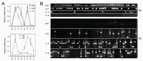
FIG. 8.8. A: Profiles of immunoglobulin (Ig) rearrangement-related gene expression. Cells isolated using fractionation scheme in Figure 8.3 for multilineage progenitors and Fr. A; in Figure 8.5 for Fr. B through Fr. F. Relative messenger ribonucleic acid levels assessed by performing semiquantitative reverse transcription-polymerase chain reaction using limited numbers of cycles, blotting, then probing, and quantitating the probe signal. Note the biphasic expression of Rag genes and the early expression of sterile µ (labeled µ0) during the first wave, where heavy chain rearranges, and the upregulation of sterile kappa transcripts (κ0) during the second Rag wave, when most light chain rearrangement takes place. B: Single-cell polymerase chain reaction analysis of Ig heavy chain rearrangement in four early stages of developing B-lineage cells. Deoxyribonucleic acid prepared from individual cells isolated following the scheme shown in Figure 8.3 was divided into two aliquots and analyzed for retention of a deoxyribonucleic acid segment lost upon any D-J rearrangement (labeled GL, germline) and also for D-J rearrangement. Note that D-J rearrangement initiates at the common lymphoid progenitor stage, where 30% to 50% of cells show a rearrangement.

Careful examination of B-cell development in normal mice shows that heavy chain is first expressed at a late fraction of the B220+CD43+ stage, termed Fr. C-prime (Fig. 8.9A). This fraction is also interesting because it shows a much higher proportion of cells in cycle (revealed by a high frequency of cells with greater than 2N DNA content; Fig. 8.9B), compared with any other B220+ stage in bone marrow.129 Mice unable to assemble a pre-BCR, due to inability to rearrange heavy chain (Rag-1 null), show a block in development at the CD43+ stage that can be complemented by introduction of a functionally rearranged µ heavy chain as a transgene180 (Fig. 8.9C). Analysis of several types of pre-BCR defective mutant mice shows a complete absence of Fr. C-prime stage cells, suggesting that pre-BCR signaling results in the upregulation of CD24/HSA and also entry into rapid cell proliferation. Thus, a model of pre-BCR function is that it signals the clonal expansion phase of pre-B-cell development, amplifying cells with in-frame VDJ rearrangements capable of making heavy chain protein. The precise nature of pre-BCR signaling remains to be completely understood. An early model suggested that cross-linking of heavy chain was mediated through interaction of SLC with a bone marrow expressed ligand. However, subsequent experiments showed that normal light chain could substitute for SLC, and that even a VH truncated µ heavy chain could mediate progression past this stage. Furthermore, intensive searches for the putative ligand over a 10-year period have been fruitless, leading to the model
that pre-BCR signaling is more akin to “tonic” signaling in mature B cells.231,232,233 That is, simple assembly of the complex (or possibly some degree of multimerization fostered by the self-aggregating nature of SLC234) probably is sufficient for the cell to pass this developmental checkpoint (Fig. 8.10B). One clear-cut finding is that pre-BCR signaling in a transformed pro-B-cell model system can occur in the absence of any additional cell type, suggesting that if a ligand exists, it must be expressed on B-lineage cells rather than stromal cells.235 Possibly, pre-BCR homodimerization is mediated through glycosylation at a conserved asparagine residue in the first µ constant region domain.236 Studies of the T-cell analog of the pre-BCR, pre-Tα, provide strong evidence that it signals through spontaneous dimerization, without requirement for an external ligand.237,238,239


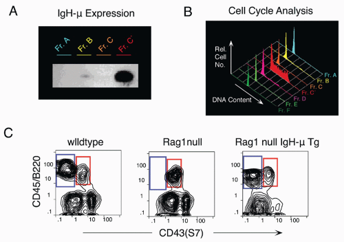
FIG 8.9. A: Western blot of immunoglobulin µ heavy chain expression showing high-level expression in Fr. C-prime. B: Cell cycle analysis of individual fractions shows most cells in Fr. C-prime are cycling. Propidium iodide staining of permeabilized sorted cells allows determination of deoxyribonucleic acid content per cell using flow cytometry. C: Block in B-cell development in Rag-1-deficient mice can be overcome by introduction of an immunoglobulin µ heavy chain transgene.


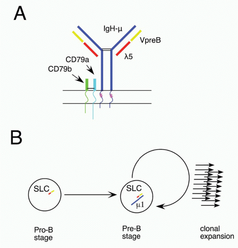
FIG 8.10. A: Diagram of the pre-B-cell receptor (BCR) µ heavy chain with surrogate light chain (λ5 and VpreB) in place of conventional light chain. As in the BCR, immunoglobulin (Ig)-α and Ig-β serve to couple signals between the receptor and cytoplasmic components, such as BLNK and Syk. Starred m transmembrane residues are important in mediating interaction with Ig-α/Ig-β, as mutation of these diminishes BCR function. B: Clonal expansion mediated by pre-BCR assembly. Association of newly generated µ heavy chain with pre-existing surrogate light chain leads to a burst of proliferation at the pre-B stage.

Mutations in other molecules in the pre-BCR signaling pathway have been shown to affect B-cell development and pre-B-cell clonal expansion. Although the Btk mutation is less severe in mouse than in human, there nevertheless is an alteration in pre-B-cell expansion in Btk-deficient mice.240 Also, X-linked immunodeficiency (xid) B cells (deficient in Btk) have been reported to proliferate more in stromal cell cultures, possibly due to decreased differentiation to later nonproliferative stages.241,242 The role of Btk is thought to modulate BCR signaling strength,243 and this is probably also the case for pre-BCR signaling, allowing only strongly signaling pre-BCRs to progress in the mutant mice. BLNK/SLP65 serves to link the pre-BCR to the Syk kinase critical in BCR signaling.244,245 Mutant mice lacking BLNK show a partial block in B-cell development at the pro-B to pre-B transition.246 Curiously, whereas pre-BCR signaling is thought to mediate allelic exclusion (expression of a single heavy chain allele), this remains intact in BLNK-deficient mice.247 Animals deficient in both BLNK and Btk develop an extensive pre-B expansion that progresses to lymphoma, leading to study of such mice as a model for human pre-B acute lymphoblastic leukemia (ALL).248,249

Syk-deficient mice show a more severe block at the pro-B to pre-B transition and a lack of allelic exclusion.229,230,247 There is also evidence that pre-BCR signals through Erk to induce proliferation.250

Outcomes of pre-BCR signaling, in addition to pre-B proliferation, are downregulation of the Rag genes,251 down-regulation of TdT,235 and transcriptional activation of the kappa locus, detected as upregulation of sterile kappa transcripts.252 A control element for regulating Rag expression has been identified.253,254 Extinction of recombinase activity is probably important for chromosomal stability during the clonal burst period of B-cell development255,256,257 and is also at least a part of the mechanism that assures allelic exclusion, the expression of a single heavy chain by any given B cell.258 There is evidence that pre-BCR selection requires low levels of IL-7259 and probably occurs naturally as the developing precursors migrate through different stromal cell microenvironments in bone marrow.

The function of the pre-BCR may be more complex than simply to sense whether an in-frame VDJ rearrangement has occurred. This possibility is suggested by the observation that heavy chains with different VDJ segments vary in their capacity to assembly with SLC components.260,261,262,263 V regions are classified into families based on sequence homology, and many members of two of these families, the 7183 and Q52, appear to frequently generate heavy chains that assemble poorly with SLC.262 A consequence of this will likely be poor pre-BCR signaling and little clonal expansion; such cells will become underrepresented at later stages of B-cell development relative to cells containing heavy chains that signal effectively. One explanation for this SLC assembly-mediated clonal expansion is that it serves a quality control function to test heavy chain V regions for their potential to fold with real Ig light chain, a critical requirement if the cell is to express a complete BCR. An alternative (not necessarily mutually exclusive) explanation is that making pre-B-cell proliferation dependent on pre-BCR expression provides a simple mechanism for regulating the extent of clonal expansion, as an immediate consequence of pre-BCR signaling is to terminate SLC expression. Thereafter, SLC protein levels decay and are diluted by cell division so that after several rounds of proliferation, pre-BCR levels will decrease to below the threshold required to provide the signal to maintain the cell in cycle. Figure 8.11 illustrates a model for bone marrow B-cell development, showing the pre-BCR checkpoint.


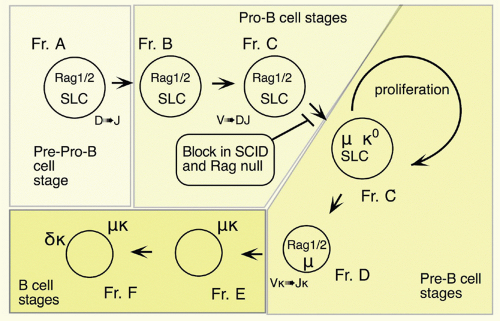
FIG. 8.11. Model of Mouse Bone Marrow B-Cell Development Showing Relationship of Immunoglobulin Rearrangement with Progression and Proliferation.

One of the most striking examples of pre-BCR selection is seen with the D-proximal VH gene, VH81X, where early precursors show biased overutilization due to preferential rearrangement of this VH gene.11,264,265 In fact, regulatory sequences interspersed throughout the distal VH region, termed PAX5-activated intergenic repeat elements, appear to enhance utilization of distal J558 family VH genes.266 Furthermore, a recombination regulatory region, the intergenic control region 1, which lies between the VH and D clusters, has been shown to inhibit proximal and promote distal VH gene utilization.267

Curiously, although it is abundant in early B-cell precursors, VH81X is rarely seen in the mature B-cell compartment. The demonstration of the decrease in representation of cells with VH81X rearrangements at the pre-B clonal expansion phase in bone marrow,268,269 together with the finding that heavy chains utilizing VH81X frequently fail to assemble functional pre-BCRs,260,261,270 explained this paradox. Models of pre-BCR three-dimensional structures based on x-ray crystallography may help to explain variation in the ability of different heavy chains to assemble with SLC.271

However, it is still puzzling that the most frequently rearranged VH gene is so strongly selected against at the clonal expansion stage. A possible explanation may lie in comparisons of VH utilization during fetal development. That is, in contrast with bone marrow precursor cultures, the ratio of productive/nonproductive VH81X does not decrease during cultures of fetal precursors.272,273 Furthermore, the proliferative burst that pre-BCR assembly provides to bone marrow pre-B cells may instead result in exit from cell cycle in fetal precursors,263 leading to selection of very different BCR repertoires during fetal and adult B lymphopoiesis. The possible significance of this is discussed subsequently in the section on B1 B cells. Leaving aside such developmental difference, the role of the pre-BCR in B-cell selection remains subject to debate, with evidence suggesting it functions to eliminate274 or enrich275 self-reactivity.


Light Chain Rearrangement and Generation of Immature B Cells

Besides termination of TdT and SLC gene expression, pre-BCR signaling also results in the downregulation of Rag-1 and Rag-2 expression, mediated by activity of Gfi1b on Erag, the B cell-specific Rag regulatory element,254 and the repression of FoxO1.276 It appears that induction of Bcl-6 by pre-BCR signaling functions in this progression by repressing Myc and Ccnd2, thereby promoting exit from cell cycle.277,278 As mentioned previously, there is evidence that the pre-BCR signals through ERK and this Ras-MEK-ERK signaling acts to silence transcription of Ccd3, encoding the cell cycle protein cyclin D3, thus promoting cell quiescence.279 Opposing cell cycle exit, IL-7R signaling activates STAT5, maintaining Ccd3 expression. Eventually, the pre-B cell escapes from
IL-7 signaling, exits the cell cycle, and the Rag genes are reexpressed at high levels. Sterile kappa transcripts become detectable during the cycling stage, likely reflecting chromatin remodeling to make the kappa light chain locus accessible,252 so induction of the recombinase machinery can initiate kappa light chain V to J rearrangement.

An interesting feature of the Vκ locus is that the approximately 100 genes are in both transcriptional orientations, and so these genes can rearrange either by deletion (generating an extrachromosomal excision circle) or by inversion.280 The absence of intervening D segments also means that it is possible for upstream V kappa genes to rearrange to downstream J kappa segments, “leapfrogging” the initial rearrangement, assuming it was to any Jκ other than Jκ5. The successive association of different kappa chains with the same heavy chain in a B cell is referred to as BCR “editing” and was originally observed in the context of autoreactivity, which maintains Rag expression even at the B-cell stage281,282 (see following section on B-cell tolerance). Because assembly and expression of a complete BCR (that is not self-reactive) terminates Rag expression, an additional reason for light chain editing in the bone marrow may be to replace an initial light chain that fails to assemble effectively with the particular heavy chain present in that pre-B cell. This is probably the explanation for multiple light chain rearrangements detected in single early B-lineage cells.283 Complete failure of kappa rearrangement, possibly after receptor editing to avoid self-reactivity, leads the pre-B cell to a second phase of light chain rearrangement, dependent on IKK-mediated NF-κB signaling, where the λ light chain locus rearranges.284

Newly formed B cells can be distinguished from mature B cells on the basis of their inability to proliferate in response to BCR cross-linking (ie, they are functionally immature). This is also the stage where negative selection is reported in transgenic models of autoreactivity.285,286 Cells at this stage have a short half-life of only a few days, compared to mature follicular B cells with a half-life measured in months. They can be distinguished by surface phenotype from other B cells based on expression of certain combinations of markers, such as IgM+IgD−, absence of CD23, and high-level expression of CD24/HSA.287 Recently, there have been reports of single markers that are useful in distinguishing newly formed cells from any mature subset, such as the molecules recognized by monoclonal antibodies 493288 and AA4.1.289 The AA4.1 target molecule has been cloned and identified as the mouse ortholog of a component of the human C1q receptor.290

Only gold members can continue reading. Log In or Register to continue

Stay updated, free articles. Join our Telegram channel

Aug 29, 2016 | Posted by in IMMUNOLOGY | Comments Off on B-Lymphocyte Development and Biology

Full access? Get Clinical Tree

Get Clinical Tree app for offline access