Autoimmunity and Autoimmune Diseases


Autoimmunity and Autoimmune Diseases


Ken T. Coppieters

Matthias G. von Herrath

Dirk Homann


INTRODUCTION

The concept of autoimmunity (ie, the phenomenon of immune reactivity directed against an organism’s constituent organs, tissues, cells, and/or extracellular factors) comprises both physiologic and pathophysiologic components that attain practical relevance in the context of autoimmune diseases, a collection of more than 80 disorders with circumscribed pathologic features that are thought to result primarily from the induction and perpetuation of aberrant immune reponses. The precise etiopathology for most human autoimmune diseases remains incompletely defined but appears to be promoted by the complex interplay of genetic predispositions, environmental insults such as viral infections and, in an acknowledgment of the many questions yet to be answered, bad luck. However, a role for immune-mediated processes and pathologies has by now been documented in at times compelling detail and provides important direction for novel and improved diagnostic, prophylactic, and therapeutic modalities. Herein, we discuss the evolution of major conceptual and practical advances and challenges in autoimmunity and autoimmune diseases in general, and consider individual autoimmune disorders regarding the contribution of genetic and environmental components, specific pathology and autoimmune features, experimental models and ongoing research efforts, as well as current and potential future therapeutics.


GENERAL CONCEPTS IN AUTOIMMUNITY


Immunity and Autoimmunity


“… weil, so schliesst er messerscharf, nicht sein kann, was nicht sein darf”

(“… for, he reasons pointedly, that which must not, can not be”)

—Christian Morgenstern, The Impossible Fact (1910)

Autoimmunity is “the Other”1 of immunity. And in true dialectical fashion, immunity’s inception as a scientific discipline encapsulates the conceptual problem that was to shape the immunologic debates in the first 15 and last 50 years of the past century. As much as the prefix auto assigns a specific place apart from immunity proper, the idea of immunity is not conceivable, for better or worse, without the notion of the self.2 Immunity’s distinct association with the individual self stretches from its etymologic roots in the Roman legal concept of an individual’s exemption from duty, service, or tax; to its official induction into the canon of medical terminology (defined as “idiosyncratic condition” in the 1878 edition of Littré’s Dictionnaire de Médecine3); and beyond. However, with the dawn of a new century, in the wake of the seminal discoveries of immune protection by active immunization with attenuated pathogens4 or passive transfer of convalescent serum5 and the seemingly unstoppable success of the “New Immunology,” the notion of the immunologic self underwent a dramatic reconfiguration. The price for immunity, it appeared, was autoimmunity, a concept so problematic that its existence had to be relegated to the realm of the almost unspeakable, that is, the Greco-Latin neologism of a “horror autotoxicus.”6 Paradoxically, the time that witnessed the concept of horror autotoxicus acquire quasi-dogmatic status as the “law of immunity research”7 also became what A. Silverstein8 has termed the “classical period” of autoimmunity research. Though declared anathema, autoimmune phenomena were reported in a quick succession of widely publicized observations. In 1902, Portier and Richet9 reported the phenomenon of anaphylaxis (as opposed to pro-phylaxis); Maurice Arthus10 characterized the local inflammatory response that was to bear his name in 1903. Donath and Landsteiner11 described the first human autoimmune disease (paroxysmal cold hemoglobinuria) in 1904, and the term allergy (“altered reactivity”) was coined by von Pirquet and Schick12 in their analysis of serum sickness in 1905. The
idea that tissue destruction may lead to expanded immunopathology (nowadays referred to as “determinant spreading”) was proposed by Weil and Braun in 1907: In the course of an infectious disease, “tissue destruction results in the generation of antibodies that […] will be directed not only against the degraded products of the destroyed cells but also against human cell products as such. […] These autoantibodies could attack cells, [and] liberate antigen which in turn could induce the generation of autoantibodies.”13 Even the concepts of organ specificity,14 immunoprivilege, and a breakdown of regulatory mechanisms as a cause for autoimmunity7 were developed in the early days of autoimmunity research.

This period of extraordinary productivity was followed by an almost 40-year hiatus, “the dark ages of autoimmunity research.”8 The reasons for a generalized disinterest in autoimmune phenomena are manifold and include political reconfigurations after World War I, the death of both Ehrlich (1915) and Metchnikoff (1916), a misconception of horror autotoxicus as the immune system’s inability to generate responses against “self,” as well as a paradigm shift in the field of immunology in favor of immunochemical approaches.8 The renaissance of autoimmunity research had to await observations about immunologic tolerance of mice congenitally infected with lymphocytic choriomeningitis virus (LCMV),15 description of tolerance in chimeric cattle twins,16 and Peter Medawar’s work on skin transplant rejection,17,18,19,20 as well as the integration of these findings into a conceptual framework of self and nonself as determinants for immunologic reactivity.21 Burnet subsequently developed and extended these ideas into the “clonal selection theory of antibody formation,”22 thereby establishing the conceptual centrality of self, nonself, and immunologic tolerance. Although the dogmatic reading of horror autotoxicus was still prevalent (Ernest Witebsky delayed his publication about thyroid antibodies23 for several years assuming an experimental error8), Ehrlich’s original conception as “regulatory contrivances” that prevent autoimmunity was now validated within the context of the clonal selection theory.

Again, however, the usefulness of self and nonself as distinguishing parameters was challenged at the very time they began their rise to prominence. Ludwik Fleck, in his singular study Genesis and Development of a Scientific Fact, questioned the capacity of an immune system that only interacts with structures that are strictly nonself: “… it is very doubtful whether an invasion in the old sense is possible, involving as it does an inference by completely foreign organisms in natural conditions. A completely foreign organism could find no receptors capable of reaction and thus could not generate a biological process.”24 This view is echoed and elaborated upon in the work of Jerne25 and Coutinho et al.,26 and even the possibility of a beneficial role for autoimmune processes was postulated by concepts such as “physiologic autoimmunity,”27 “positive autoimmunity,”28 and “protective autoimmunity.”29 The importance of autoreactivity as an integral aspect of immunity is furthermore demonstrated by the process of “positive selection” of developing T cells in the thymus and the T cell-mediated destruction of transformed or infected tissues that is based on the recognition of “foreign” (eg, viral peptides or even self as in the case of some tumorderived antigens) in the context of self (major histocompatibility complexes [MHCs]). In fact, events associated with “danger” or the preservation of “tissue integrity” rather than the discrimination between “self/nonself” have been postulated as a primary driving force that engages the immune system.30,31 More recently, the notion that immune reactivity is generated by the introduction of strong antigenic discontinuities (“criterion of continuity”) has been proposed and may offer an elegant solution to some of the conceptual problems outlined previously.32,33 Still, the idea of “recognition”2,34,35 remains a central element common to all these models as the immune system’s principal task is cast as a differentiation between organismal states (self vs nonself, safety vs danger, integrity vs damage, continuity vs discontinuity, etc.). Such operational distinction is indeed a powerful tool to conceptualize, with both impressive success and some obvious shortcomings, the functions of the immune system. Yet, it may also insinuate a proximity of logical and functional categories that can culminate in the postulate of a principal objective, a veritable raison d’être for the immune system, and thus reintroduces a tinge of teleology. Rather, we favor an evolutionary perspective that conceives of the immune system, devoid of a particular purpose,36 as “the cause of its own necessity.”37

The notion of autoimmunity as an aberrant phenomenon has informed much of our current understanding about the immune system and its functions, and there appears to be a growing awareness that immunity and autoimmunity are both historically and conceptually inextricably intertwined. An emerging consensus indicates that the anthropomorphisms of “self” and “non-self” should be overcome (eg, as suggested in the respective forewords to two major autoimmunity textbooks38,39), and that autoimmunity is likely a universal phenomenon in the evolution of the vertebrate immune system. As part of the evolving organism, the immune system processes antigen stimuli in a deterministic fashion restricted by genetics, previous antigenic experience of the host, nature of the antigen, and the conditions of its presentation.38 However, imbuing the immune system’s function with an overriding purpose, no matter how important for our conceptualization and experimentation, has to consider that evolution is ignorant to teleology. In this respect, the remark by the Darwinist Paul Ehrlich that production of autoantibodies is “dysteleological in the extreme”6 may be extended to the functionality of the immune system as a whole: There is no teleology in autoimmunity nor immunity, just the workings of a complex system under evolutionary constraints. The rules that inform immunity are the same ones that govern autoimmunity.


The Burden of Autoimmune Diseases

The existence of autoimmune diseases in humans has been known for 100 years. By now, autoimmune pathogenesis has been attributed to more than 80 human diseases,40 yet it is still far from clear which features can conclusively prove an underlying autoimmune pathogenesis. It has been suggested, somewhat provocatively, that with knowledge about
an infectious origin, diseases are called immunopathologically mediated, whereas lack of such knowledge results in reference to such diseases as autoimmune.41 While this argument is akin to the medical taxonomy where diseases of unknown origin are assigned to the domain of the “endogenous,” “idiotypic,” “essential,” or “primary,” a “positive” definition for autoimmune diseases is needed to provide a specific diagnostic framework that allows for unequivocal identification of distinct autoimmune disorders, yet remains flexible enough to accommodate new insights in etiologic and symptomatologic processes. A first attempt to provide such a basis for the establishment of the autoimmune origin of human diseases was formulated by Witebsky et al.42 who modeled their postulates on those of Koch: recognition of an autoimmune response (autoantibody or cell mediated), identification of a corresponding autoantigen, as well as induction of an analogous autoimmune response and disease in experimental animals. A timely update for these criteria has been proposed by Rose and Bona,43 who suggested a combination of direct evidence (transfer of pathogenic antibodies or T cells), indirect evidence (reproduction of disease in experimental animals), and circumstantial evidence (clinical clues) to determine an underlying autoimmune etiology for human diseases. However, it is important to note that any specific guidelines have to be tailored to individual autoimmune disorders. An example for a catalog of diagnostic criteria to be evaluated in a scoring system for identification of patients with a specific autoimmune disease is the report of the International Autoimmune Hepatitis Group.44 This report also illustrates the importance of distinguishing between an autoimmune and infectious origin for hepatitis44: Immunosuppressive therapy has a beneficial effect on the course of autoimmune hepatitis (AIH); responsiveness to such therapy is in fact one of the diagnostic criteria for AIH but may be detrimental when employed for treatment of virus-induced hepatitis.

As a first of its kind, a meta-study by Jacobson et al.45 provided the comprehensive evaluation of prevalence and incidence studies conducted for 24 autoimmune diseases between 1965 and 1995. Overall, 1 in 31 Americans (˜8.5 million people, 3.2% prevalence) were estimated to be afflicted by autoimmune diseases, and the most common disorders, divided into organ specific and systemic conditions, and ranked in order of prevalence, are listed in Table 44.1. More recent epidemiologic studies have provided even higher estimates for the comtemporary burden of autoimmune diseases. Eaton et al.,46 using national hospitalization registry data in Denmark from 1977 to 2001, arrived at a prevalence of 5.3%, and Cooper et al.47 calculated a global prevalence rate of 7.6% to 9.4% for the period from 1989 to 2008. Although clearly only approximations, it therefore appears that autoimmune diseases are much more frequent than previously thought. Walsh and Rau48 approached this subject in a different manner by
determining the relative ranking of autoimmune diseases in terms of mortality risk among women under the ages of 65. Remarkably, the collection of 24 autoimmune diseases specified by Jacobson et al. ranked within the top 10 causes of death.48 Thus, autoimmune conditions not only decrease the quality of life among afflicted individuals but also due to their high prevalence constitute a major public health burden. In addition, a trend toward rising incidence rates among most autoimmune disease has been noticed over the past few decades. For example, the incidence of multiple sclerosis (MS) in Italy has doubled between 1981 and 2002,49 and in the United States, the incidence of celiac disease increased fivefold in 15 years.50 These trends are not likely to abate, and predictions for the number of new type 1 diabetes (T1D) cases in Europe are as high as 24,400 by 2020, whereas while the prevalence under the age of 15 years is estimated to rise from 94,000 in 2005 to 160,000 in 202051; in Finland, the country with the highest incidence of T1D, the number of new cases diagnosed at or before the age of 14 years will likely double in the next 15 years.52 The economic challenges associated with these developments are indeed staggering as shown by the health care-related expenses in the United States and provided by the American Autoimmune Related Diseases Association for Crohn disease ($10.9 to $15.5 billion in 2008), rheumatoid arthritis (RA; $19.3 billion in 2005), or psoriasis ($11.2 in 2010). Treatment costs are expected to increase even further due to the nature of some of the more succesful therapeutic modalities developed for chronic autoimmune conditions, for example, the introduction of antitumor necrosis factor (TNF) biologics. While clearly an important advance and of great benefit to patients, these drugs do not promote a cure for the underlying disease, and the need for continuous treatment will exacerbate associated health care costs.


TABLE 44.1 Prevalence of Autoimmune Diseases


Autoimmune Disease


Organ


Weighted Mean Prevalence Rate/100,000


Weighted Mean Incidence Rate/100,000


Thyroiditis/hypothyroidism


Thyroid


1323.8


21.8


Grave disease/hyperthyroidism


Thyroid


1151.5


13.9


Rheumatoid arthritis


Joints, lung, heart, other


860.0


23.7


Vitiligo


Skin


400.2


Type 1 diabetes


Pancreatic B cells


192.0


12.2


Pernicious anemia


Stomach


150.9


a


Multiple sclerosis


Brain/spinal cord


58.3


3.2


Glomerulonephritis (primary)


Kidney


40.0


3.6


Systemic lupus erythematosus


Skin, joints, kidney, brain, lung, heart, other


23.8


7.3


Glomerulonephritis (immunoglobulin A)


Kidney


23.2


2.4


Sjögren syndrome


14.4


a


Addison disease


Adrenal


5.0


a


Myasthenia gravis


Muscle


5.1


0.4


Polymyositis/dermatomyositis


Muscle, lung, heart, joints, other


5.1


1.8


Scleroderma


Skin


4.4


0.8


Primary biliary cirrhosis


Liver bile ducts


3.5


0.9


Uveitis


Eye


1.7


18.9


Chronic active hepatitis


Liver


0.4


0.7


Data taken from Jacobson et al.45


Organ-specific autoimmune diseases in bold, systemic autoimmune diseases are in regular type face.


The prevalence/incidence rate from each study within a disease category contributed proportionately to the mean prevalence/incidence rate based on the population size of that study. The proportion or weight was calculated by dividing the study population denominator by the total of all the study population denominators for each disease.


For details see Jacobson et al.45


a No studies on disease incidence available.


These epidemiologic studies also permit several additional, if not entirely unexpected, conclusions. Many autoimmune conditions are clearly understudied, and some of the most frequently studied diseases exhibit comparatively low prevalence rates. The cause for the seeming imbalance between the public health burden posed by some autoimmune disorders and their attraction as objects for scientific study remains to be elucidated but will likely include the presence or absence of effective therapy. Pernicious anemia, the sixth most common autoimmune disease in the United States, can be effectively managed, and therefore elicits only limited epidemiologic interest. In contrast, some rare conditions may pose a pronounced burden to afflicted individuals and thus warrant continued efforts to develop more effective prophylactic and therapeutic interventions. Further, the availability of certain models for autoimmune diseases, again not necessarily a reflection of the epidemiologic importance of the corresponding human autoimmune disease, will have an impact on choices made by researchers charting their field of study. Additionally, as in other areas of research or clinical medicine, the funds and resources available are the result of multiple factors that may or may not include the public health burden exerted by a particular autoimmune disease. Balancing these aspects to appropriately appreciate and address the burden of autoimmune diseases, based on both the afflicted individual and society at large, is a challenge that will require our continued efforts to identify, investigate, inform, and, hopefully, improve the therapies for many autoimmune diseases.


Spectra and Continua: Organ-Specific and Systemic Autoimmune Disorders, Autoinflammatory Diseases, and the Challenges of Taxonomy

A perennial approach in our quest to make sense of the complex phenomena we encounter is the establishment of dichotomies, however, fraught with shortcomings, inconsistencies, and exceptions to the rule. Steeped in clinical traditions and immediately intelligible, the distinction between systemic and organ-specific autoimmune diseases is as useful as it is inadequate. Given that our evolving understanding of autoimmune diseases requires a constant reevaluation of our concepts pertaining to etiopathogenesis and effective treatment modalities, it would be premature to abandon such a simple and still useful classification. Rather, that porous juncture between systemic and organ-specific disorders may reveal hitherto unappreciated aspects of pathogenesis. On the surface, the patterns of pathology result from the distribution of anatomic niches that provide a suitable environment to “interface” antigens and immune effectors. Leaving for the moment aside the difficulties pertaining to the identification of initiating autoantigens in many human autoimmune diseases and the challenging task to correlate markers of immunologic activity (eg, autoantibodies) with cause or consequence of tissue destruction, a particularly puzzling phenomenon is the seeming organ specificity of some disorders in the face of autoimmune responses that target ubiquitous antigens. For instance, the ribonucleoprotein antigens implicated in Sjögren syndrome or the transfer ribonucleic acid (RNA) synthetases targeted in polymyositis are widely expressed intracellular antigens, yet the pathology of these diseases is relatively circumscribed. Another intriguing example is the K/BxN arthritis model in which pathogenic antibodies recognize the ubiquitous cytoplasmic enzyme glucose-6-phosphate isomerase. Here, the preferential involvement of the joints apparently results from unique properties of the regional vasculature that allow for an antibody-mediated increase of vasopermeability and amplification of pathology by extracellular glucose-6-phosphate isomerase deposition in the articular cavities.53,54 The observation that autoimmune damage is critically dependent on aspects of the local microanatomy emphasizes the importance to consider autoimmune processes in the larger context of interdependent organ systems.

In addition, an examination of some animal models used for the study of particular organ-specific autoimmune disorders further challenges the simple notion of restricted pathology and may provide clues about etiologic commonalities of ostensibly disparate clinical autoimmune syndromes. The nonobese diabetic (NOD) mouse is the most widely used animal model for the study of T1D, a severe condition caused by autoimmune destruction of
the insulin-producing β cells in the pancreas.55,56 However, NOD mice also exhibit aspects of type 2 diabetes57 and are prone to autoimmune sialitis, thyroiditis, peripheral neuropathy, prostatitis, a lupus erythematosus-like syndrome that develops after exposure to killed mycobacteria, as well as, under certain circumstances, exocrine pancreatitis.56 Similar to the etiology of T1D, specific T cells are involved in the pathogenesis of all these disorders, although antigenic targets and requirements for costimulatory interactions are distinct. Thus, as in human T1D, the NOD mouse combines a generalized genetic susceptibility to multiorgan autoimmunity that is focused on pancreatic β cells but not limited to endocrine organs.

While the preceding considerations argue against a rigorous opposition of systemic versus organ-specific autoimmune diseases and rather support the notion of a spectrum of clinicopathologic features that variously emphasize prominent organ-specific and systemic features, even the conceptual distinction of autoimmune disorders at large is suffused with certain limitations (eg, absence of MHCs or autoantibody associations with some diseases tentatively labeled “autoimmune”) that have prompted new taxonomic proposals for immunologic diseases in general. Perhaps most prominently, McGonagle and McDermott introduced the concept of autoinflammation defined as “self-directed tissue inflammation, where local factors at disease-prone sites determine the activation of the innate immune system.”58 Accordingly, a continuum of immunologic diseases is demarcated by rare monogenic disorders that are purely autoimmune and demonstrate preferential involvement of adaptive immune responses (eg, autoimmune polyendocrine syndrome-1 resulting from mutations of the AIRE gene), and purely autoinflammatory conditions defined by mutations in cells or molecules of the innate immune system and localized pathologies that escape the traditional purview of “classical” autoimmune mechanisms (eg, TNF receptor-associated periodic syndrome). Between these boundaries, the vast majority of other immunologic disorders can be organized along a continuum of pathologies that range from classic polygenic autoimmune diseases to mixed pattern diseases and to polygenic autoinflammatory diseases.58 The major appeal of this expanded notion of autoimmunity lies in the recognition of innate immune mechanisms as an important component of autoimmune disorders (with inevitable echoes of the Ehrlich-Metchnikoff rivalry informing the current discussions) and in the provision of an inclusive classification of immunologic diseases.


Central and Peripheral Tolerance: Implementing an Operational Concept

A detailed historical discussion of the concept of tolerance is beyond the scope of this chapter, but some aspects of the usage of the term tolerance require clarification at the outset. Tolerance in adaptive immunity, sensu stricto, is the absence of specific lymphocyte activity, the consequence of physical deletion, or functional silencing of specific T and B cells. Some researchers refer to these tolerance mechanisms as “passive” or “recessive” tolerance to explicitly distinguish them from “active” or “dominant” tolerance. While the latter mechanisms constitute bona fide immune responses (therefore, other researchers do not categorize them as a mode of tolerance), their particular nature results in a phenotype that is comparable to that achieved by means of passive/recessive tolerance. Distinct effector mechanisms (eg, immunosuppressive cytokines) and possibly dedicated classes of immune cells (eg, “regulatory T” [Treg] cells) assure that local or systemic autoimmunity is avoided.

The concept of T-cell suppressors, first proposed in the early 1970s by Gershon and Kondo,59 was resurrected in the form of “professional” cluster of differentiation (CD)25+FoxP3+CD4+ and “adaptive” Tregs cells (TR1 and other Tregs) and has since attracted considerable attention. However, while there is indeed a CD25+ lineage of T cells committed to regulatory activity in naive, nonimmunized mice, we wish to underscore that regulatory functions, including those that limit autoimmunity, are a feature of the immune system as a whole and can be exercised by other classes of immune cells as well (eg, CD8+ T cells, γδT-cell receptor (TCR) T cells, natural killer T cells, etc.). Thus, while CD25+CD4+ Treg cells occupy a distinct and important niche in the complex dynamic network of immune functions, not all T-cell regulators are CD25+CD4+ nor do all CD25+CD4+ T cells function as suppressors. Indeed, novel markers might characterize regulatory function better; among these are expression of the transcription factor FoxP360,61,62,63,64,65,66 and secretion of cytokines with regulatory function such as interleukin (IL)-1067,68 or transforming growth factor (TGF)-β.69 The multiplicity of current efforts to understand the nature of CD25+CD4+ Treg cells has been expertly reviewed elsewhere,70,71 and we note in particular more recent observations on the transcriptional, phenotypic, and functional instability of Treg population.72,73 This has important repercussions in the context of autoimmunity, as Tregs can lose their regulatory capacities when exposed to local conditions of autoimmune inflammation. On the one hand, these results suggest that aberrant conversion from regulatory to effector phenotype may partly underlie progression to overt autoimmune disease in susceptible individuals. On the other hand, such propensity may jeopardize the long-term efficacy of therapeutic strategies based on the induction or adoptive transfer of autoantigen-specific Treg, as these cells could potentially convert to effectors once they arrive in the inflamed tissue. Therefore, these recent insights clearly warrant caution regarding the popular endeavor of assigning T cells to defined subpopulations based on their “signature” transcription factors and functionalities and emphasize the importance of plasticity and mutability the mechanistic foundation and relevance of which remains to be explored in further detail. Lastly, we emphasize that changes in Treg functionality rather than a mere numerical decrease appear to constitute a correlate for some human autoimmune diseases.74 Thus, the simple quantification of peripheral Treg numbers as a measure of risk in autoimmunity-prone indviduals or as a biomarker in clinical studies will have to be complemented with functional assays to generate relevant data.


T-Cell Tolerance

Autoreactivity, by definition, designates a specific immune response to self-antigens. Antigen nonspecific responses such as inflammatory and innate immune processes should not be considered autoimmune in the strict sense, although they may accompany, enhance, or even trigger autoimmune processes proper. Thus, antigen-specific T- or B-cell immunity will have to underlie a genuine autoimmune disorder. Furthermore, for organ-specific autoimmune diseases, antigen specificity of primary effector lymphocytes must be largely restricted to autoantigens derived from defined organs or tissues. Once initiated, specific responses that precipitate or “drive” the localized autoimmune reaction may diversify to comprise additional specificities (determinant spreading) and pathogenic mechanisms.

How does the adaptive immune system restrict generation and activation of autoreactive lymphocytes? The central process by which the generation of TCR diversity is limited is called thymic selection. Thymic selection is a developmental process that selects T cells with a biased repertoire for export into the periphery.75,76,77,78,79 T cells that interact at least weakly with self-peptides presented in the context of MHC molecules are chosen in the course of positive selection,80,81 while those that do not effectively interact with MHC/peptide complexes die “by neglect.” However, interactions above a certain avidity threshold result in elimination by negative selection and constitute the basis for “central tolerance.”75,79 Thus, central tolerance prevents widespread autoimmunity as a function of lymphocyte/antigen-complex avidity and preferentially selects T cells with specificity for antigens not expressed in thymic epithelium for export into the periphery. However, central tolerance is not a complete mechanism, and a sizable pool of T cells with intermediate avidity can escape negative selection and constitutes most autoreactive T cells found in the peripheral immune system. The presence of these autoreactive T cells can be considered physiologic, and they are not noxious for two reasons: either they are usually not activated and exhibit a “naive” phenotype or they exhibit regulatory effector functions and act as adaptive Tregs following activation. Thus, not all self-reactive lymphocytes need to necessarily exhibit an aggressive phenotype. Depending on their specific effector functions, autoreactive T cells may exhibit regulatory functions and may critically modulate or even abort local autoimmune processes. Such autoreactive regulators might occur physiologically and constitute most autoimmune responses present in healthy individuals. Only an encounter under appropriate stimulatory conditions (ie, presentation of autoantigen-derived peptides presented in the context of MHC class I or II molecules accompanied by antigen-nonspecific costimulatory interactions and strong inflammatory signals) can lead to their full activation and detrimental effector functions in the periphery. As such “armed effectors,” autoreactive T cells are now potentially very dangerous and may initiate specific autoimmunity, if they recognize the autoantigens or closely related proteins in a defined tissue. It is thought that a few autoaggressive “driver clones” with highly detrimental effector function can sustain a localized autoimmune process. High receptor/MHC/self-peptide avidity likely, but not necessarily, predisposes to this phenotype.82,83,84

The presence of autoreactive T cells in the periphery might suggest that detrimental autoimmunity should occur quite frequently if organ-specific autoantigens are not expressed in the thymus, or alternatively or in addition, such physiologically occurring autoimmunity is not of a regulatory nature. Yet, there are several additional mechanisms that maintain tolerance in the periphery. “Peripheral tolerance” involves a set of mechanisms that ensures that autoreactive T lymphocytes are not activated in the periphery. It should be noted that these mechanisms pertain to both autoreactive and “heteroreactive” T cells and involve the following pathways. First, it has been observed that naive T cells triggered by a strong signal through the TCR alone may lose the ability to proliferate, and some but not all effector functions become “anergic.”85,86,87 Presence of certain cytokines or costimulatory interactions can avoid the induction of anergy or may reverse an anergic state. Second, highly activated T cells will eventually undergo activationinduced cell death (AICD).88 AICD is thought to be essential for the downmodulation of immune responses and the reestablishment of immune homeostasis. Impairment of AICD may lead to continued immune activation and generalized autoreactivity. For CD4 lymphocytes, AICD is FAS/FAS-L dependent89,90; it is not clear which interactions precisely control AICD in CD8 cells. Third, molecules that can deliver specific negative signals, such as the B7-binding cytotoxic T-lymphocyte antigen (CTLA)-4 are involved in “turning off” of antigen-specific T cells.91,92 Finally, other factors such as regulatory lymphocytes and regulatory antigen-presenting cells (APCs) might play important roles in maintaining peripheral tolerance.59,70,93


B-Cell Tolerance

Although they are not selected in the thymus, similar paradigms as those for T cells apply to autoreactive B lymphocytes. Clonal selection occurs after recognizing antigens, is avidity dependent, and allows the B cells to undergo further receptor editing. Both central (ie, bone marrow) as well as peripheral tolerance mechanisms are in place to avoid generating mature B cells with specificity for self-antigens. After B cells mature in the bone marrow, they clonally expand after recognizing antigens in the periphery. T-helper (Th) lymphocytes are needed for this process in response to most protein antigens, and these B-cell responses are therefore termed “thymus dependent” or, in other words, require T cells with specificity linked to an epitope on the antigen they are reacting with. Th cell-independent B-cell responses occur mostly to bacterial and lipid antigens, for example, to lipopolysaccharide, and are therefore rarely autoaggressive. Thus, T-cell tolerance directly controls B-cell reactivity to autoantigens. In general, systemic autoimmune disorders such as systemic lupus erythematosus (SLE) are B-cell dependent, and organ-specific diseases such as MS and T1D are less dependent on autoantibodies, although
B cells can play important roles as APCs, and antibodies can possibly enhance disease pathogenesis. In both diabetes and MS, autoantibodies correlate with disease progression. In thyroiditis, autoantibodies are instrumental for causing disease and in AIH, their role is thought to be crucial.


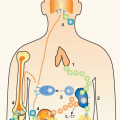
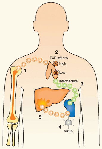
FIG. 44.1 Events Important for the Pathogenesis of Autoimmunity. 1: Lymphocyte precursors migrate from the bone marrow to the thymus to undergo maturation. 2: In the thymus of susceptible individuals, autoreactive lymphocytes can escape thymic selection. Whereas cells with high or low T-cell receptor affinity are generally deleted; “intermediate” lymphocytes fail to undergo negative selection. 3: Such initially naive cells migrate to peripheral organs such as the spleen and may remain unresponsive for many years. 4: Certain environmental triggers have the ability to activate these naive cells by bystander activation, molecular mimicry, or antigenpresenting cell cross-presentation. 5: As a consequence, activated autoreactive lymphocytes migrate to target organs such as the liver and mediate inflammation and tissue damage, leading to clinical autoimmune disease symptoms.

In conclusion, central (thymic) and peripheral tolerance mechanisms will effectively control the vast majority of autoreactive lymphocytes, which assures that autoaggressive immune responses are relatively rare (< 5% overall population). Some of the considerations described in this paragraph are illustrated in Figure 44.1.


Organ-Specific Tuning—Regulatory and Destructive Autoimmunity

As indicated previously, while autoimmune disorders must conform with a set of general criteria, organ-specific autoimmunity must be considered in the context of the target organ affected. Certain effector functions exerted by autoreactive lymphocytes will be detrimental only to particular cells or tissues. For example, the pancreatic β cells are more sensitive to the damaging effect of inflammatory cytokines than neighboring α cells (both cell types are part of the islets of Langerhans) and other cells in their vicinity (eg, fibroblasts or acinar tissue of the pancreas).94 On the other hand, some organs provide a microenvironment that suppresses inflammatory responses. For example, both the gut and the central nervous system (CNS) contain relatively large amounts of TGF-β, which can have direct anti-inflammatory effector functions on T cells and APCs, and activate Tregs, unless IL-6 is present.69,95,96,97,98 Also, certain large organs such as the liver may better tolerize lymphocyte responses directed toward them because they contain a high number of cells that are incapable of costimulation (eg, hepatocytes) and will, therefore, more likely shut down naive autoreactive lymphocytes that stimulate them.30 Finally, the precise activation state, phenotype, and effector function of an autoreactive cell is critical in determining their impact on tissues expressing their cognate antigen. Some molecules will exert beneficial functions, for example, IL-4 and probably IL-10 and TGF-β in T1D.99,100,101 Antigen-specific adaptive and professional regulatory cells are likely participants of every local autoimmune process and can therefore be essential for delay or prevention of clinical disease altogether. While their induction is a clearly therapeutic goal, phenotype and mechanistic aspects essential for regulatory function are not yet understood in complete detail. Likely, professional and adaptive Tregs may exert their function by targeted alteration of APC function. Recent evidence even suggests that Tregs may safeguard against autoimmunity by specifically killing APCs that present autoantigens to effector T cells.102 They may also act as true bystander processors if their suppressive action via APC modulation is extended to aggressive immune responses regardless of specificity found in the microenvironment of the affected organ (Fig. 44.2). Indeed, such cells have been found in several animal models,100,103,104,105 but their existence and function in humans is not well defined. Alternatively, regulatory lymphocytes may directly affect activated or naive aggressive lymphocytes and induce anergy or apoptosis. Lastly, by changing the overall cytokine milieu of a given inflammatory process, the number and function of aggressive cells and cytokines/chemokines may be dampened in a localized area.106 Probably, the best understood balancing circuit employed by autoreactive adaptive Tregs are the TH1/TH2 paradigm107,108,109 and signaling by IL-1068 and/or TGF-β.69 In light of these observations, autoreactivity is not necessarily detrimental and maybe even essential in the sense of Tregs.

Last, it might be difficult or even impossible to define a general phenotype of autoaggressive lymphocytes that will cause damage at any site or organ. Rather, autoaggression has to be defined in relation to the target organ or cell that is under
attack, and lymphocytes detrimental in one organ/disease will not necessarily be detrimental to other organs, and ubiquitous autoimmunity will therefore occur very rarely.


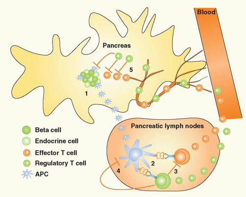
FIG. 44.2. The Concepts of Bystander Suppression and Infectious Tolerance. 1: Autoantigens are taken up by professional antigen-presenting cells (APCs) in the target organ, here depicted as the pancreas in type 1 diabetes. Upon maturation, APCs migrate to the draining lymph nodes. 2: In the lymph nodes, lymphocytes specific for distinct β cell antigens are activated by the APCs. 3: As a consequence, aggressive T cells directed toward one antigen could be regulated by regulatory T (Treg) cells specific for another autoantigen, a process termed “bystander suppression” by Howard Weiner.561 4: In theory, Tregs could also modulate APC function and foster induction of more Tregs to the same or other antigens, a process termed “infectious tolerance” by Hermann Waldman.562 5: Identical mechanisms may then regulate autoimmune responses in the target organ, after both effector cells and Tregs gain access via the circulation.


Initiating Autoimmunity: Antigens, Genes, and Environment


Antigens

A central challenge in most human autoimmune disorders is the identification of an initiating autoantigen or autoantigens. For example, several candidate autoantigens have been identified in T1D. However, to date, it remains unclear and controversial whether any are actually involved in initiation of the disease. Similar situations exist for multiple sclerosis and arthritis. Thus, distinguishing features that constitute an ideal candidate autoantigen and parameters by which potential candidates can be searched for and identified as the initiating autoantigen antigen for a given organ-specific disease must be determined with much greater precision to yield insights applicable to preventative or therapeutic interventions.

The concept that “processing of antigens determines the self” has been introduced by Sercarz.110,111 Autoantigens that are not expressed in the thymus or are of cryptic nature110,112,113 may encounter a more extensive T-cell repertoire that is likely of higher avidity. Such antigens appear to be better targets. Low levels of thymic antigen expression or limited numbers of thymic medullary cells may lead to partial tolerance.114,115 In addition, the precise timing of antigen expression during embryonic development may play a major role for the establishment of tolerance. Antigens that are cryptic or not expressed during early embryonic development might become better targets later because more autoreactive T cells will be present in the periphery. It should be considered that certain “embryonic antigens” are involved in initiating autoimmune diseases.116

Of further importance are the constraints with which particular antigens can be presented to the immune system in a given target organ. Exogenous uptake of soluble antigens usually results in processing via the MHC class II presentation pathway. Such antigens may also be processed through the MHC class I pathway by dendritic cells, a mechanism termed cross-presentation, if the antigen itself was originally expressed by a cell different from the APC.117,118 Cross-presentation appears to be less efficient than direct endogenous presentation of antigens through the MHC class I pathway. At this point, it is not clear which properties will make an antigen a more efficient candidate for cross-presentation. The class I pathway may be important in the effector phase for many autoimmune disorders because CD8+ T cells with cytotoxic function (cytotoxic T lymphocyte [CTL]) can be induced in this fashion, and CTLs have profound detrimental effector functions (interferon [IFN]γ and TNF-α secretion, lysis of target cells).119 Clearly, cross-presentation can lead to immunity, as well as tolerance, and in this way propagates or, alternatively, halts autoimmune disease.117,120 It is less likely that autoantigens that are not presented by professional APCs or within lymphoid organs after their release from target cells/organs can initiate autoimmunity. Only under special circumstances (ie, extremely high-density presentation of an antigen on a target cell or highly inflammatory conditions including chemotaxis) would this appear possible. This might occur during localized or systemic viral infections and could also be an important event in lymphocyte entry into target organs, especially if chemotaxis is involved and endothelial cells present autoantigens.121 In addition, experienced or primed autoaggressive lymphocytes will be capable of becoming activated after seeing antigen in the absence of costimulation; thus, accumulating a memory pool of autoaggressive T cells might be inherently dangerous to an organism.

Thus, the type of autoantigen will define its own potential “candidacy” in an autoimmune process. Autoantigens
that are not expressed during development, or were cryptic for an extended period of time, as well as antigens secreted and cross-presented in lymphoid organs by APCs, appear to be better suited for assuming the role as primary culprits.


Genes

An abundance of empiric evidence indicates the association of many organ specific autoimmune diseases with certain human leukocyte antigen (HLA) haplotypes as well as other susceptibility or protective genes. The principal specific genetic linkages will be discussed separately for each condition in part II. In general, MHC class I (such as HLA-B27)122,123 or class II (such as DR4) genes might predispose to a certain disease by enhanced presentation of pathogenic peptides in the periphery or inefficacious presentation of autoantigenderived peptides in the thymus. For example, the human HLA-DQ8 molecule has a striking structural similarity to mouse I-Ag7 class II that predisposes NOD mice to spontaneous autoimmune diabetes.124,125 However, the link to disease appears not to be as simple as reasoned previously, and more complex mechanisms might be in place. Presentation of the pathogenic peptide in gluten-induced celiac disease involves acidic modification of the protein to generate the peptide ultimately presented by MHC class II,126 and other chemical modifications can be expected to alter peptide binding to MHC and the resulting conformation of the peptide/MHC complex.

Other genes that encode immunoregulatory or inflammatory proteins may be involved in the disease process, and, finally, genes that support tissue or wound repair (eg, islet cell regeneration in T1D) may be of help in preventing disease development.127 For most autoimmune disorders, the genetic links are complex, not absolute, and many susceptibility and resistance genes act in concert to modulate the clinical phenotype. Noteworthy, newly emerging themes in the genetics of autoimmunity are the fact that small nucleotide polymorphisms might result in alternate forms of the same gene and the fact that the character of the full array of MHC region genes is becoming understood.128 A striking example for the former is the existence of an alternate spliced form of the inhibitor of cellular function, CTLA-4. Kuchroo and colleagues discovered that the presence of a secondary CTLA-4 lacking the B7.1/B7.2 binding motif can be instrumental in the genetic penetrance of diabetes pathogenesis.129 An important area still largely unexplored is the nature of genes responsible for transcriptional control of other proteins. Experimental models indicate that transcription factors can have profound effects on the development of autoimmune disease, and variations in expression and activity levels may be found between susceptible and protected individuals. In summary, a complex interplay of many genes will predispose for a certain autoimmune disease, but the concordance of clinical manifestation is frequently not higher than 30% to 40% in monozygotic twins.130,131,132,133,134,135,136 For this reason, other factors are to be considered in triggering or propagating the pathogenic autoimmune process.

Our understanding of the genetic component to autoimmunity has greatly benefited from the arrival of a first wave of so-called genome-wide association studies (GWAS) in the past 5 years. Prior to the use of this technology, genetic contribution was analyzed by two different approaches: linkage analysis and association studies. Linkage analysis tests for correlation between a genomic region and phenotype in families, whereas association analysis searches for correlations in the entire population.137 In a genetic association study of autoimmune disease, sequence variants are genotyped in patients, and controls and the frequencies of these genotypes are compared. It is then evaluated whether certain alleles are significantly more associated with patients than with controls. Most autoimmune conditions are polygenic diseases, meaning that they are caused by (a range of) combinations of polymorphisms within different genes. As mentioned previously, HLA-associated risk was discovered early in many autoimmune disorders and turned out to be reproducable because of its highly significant association. The vast majority of common genetic variants in autoimmunity, however, display modest effects. Even extremely large linkage studies have usually been unsuccessful in discovering susceptibility genes for polygenic diseases that met stringent statistical thresholds. Likewise, most common variants require large association studies for reliable detection. Advances in the development of high-throughput genotyping, statistical correlation methods, and better insight into the organization of the genome offered the opportunity to design approaches for the unbiased interrogation of the entire genome.138 Sufficiently large case-control GWAS thus have the potential of identifying disease-associated mutations, even if the contribution of that polymorphism is minor. It should be noted here that GWAS often cannot unequivocally identify gene associations (at best “candidate genes”), as risk loci contain multiple genes, and, therefore, follow-up fine mapping or functional studies are generally required. Although GWAS have rapidly increased the list of known risk loci for many autoimmune disorders, some have questioned whether this knowledge substantially aids our understanding of their etiology and offers clues for rational intervention. It could be argued that all associations are often eclipsed by the major contributing factor, HLA polymorphisms, and essentially pinpoint genes with key roles in the immune system that could be expected to be involved to some degree. In T1D, for instance, minor associations with important genes encoding molecules involved in immunity such as CTLA-4 and IL-2 were discovered. From a therapeutic perspective, such findings may be hard to translate as, based on the relatively small association rates, these genes and their products may not necessarily represent pivotal targets in a diverse human patient population. In summary, it can be concluded that most likely all major susceptibility genes associated with the most common autoimmune disorders as discussed in the following text are currently known. Future years will tell whether this comprehensive genetic dataset translates to a better understanding of the origins of autoimmunity and if this will open new avenues toward better treatements.


Environment

Viral Infections. For many years, viral infections have been discussed as potential candidates to trigger autoimmunity in susceptible individuals because of their capacity to directly
infect target tissues and induce strong inflammatory responses and immune activation. While the association between viral infections and organ-specific autoimmune disorders is an intriguing possibility, it has been exceedingly difficult to demonstrate a causative role for specific viruses in human autoimmune diseases. Among the many obstacles are the fact that 1) all individuals undergo a multitude of viral infections during their lifetime; 2) one has to assume that viruses are frequently cleared at the time of diagnosis and viral footprints can be difficult to find in individuals affected by an autoimmune disease (“hit and run” event); 3) the precise viral strain, infection kinetics, and number of T cells and type of effector functions induced may play an instrumental role in determining its effect in an individual genetically at risk, necessitating a very detailed immunologic profiling139; 4) due to MHC polymorphism, there is a significant variation in specificity of the antiviral response; 5) viral infections might not, per se, trigger autoimmunity but affect an ongoing autoimmune process in a detrimental way; and 6) there now is increasing evidence that viral and other140 infections may rather prevent than enhance ongoing autoimmune responses, either by apoptosing aggressive T cells or by augmenting the Treg pool.141,142 Thus, a successful approach should be to first explore the underlying mechanisms in virally induced autoimmunity and then apply the precise insight and paradigms developed specifically to the human situation.

The most important mechanisms to be considered are listed here and are summarized in Figure 44.3.


  • Molecular mimicry, which implies the cross-reactivity between viral and self-determinants as a principle cause or mechanism to enhance autoimmunity143,144


  • Bystander activation, which postulates that APCs and autoreactive lymphocytes will become activated indirectly as a consequence of the cytokine/chemokine by virus infection of a particular organ145


  • Virally induced determinant spreading, which involves the presentation of autoantigens (possibly previously cryptic) by virus-activated APCs146,147,148

The experimental evidence for all of these three scenarios obtained in different mouse models in vivo is well documented. However, none of these have been proven for any human autoimmune disorder due to the large size of human trials and the invasive nature of in vivo diagnostics required at the present stage. Thus, in the near future, we will continue to depend on animal models until noninvasive human in vivo diagnostic strategies have advanced and allow for imaging of trafficking of antigen-specific lymphocytes and high-resolution definition of immune process present in a specific organ. A final remark should be made for the existence of a negative association between viral infections and autoimmune disease found in several experimental models.149 These observations are in support of epidemiologic findings that the incidence of many autoimmune disorders is decreased in equatorial countries, where the presence of certain infectious diseases is significantly increased. However, no firm associations have been established to date.

Other Environmental Causes. Similar to viral infections, other inflammatory stimuli may trigger or enhance autoimmunity. The gut deserves particular attention in this respect. At this site, each individual harbors thousands of different bacterial strains, and viral infections are common. Furthermore, the mucosal lining is permeable for nutrients and constitutes a very large interactive surface with the environment. Again, the complete absence of all bacteria results in severe immune dysfunction and possibly autoimmunity. However, it would be incorrect to conclude that infections are therefore always protective. Indeed, the commensal flora appears crucial in maintaining proper immune activation and function, but certain pathogens could definitely elicit strong gut immune responses that lead to autoimmune disease.150,151


Regulatory Circuits in Autoimmune Processes


Cytokines

Cytokines and chemokines are essential regulators of cellular and humoral immune responses and lymphocyte trafficking.152,153,154 They play a central role in orchestrating autoimmune processes and constitute a multitude of positive as well as negative feedback loops.155 It is well established that certain cytokines can negatively or positively influence the production of other cytokines (ie, the Th1/Th2/Th17 paradigm) and thus determine the balance between proinflammatory and anti-inflammatory factors in the local environment. Furthermore, autocrine production feedback can augment or shut down production of a given cytokine by one cell. Cytokine networks operate with a fair amount of redundancy, and many cytokines and chemokines share common receptors. They are the most likely mediators of “bystander activation” and “suppression” processes and also offer an effective and versatile therapeutic target via the temporally restricted use of cytokine- or chemokine-blocking antibodies. Their precise function can vary quite dramatically in respect to the autoimmune disease under investigation and will be discussed in the section on Individual Autoimmune Disorders. Further, their level and timing of expression during an ongoing disease process will determine whether they have a positive or negative effect (or any at all; see the following discussion).

Many autoimmune disorders have traditionally been classified as “Th1-” or “Th2”-driven disorders based on, among other factors, the pathogenic role of cytokines such as IFNγ and IL-4.107 Recent years have challenged this perspective with of the description the Th17 helper subset, characterized by the production of IL-17.156,157,158 This class of T cells is considered highly proinflammatory, and its discovery was found to be of importance in several autoimmune disorders in both animal models and humans.159 One example of an autoimmune disease where Th17 cells have taken center stage is RA. The disease’s principal animal model was long referred to as a Th1 driven, but the field struggled with the observation that mice treated with neutralizing antibodies to IFNγ- and IFNγ receptor-deficient mice developed more severe arthritis.160,161 Dominance of Th17-directed immunity was put forward when IL-17 knockout mice were found
to develop less arthritis,162 and treatment with neutralizing antibodies to IL-17 or soluble IL-17 receptor alleviated joint inflammation.163 In humans, it was found that IL-17 is increased in RA sera and synovial fluid and is present in the T-cell rich areas of the synovium.164 Furthermore, IL-17 has profound inflammatory effects on a range of cell types within the joint. Several clinical trials are underway to either block IL-17 or the cytokines that govern the generation of Th17 cells such as IL-6.


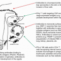
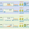
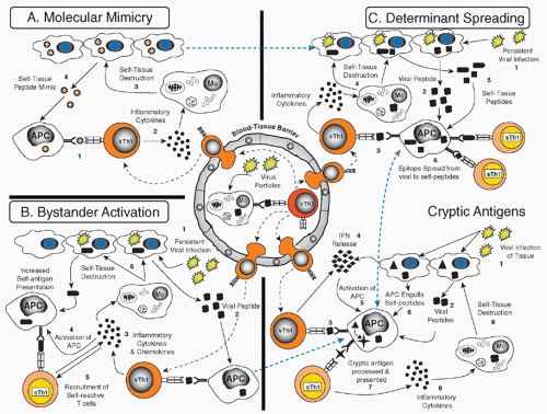
FIG. 44.3. Potential Mechanisms of Autoimmune Disease Induction. After a viral infection, activated virus specific T-helper cells (vTH1) and cluster of differentiation 8 cytotoxic T lymphocytes will migrate more readily through blood-tissue barriers to infected and noninfected organs. A: Molecular mimicry describes the activation of cross-reactive lymphocytes that recognize the original viral epitope and a self-epitope (1), which leads to the release of cytokines and chemokines (2), which enhance local inflammation, activate antigen-presenting cells (APCs), and indirectly or directly cause tissue destruction (3) and spreading of the autoimmune process (4). B: In the epitope spread model, persistent viral infections (1) could result in the activation of virus-specific T cells (2,3), which cause tissue damage by killing virally infected cells (4), leading to the release (5) and (cross-)presentation of more autoantigens (6). C: The bystander activation model describes the nonantigenspecific activation of autoreactive T cells. Infiltration by virally specific T cells (1,2) leads to inflammation and upregulation of immunity throughout the tissue (3,4), involving the activation of APCs, which now differentially process autoantigens (also de novo or previously cryptoic antigens). This can lead to activation of lymphocytes by T-cell receptor (TCR)-specific or TCR-independent mechanisms (5), which can then cause tissue damage (6). The cryptic antigen model describes the initiation of autoimmunity by differential processing of self-antigen/peptides, which can occur under inflammatory conditions. After viral infections, interferons (1) are secreted by antiviral T cells and infected cells (2-4). APCs are activated in this way (5), which enables them to engulf self-peptides (6, triangle) or to differentially process endogenous autoantigens. Cytokines can activate proteases more strongly, which might result in the presentation and processing of previously cryptic autoantigens (7-9). These displayed pathways are not mutually exclusive and are probably operational at different levels in many autoimmune responses. Currently of high interest is the presentation of neoantigens and strategies to define them. Courtesy of Steve Miller and Ludovich Croxford, Northwestern University, Chicago, IL.


Apoptosis

It appears to be a general paradigm of great functional consequence that activation of the immune system is followed by a process that reverses the activation and reestablishes
homeostatic baseline levels of immunity.88,90 In the absence of such regulatory mechanisms, immune responses will overshoot their goals and excessive immunopathology will occur. Thus, AICD is believed to play an important role in regulating autoimmunity. Apoptotic lymphocytes, for example, are easily detected in islet infiltrates in T1D,165 and targeted induction of limited apoptosis may even prevent onset of autoimmune disease.166 While increased apoptosis of aggressive lymphocytes that exceeds the “supply” of newly activated cells may directly limit an ongoing autoimmune process, limited apoptosis of target tissues may indirectly facilitate induction of protective regulatory responses. On the other hand, while apoptosis of target cells should at best be limited, decreased apoptosis of autoreactive aggressive lymphocytes will propagate autoimmunity. In RA, for instance, insufficient apoptosis of synovial macrophages, fibroblasts, and lymphocytes is one mechanism that might contribute to persistence of the disease and lead to synovial lining hyperplasia.167 It is therefore important to consider precisely which cells undergo apoptosis in order to predict the possible outcome.


  • If too many target cells die by apoptosis, organ destruction occurs more rapidly; however, at the same time, antigens releases from apoptotic cells appear to propagate tolerance rather than immunity.166,168


  • If Treg cells die by apoptosis, autoimmunity will be enhanced.169


  • If aggressive lymphocytes die by apoptosis, disease should be ameliorated. However, because they have to first be activated, they might induce organ damage during their activation phase.

Thus, an ongoing autoimmune process can be viewed as a rather fine-tuned and fragile equilibrium of aggressive and regulatory components, and the precise activation kinetics and survival times of all lymphocyte types implicated in the process will determine the outcome. We are, at present, unable to delineate the precise in vivo cellular kinetics, and a more thorough understanding will require improved noninvasive diagnostic techniques.


Kinetics

One of the most important emerging areas for an improved understanding of the pathogenesis of autoimmunity is concerned with the kinetics of immune responses. The pathophysiologic or therapeutic effect of a given lymphocyte population depends not only on specificity, activation state, and effector functions but is also a function of the timing during which phase of an ongoing disease process it is present. Indeed, inflammatory cytokines such as IFNγ or TNF-α exhibit opposing effects in T1D depending on the precise time point of generation.170 Early expression enhances islet destruction and disease development, whereas late expression ameliorates disease by inducing apoptosis of autoaggressive cells. These kinetic issues constitute a major obstacle for successful immune intervention because they preclude the use of specific blocking agents or administration of cytokines without precise knowledge of their kinetically differential role in the disease process. Figure 44.4 illustrates these kinetic considerations in relation to target cell destruction. A better understanding of the underlying “autoimmune kinetics” is essential and treatments will likely have to be individualized, in particular for antigen-specific immune-based interventions.


Therapeutic Considerations


Efficacy, Specificity, and Undesired Effects

Treatment of autoimmune disorders is not that different conceptually from cancer therapy. A fine balance must be found between efficacy of the intervention and acceptable undesired effects. The main goal of autoimmune disorder therapy is suppression of the pathologic autoimmune response. Therapeutic options range in principle from continuous immunosuppression of the entire immune system to specific, targeted, temporally limited, and local immunosuppression. Systemic immunomodulation or anti-inflammatory therapy will affect the entire immune system and may compromise the immune status of the individual. One of the more succesful examples for effective systemic treatments is the blockade of TNF-α to ameliorate RA.171,172 This new class of drugs is especially valuable because TNF blockade effects inflammatory pathways distinct from the targets of conventional anti-inflammatory therapy with corticosteroids or nonsteroidal anti-inflammatory drugs. TNF inhibitors can thus be used in patients that are refractory to the latter treatments with the added advantage of considerably improved outcome in terms of structural bone erosions. The remarkable therapeutic success of these drugs has since been replicated in other autoimmune conditions such as Crohn disease, ulcerative colitis, ankylosing spondylitis, and psoriasis. Whereas these diseases target different organs, common therapeutic improvements upon TNF blockade thus indicate shared mechanisms in terms of cytokine signaling. While undesired effects are relatively low, SLE-like symptoms have been observed in a few patients, as has the enhanced susceptibility to tuberculosis. Another important disadvantage, in addition to the high cost of these biologics, is that not all patients respond to TNF blockade. This has spurred a search for more potent blockers of inflammation, with the most promising developments stemming from the design of Janus kinase inhibitors for treatment of RA.173 This class of small molecular drugs that interrupt signaling downstream of multiple rather than individual cytokines has recently cleared phase III trials in RA and is expected to revolutionarize the therapeutic landscape. While indications exist that this approach is at least safe in the short-term and that it outperforms anti-TNF biologics, long-term safety profiles obviously remain to be determined.

Autoantigen-specific immune interventions, in contrast, bear the promise of lower systemic side effects, as they can be targeted to antigens that are exclusively expressed in the diseased organ.174,175 However, the efficacy might be lower and suitable target antigens have to be chosen carefully, because enhancement of autoimmunity is an important concern. The goal is either deletion of aggressive autoreactive T cells or induction of regulatory cells.176 To achieve the latter, response modifiers are probably required at the time
of immunization in order to skew the resulting immune response to exert regulatory effector functions. Deletion of autoaggressive lymphocytes or anergy induction is even more risky, as only suboptimal immunization (ie, in the absence of costimulators) will result in this outcome. To control this in vivo is rather difficult. Ultimately, antigen-specific therapy will likely have to be individualized due to MHC polymorphism and distinct T-cell repertoires between individual patients and should be combined with other systemically acting agents, for example, antibodies against CD3 or CD4 or costimulation blockade. Indeed, there is intriguing evidence along these lines in animal models: Combination of a non-Fc-binding anti-CD3 antibody with mucosal immunization of insulin or insulin-derived peptides exhibited clear synergy and enhanced efficacy in reversing recent-onset diabetes in the NOD.177 In this case, induction of insulin specific adaptive Tregs and their effector function were enhanced and protection by these Tregs was transferable and highly effective in that it reversed recent-onset T1D.


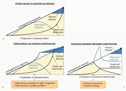
FIG. 44.4. Kinetic Issues in Autoimmune Diseases. A: Clinical manifestation of autoimmunity is the consequence of a dysequilibrium between protective (regulatory) and aggressive (effector) responses. An important consideration is that the destruction of the target cell or organ will usually lag somewhat behind the peak of the aggressive responses, because organ regeneration is common. B: Inflammatory stimuli will augment the effector arm of the autoreactoive response resulting in more rapid disease development. C: In contrast, induction of regulatory T cells can delay or dampen organ destruction.


Promising Targets

For anti-inflammatory interventions, the factor to be targeted should be as disease specific as possible. Therefore, blockade of TNF works well for RA but not for diabetes or MS.178 Experimental evidence supports this observation because TNF is a crucial mediator found to be elevated in affected joints,172 but has clearly positive, as well as negative effector functions in murine models of MS and diabetes.179,180 Targeting ubiquitously present chemokines or cytokines will likely not bear much success because of the resultant generalized immune modulation or suppression.

For antigen-specific interventions, antigens that are already targeted by regulatory autoreactivity are likely to constitute good targets to augment such a preexisting response.181 In contrast, these antigens should not be selected to delete autoreactive cells, which results in a loss of regulation. For anergy or apoptosis induction, antigens targeted by a primarily aggressive response will be better suited. The fact that autoantigenic and epitope spreading occurs during each ongoing autoreactive process makes such interventions difficult to design, and individualization will likely be necessary.


Reestablishment of Tissue-Specific Immune Regulation

One of the factors that pose a challenge to understanding the pathogenesis of distinct autoimmune diseases may also hold a clue to developing effective and specific treatment strategies. Each target cell, tissue, or organ exhibits specific features that distinguish it from other sites of the body. These site-specific features of autoimmunity will
likely offer unique target sites for interventions with lower systemic side effects. However, one concern is that reestablishing proper immune homeostasis and regulation in one organ may still affect homeostasis systemically or at another site. Therefore, thorough preclinical evaluation and careful monitoring of undesired effects is urgently needed. Ideally, treatments have to be administered before complete organ destruction has occurred in patients identified by genetic or other screening to be at risk to develop full-blown clinical disease. During the preclinical state, frequently regulatory autoreactive responses are still strong and their augmentation can result in protection (Fig. 44.5). During advanced stages of autoimmunity, Treg function or susceptibility of effector T cells to regulation might decrease.182 The goal to reestablish homeostasis and proper regulation after an initial insult that caused organ-specific inflammation appears a natural countermeasure to which the specific immune system may be successfully harnessed. An intriguing example is the emerging concept of organ repair that can potentially be enhanced by drug therapy. In T1D, regeneration and replication of β cells can possibly occur after giving exenatide, a GLP-1 agonist, which is an established intervention in type 2 diabetes and under investigation in combination with other drugs such as anti-CD3 in T1D.


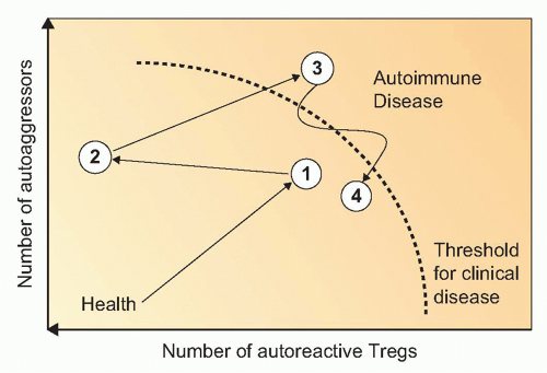
FIG. 44.5. Development of Autoimmune Disease as a Function of Aggression versus Regulation. Following induction of disease (1), distinct numbers of autoaggressive and autoreactive regulatory T cells will be generated and activated. Undulations between protective states and diseases states follow (2, 3) (ie, the so-called honeymoon phase in T1D), which can eventually lead to an irreversible state of clinical manifestation of disease (4). An additional important component to factor in is the ability of the target organ to regenerate.


INDIVIDUAL AUTOIMMUNE DISORDERS

A comprehensive and balanced discussion of autoimmune and autoinflammatory diseases at large is well beyond the scope of this chapter. Our choice of individual diseases for this section of this chapter was guided by their relevance to human health (ie, disease prevalence and severity), recent insights into pathogenetic mechanisms, as well as the development of novel treatment modalities. Given these limitations, we extend our apologies to all those scientists and clinicians whose work on autoimmune disorders is not mentioned in our discussion.


Endocrine Autoimmune Diseases


Thyroid: Graves Disease and Hashimoto Thyroiditis

Introduction and Disease Description. The year 1956 was a seminal year for the field of human autoimmunity given the discoveries of Hashimoto thyroiditis as an autoimmune disease and of Graves disease as caused by an autoantibody. These discoveries have prompted some straightforward and relatively uncomplicated treatments.183,184 In Graves disease, autoantibodies directed against the thyroid-stimulating hormone receptor on thyroid cells stimulate excessive production of thyroid hormone, which is normally controlled by feedback regulation. In contrast, in Hashimoto thyroiditis, autoantibodies to thyroid peroxidase and thyroglobulin are present over years and likely able to fix sublytic doses of complement to cells of the thyroid. The result is an inflammatory reaction, which is also associated with T cell-mediated cytotoxicity. Thyroid damage due to painful Hashimoto thyroiditis may be associated with the development of Graves disease, indicating that there is a tendency for spreading of the autoimmune reaction in humans. Treatment of thyroiditis is relatively straightforward with antithyroid drugs (methimazole) and radioactive iodine.

Autoimmune Features. Autoantigens targeted in thyroiditis are thyroid peroxidase, a cell surface protein (Hashimoto thyroiditis), and the thyroid-stimulating hormone receptor (Graves disease). Autoimmune responses to thyroglobulin are also seen in animal models. The B-cell epitopes to these autoantigens have been mapped relatively well; however, as it is the case for other human autoimmune disorders, T-cell responses, their tracking and specificity, as well as their eliciting antigens in humans have remained largely elusive.183 An interesting genetic link has been established to the CTLA-4 region, which is supported by the finding that NOD mice that also exhibit a link to this gene may present with thyroiditis. The usual suspects (ie, APC dysfunction, autoaggressive lymphocytes, and links to viral infections) have been examined, but no conclusive etiologic or mechanistic evidence has been obtained to date. It is noteworthy that a role for Treg cells has been established in Mason’s animal model for thyroiditis.185

Genetic Features and Environmental Factors. Interestingly, numerous viruses have been implicated in the pathogenesis of different thyroid diseases, but firm evidence for a direct involvement of viruses or virus-induced immune responses leading to clinically manifest disease is scarce. Subacute thyroiditis is a clinical and pathologic form of thyroid involvement that appears after infection with viruses such as measles, influenza, adenovirus, Epstein-Barr virus, and Coxsackie virus.186 Again, however, a causative role in vivo has not been shown for any single infectious agent.187 Presence of viral material in the thyroid and elevated virus-specific antibody titers were found to correlate with subacute thyroiditis.
In other instances, direct virally induced thyroiditis has been documented epidemiologically with thyroid or parathyroid disease.

Retrovirus, in particular human immunodeficiency virus (HIV) infections have generated much interest. Although HIV infection and acquired immunodeficiency syndrome (AIDS) may affect multiple endocrine organ systems,188,189,190,191 thyroid dysfunction usually reflects weight loss, anorexia, and cachexia of advanced HIV disease, rather than a direct viral effect on the thyroid.192,193 Thus, direct involvement of the thyroid by HIV or by opportunistic infections is uncommon and may include subclinical hypothyroidism and “euthyroid sick syndrome.” The clinical relevance is probably limited, as overt hyperthyroidism or hypothyroidism does not occur with greater frequency in patients with HIV and AIDS as compared to patients with other nonthyroidal illnesses (HIV and the thyroid gland have been reviewed in Heufelder and Hofbauer192). Involvement of the parathyroid in patients with AIDS could be shown by reduced basal and maximal parathyroid hormone levels, but the mechanisms underlying these findings have not yet been elucidated.194 To account for a possible role of HIV in thyroid autoimmunity, a 66% homology between the HIV-1 nef protein and the human thyroid-stimulating hormone receptor has been noted. However, reactivity of sera of patients with Graves disease against a nef peptide showed no significant differences as compared to normal controls.195 This does not rule out the presence conformationally shared T- or B-cell epitopes with HIV proteins. In analyzing mechanisms of molecular mimicry, studies of potential antigenic surfaces have emerged as an important supplement to analysis of sequence similarity.196

In addition, antibodies from a patient with Graves disease showed reactivity to the gag proteins of another retrovirus, human foamy virus.196 The association of human foamy virus with Graves disease or subacute thyroiditis is controversial. Whereas one study demonstrated human foamy virus-related sequences in the deoxyribonucleic acid (DNA) of peripheral blood in two-thirds of patients with Graves disease but none in normal controls,197 another study could not confirm these findings.198

In other studies, an association between human T-lymphotropic virus I and II and the occurrence of autoimmune thyroiditis or Graves disease was reported.198,199,200,201 Further, as in the case of HIV, hepatitis C virus was shown to lead to a wide variety of autoimmune disorders including involvement of the thyroid gland.202,203,204,205,206 Moreover, treatment of chronic hepatitis B and C with IFNα leads to induction or enhancement of autoimmune disease.207,208,209 For congenital rubella infection, which has been associated with diabetes mellitus, Addison disease, growth hormone deficiencies, as well as thyroid disorders,210 it is not clear whether thyroid involvement is the result of a direct viral effect or a more generalized dysfunction of the immune system.187

Animal Models. Animal models in mice and chicken have been used to study virus-induced thyroiditis.187 Mice persistently infected with LCMV showed reductions of thyroglobulin messenger RNA and circulating thyroid hormones in the absence of thyroid cell destruction.211 Thyroiditis characterized by focal destruction of the follicular structures, inflammatory infiltration, and generation of antibodies against thyroglobulin and thyroid peroxidase was observed in a reovirus type 1 mouse model of thyroiditis.212 The reovirus gene responsible for autoantibody induction was identified and the encoded polypeptide shown to bind to tissue-specific surface receptors.213 Spontaneous lymphocytic infiltration of the thyroid is observed in the obese strain of chickens. Such chickens express an endogenous retrovirus, avian leukosis virus (ev22), not found in healthy normal inbred strains.214 Although ev22 appears to be a genetic marker rather than cause for thyroiditis, infection of normal chicken embryos with avian leukosis virus can cause hypothyroidism.215 Moreover, aberrant MHC class II expression is demonstrated in obese strain chickens and elevated levels of 2.5-oligoadenylate synthetase as well as 2,5 oligoadenylate-polymer levels in the cytosol of thyroid epithelial cells occur, suggesting viral involvement.216 Again, not all cross-reactivities with self-ligands need to increase autoimmunity,217 and regulatory cells also play a major role in modulating autoimmune thyroiditis.185


Endocrine Pancreas: Type 1 Diabetes

Introduction and Disease Description. While the distinct symptoms of diabetes mellitus have been known since antiquity, the underlying pathophysiologic processes were only identified in the late 19th and early 20th centuries. The proof of the involvement of the pancreas in diabetes etiology was conducted by von Mering and Minkowski,218 who demonstrated in 1890 that extirpation of the canine pancreas results in the classic symptomatology of hyperglycemia, abnormal hunger, increased thirst, polyuria, and glycosuria. Subsequently, inflammatory changes in the endocrine pancreas (ie, the islets of Langerhans) were correlated with diabetes by Schmidt219 in 1902; two decades later, Banting and Best220 identified insulin as a pancreatic hormone, thereby providing the basis for insulin substitution therapy, which remains to this day the cornerstone for T1D management. In a classic 1965 paper, Gepts221 noted the histopathologic similarity between thyroiditis and insulitis and suggested an immune basis for the disease. By 1974, the concept of T1D as an autoimmune syndrome was firmly established by the discovery of islet cell antibodies and an association between T1D and certain HLA genes.222,223,224

Autoimmune Features. Today, a quarter century later, the possible autoimmune origin of T1D is understood in much greater detail. However, the lymphocytic infiltration of the islets of Langerhans (Fig. 44.6) and the presence of antibodies specific for β-cell antigens associated with the progressive destruction of insulin-producing β cells225,226 still constitute the cardinal evidence for an autoimmune etiology. While there is a reasonably strong genetic linkage to certain HLA molecules, the disease has to be considered polygenic in nature,130 and a significant discordance of disease among monozygotic twins suggests that environmental factors contribute to trigger and/or exacerbate the disease. Furthermore, it remains unclear which islet antigens are the
primary targets. The earliest islet cell-specifc antibodies in human individuals at risk are directed to insulin,227,228 and evidence from relevant animal models and human studies points toward insulin as a primary antigenic target,229 islet-specific glucose-6-phosphatase catalytic subunit related protein,230,231 and possibly glutamic acid decarboxylase (GAD),232 but definitive proof for a pathogenic role has only been ascertained in the NOD mouse model and not yet in humans.233 The cellular infiltrates found in the islets contain both CD4+ and CD8+ T lymphocytes and their irreducible role in β-cell destruction has been documented in several animal models.234,235,236 CD8+ T cells can exert direct cytotoxic effects toward MHC class I-expressing β cells, while CD4+ T cells secreting inflammatory cytokines and can provide help to CD8+ T cells as well as B cells. Ultimately, it is important to consider that β cells constitute about 60% of the islets, which in turn contribute only -2% to the pancreas mass and demonstrate, unlike many other tissues targeted in autoimmune disorders, an exquisite sensitivity to cytokines such as IL-1, TNF, and IFNs that will result in their apoptotic cell death after prolonged exposure.94


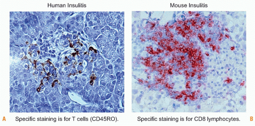
FIG. 44.6. Human and Mouse Insulitic Lesions. Comparison of human insulitis (Left), courtesy of Francesco Dotta (University La Sapienzia, Rome, Italy), and mouse insulitis (Right), from a rat insulin promoter-lymphocytic choriomeningitis virus mouse 14 days postinfection with lymphocytic choriomeningitis virus (von Herrath laboratory, La Jolla Institute for Allergy and Immunology, La Jolla, CA).

The detection of islet antigen-specific antibodies remains an essential tool in identifying prediabetic subjects and monitoring the progression of subclinical and clinical disease. Procedures for autoantibody determination have been substantially refined and standardized worldwide. Emerging data from clinical studies support the notion that with progression of the prediabetic phase, generation of islet antibodies also is increased.237,238,239 Usually, antibodies to insulin become discernible first, then to GAD, thereafter to insulinoma antigen 2. Individuals with islet antibodies to three or more distinct antigens have a greater than 90% risk of developing T1D.136 Thus, islet antibodies are an excellent marker for disease risk. However, they appear not to play a role pathogenetically, as transfer of antibodies from mothers to children does not increase the risk for T1D, and B cells are not needed for human diabetes.240,241,242 In this crucial respect, the NOD mouse appears to provide a paradigm that might not be applicable to human diabetes, as maternal antibodies are an essential factor for diabetes development in NOD offspring.243

Measurements of human T-cell responses to islet antigens are not yet standardized and can vary considerably between different laboratories. One reason for this may be the source of T cells that are generally subjected to specificity analyses: blood-borne CD4+ or CD8+ T cells may not reflect the specificity distribution and frequencies of isletspecific T cells found in the target organ (ie, pancreas and its draining lymph nodes). Even the study of spleen-derived islet-specific T cells readily obtained from NOD mice and analyzed in standardized proliferation assays has shown variations between different NOD colonies.244 Therefore, measurement of multiple effector functions (ie, cytokine production, etc.) in highly standardized assays will likely be required to assess T-cell autoreactivity on a routine basis.245 A recent study shows that while techniques such as enzyme-linked immunosorbent spot can discriminate between patients and controls, difficulties in detection of low-frequency antigen-specific T cells probably leads to the limited reproducibility of such measurements.246 Another complicating fact is that T-cell responses between individuals are expected to vary and depend on the HLA haplotype and individual trigger(s) that precipitate T1D. An emerging technology that may improve specificity and sensitivity is through the use of combinatorial HLA multimers loaded with multiple disease-associated epitopes.247 A remarkable paper248 has shown that tracking of immune responses to naturally processed peptide epitopes can discern between healthy individuals and those afflicted with T1D: Whereas the number of overall T cells reacting with the naturally
processed peptide epitopes was similar, individuals with T1D produced more IFNγ relative to IL-10, whereas the ratio was the opposite in healthy individuals.

Genetic Features and Environmental Factors. Recent data show that concordance rates among monozygotic twins are higher than previously thought.249 Still, the remaining discordance and particularly the major temporal differences in clinical onset indicate that environmental factors have to act in concert with diabetes susceptibility genes to orchestrate the autoimmune destruction of β cells. The initial hope that only a few genes would contribute to disease pathogenesis and that genetic links would help to directly understand the mechanistic aspects of T1D pathogenesis has been progressively eroded. Instead, a complex network of susceptibility and resistance genes in both humans and animals (eg, the NOD mouse) has slowly taken shape. The recent publication of three GWAS has extended the list of gene associations in T1D. So far,16 robust T1D-associated loci were discovered, some of which encompass important immunity genes (eg, IL-2, IL-2Rα [CD25], CTLA-4) or β cell-specific products (insulin), while other candidate genes do not have an apparent link to immune functions and/or β cells. The implications of this enhanced genetic characterization have so far not led to the identification of monogenic pathways that directly lead to disease. Rather, consensus is growing that disease susceptibility is the consequence of a global problem of immune regulation, with many genes involved in a variety of combinations in individual patients.250

Many of these genes exhibit direct parallels to NOD diabetes susceptibility genes.251

The association between particular HLA/MHC class II haplotypes and the occurrence of human diabetes has been of particular interest. The DRB1/04-DQA1/0301/B1 and DRB1/03-DQA1/0501/0201 strongly predispose to T1D, and more than 80% of patients carry either one or both alleles.252 In contrast, other MHC class II haplotypes can protect from disease, as evidenced by the sixfold reduced risk to T1D in DRB1/15-DQA1/B1/0602-bearing individuals. Considerable evidence indicates that other genes in the MHC region likely contribute significantly to T1D risk.128

An intriguing mechanistic hypothesis was put forth by McDevitt’s observation that predisposing HLA class II alleles appear to express small neutral amino acids at position 57 of the DQ allele of Caucasoid populations, whereas an aspartic acid is found in resistant alleles at the same position. Because position 57 is part of the peptide-anchoring pocket, amino acid substitutions in this area will affect peptide binding. Indeed, the susceptibility alleles prefer different peptides, but the contribution to T1D development is not yet clear and a mechanistic link has to be established. Both central and peripheral tolerance mechanisms have been implicated but no direct proof has been obtained. It is important to realize that the human susceptibility MHC class II alleles share amino acids at position 57 with the I-Ag7 alleles expressed in the NOD mouse and are required for NOD T1D predisposition. However, as previously mentioned, polymorphisms in the MHC class II coding region alone cannot explain diabetes pathogenesis. The amount of complexity involved in the immunogenetics of T1D has been well described by Serreze253:


Many genes contributing to T1D may contribute to dysregulation of different biochemical steps in a common developmental or metabolic pathway. For example, sequential expression of hundreds, if not thousands, of genes would be expected in the developmental and functional maturation of a macrophage or dendritic cell from stem cell precursors. This process does not occur in a vacuum, but is contingent upon cues provided by the physical environment. In the case for APC development, the microfloral and dietary environments are crucial.

Thus, diabetes susceptibility and resistance genes contribute to disease in a polygenetic/multifactorial fashion that appears to gain in complexity as it is being unraveled. The link to environmental factors will be defined to shape gene expression and disease development. Major contributors in this respect appear to be the gut and viral infections.

With more than 400 m2 of mucosal epithelium, the gut constitutes the largest interactive surface area of the human body connecting us with the environment and its pathogens.150 Therefore, exposure to antigens or pathogens through the gut, mediated by the largest outpost of the immune system, the gut-associated lymphoid tissue, will strongly affect specific and general immune functions. It is intriguing that immune tolerance to the numerous foreign protein antigens found in food, as well as bacterial antigens derived from the commensal flora, is generally well maintained.254 This may be attributable to the high levels of immunoglobulin (Ig)A and TGF-β in the gut and to the phenomenon of “oral tolerance.”255 Oral tolerance has been observed in animal models and humans and is characterized by tolerance induction to protein antigens present in the gut. It occurs via two principal mechanisms: Low amounts of antigens will induce a nonaggressive immunoregulatory response while high amounts of antigen can lead to lymphocyte anergy or deletion,256,257,258 which is likely achieved via APC modulation. In addition, the profound immune dysregulation found in the absence of a bacterial flora in both animals and humans points to an important physiologic role that foreign antigens play in immune homeostasis in the gut.259 Furthermore, NOD mice only exhibit high levels of autoimmunity when kept in a clean, specific pathogen-free environment; they do not develop T1D when housed under “dirty” conditions.260 A seminal paper by Wen et al.261 highlighted the pivotal role of the mucosal immune compartment by showing that host recognition of the digestive flora is essential in preventing T1D through engagement of a myeloid differentiation primary response gene 88-independent signaling pathway. Thus, changes in the intestinal microbiome, integrity of the mucosal barrier, infections of the gut, or certain dietary components may play a role in T1D pathogenesis.

Several reports and studies have attempted to establish a link between the introduction of cow’s milk and development of T1D in young infants. This link was not observed in the German, Australian, and American baby diabetes studies but was in a Finnish epidemiologic study.262,263 The Finnish study differed from most of the others by an
extended observation time involving infant as well as childhood consumption of cow’s milk. Therefore, a dietary link between milk feeding and T1D can be considered unlikely but not excluded after long-term exposure to cow albumin or other milk proteins. Similarly, wheat-derived gluten and milk-derived insulin have been implicated as a cause for childhood diabetes. The evidence, however, is not convincing at this point, and no firm links have been established.

Some intriguing observations have been published more recently supporting the concept of a viral etiology for T1D. The mechanistic links between viral infections and autoimmunity can be manifold and have been discussed in detail in the introductory section of this chapter. A significant association between rotavirus infection in young infants and the first occurrence of islet autoantibodies was established by Harrison’s group in Australia264 but not in Finnish populations.265 Rotavirus is a double-stranded RNA virus, infects the intestinal mucosa, and is a common cause for seasonal childhood diarrhea. It can polyclonally activate T and B lymphocytes, and might possibly harbor antigens that could immunologically mimic islet cell-derived self-proteins. However, it is not clear whether it infects the pancreas or islets directly. The most convincing case can be made for enteroviruses, and there is now robust evidence for a significant association with T1D.266 Coxsackie B4 virus has been isolated from islets of a child with acute-onset T1D,267,268,269 and Coxsackie B3 and 4 strains commonly infect the gut, pancreas, and heart.270 They lead to profound pancreatitis if they replicate at high enough titers and might harbor a mimicry antigen (P2C protein)226 cross-reactive on the T-cell level with a human GAD epitope. However, this evidence could not be replicated by other laboratories and is still controversial. It has become apparent, however, that Coxsackie B3 viral strains can effectively trigger271 or prevent142 T1D in the NOD mouse, depending on the timing of infection. Thus, diabetogenicity of a viral infection may critically depend on the preexistence of insulitis and may either act as an initiating factor or by aggravating ongoing inflammatory processes. Clinical studies have recently provided indirect support for both of these scenarios.272,273 The finding that infections under certain conditions confer protection might support the “hygiene hypothesis,” which suggests that infections protect from rather than enhance autoimmunity. It is still uncertain whether enteroviruses routinely infect or persist in pancreatic islets, although recent reports indicate that viral particles are found more frequently in islets of patients with T1D around the time of disease onset.274 Similar to Coxsackie, other enteroviruses such as polio or echoviruses have been detected in the pancreas and might therefore at least have enhancing effects on ongoing islet destruction in prediabetic individuals at risk.267 The establishment of a firm association between viral infections and T1D is difficult because the underlying mechanistic links established in several animal models allow for the virus to be cleared before autoimmunity develops (ie, in the rat insulin promoter [RIP]-LCMV model); viruses need not necessarily directly induce islet-reactive T-cell responses but can act as bystander activators and, in many cases, viral infections have been found capable of preventing autoimmunity.

Exciting new data suggest that the genetic constitution may (co-)determine whether viral infections will provoke islet autoimmunity in certain individuals. It was found that certain rare polymorphisms in the IFIH1 gene are associated with protection against T1D,275 while others confer disease risk.276 This gene encodes a helicase enzyme, IFIH1 (also known as MDA5), which triggers the secretion of IFNs in response to viral infection. Most, if not all, viral infections trigger the production of IFN by the host immune system. Being an IFN-response gene, IFIH1 allows the infected cell to sense the RNA genome of enteroviruses and increase IFN production. IFNs then primarily limit viral replication to prevent damage to the infected cell. They also, however, increase the visibility of the infected cell to the immune system by enhancing the expression of MHC molecules. A scenario could be envisioned where mutations leading to decreased IFIH1 activity are associated with milder responses against diabetogenic viruses and thus disease protection. Conversely, mutations leading to IFIH1 hyperactivity could induce inappropriate, exaggerated IFN responses and localized immunity against the infected β cells. Altogether, such a mechanism would explain why viruses that have presumed diabetogenic effects do not necessarily trigger diabetes in all infected individuals.277

Animal Models. Because the pancreas and its draining lymphoid organs are notoriously difficult to access, many important insights about diabetes immunology have been gained from suitable animal models that continue to refine our understanding of the pathogenesis and the development of potential prophylactic and therapeutic strategies. There are a multiplicity of animal models for T1D. The most commonly employed models take advantage of natural mutations that give rise to spontaneous diabetes onset or antigen-specific induction of disease using transgenic technology. Other models make use of β-cell damage initiated by treatment with specific chemicals (eg, streptozodozin) or virus infection. Encephalomyocarditis virus is diabetogeneic in mice; the incidence of disease is dependent on both virus and mouse strains used.144,268,269,278,279,280,281,282,283 Similarly, Coxsackie virus, associated with diabetes development in humans, causes extensive pancreatic tissue damage and release of sequestered autoantigens that lead to rapid diabetes development in some mouse strains.284

Models of Spontaneous Diabetes Onset. There are several animal models of spontaneous T1D.285 The two most extensively used are the biobreeding rat, introduced in 1974 at the Bio Breeding Laboratories in Canada, and the NOD mouse strain established 1974 in Osaka, Japan.285 Because the biobreeding rat is associated with leukopenia and other abnormalities,285 the NOD mouse has been the model of choice due to its genetic linkages that are reminiscent of human T1D.285 In both models, adoptive transfer of T cells can induce disease.286 Interestingly, viral infections, first shown with LCMV, can prevent insulindependent diabetes mellitus in both biobreeding rats and NOD mice.287,288

Only gold members can continue reading. Log In or Register to continue

Stay updated, free articles. Join our Telegram channel

May 30, 2016 | Posted by in IMMUNOLOGY | Comments Off on Autoimmunity and Autoimmune Diseases

Full access? Get Clinical Tree

Get Clinical Tree app for offline access