Demographics
Many large, population-based, cross-sectional studies have documented the increase in prevalence of eye disease and visual impairment with increasing age, particularly in persons over the age of 75 years. U.S. population estimates indicate that more than 26 million people over the age of 40 years are affected with some type of visual disorder and that more than 4 million individuals in the U.S. aged 55 years or older are currently experiencing severe vision loss.
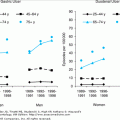
Estimates of the prevalence of visual impairment per 1000 persons in the United States demonstrate the significant increase in vision problems with age. A 2001 study estimated that 14% of persons aged 70 to 74 years have serious difficulty seeing, even with their spectacle correction, and this increases to 32% among persons aged 85 years or older. Centers for Disease Control and Prevention (CDC) and the National Centers for Health Statistics (NCHS) estimate the prevalence of severe visual impairment among persons 70 to 74 years of age at about 1% of the elderly population, but by age 85 years, nearly 2.5% of persons are severely visually impaired. After age 85 years, one in four older people cannot read a newspaper even with best-corrected vision.
Age-related visual impairment is not only challenging to the person in whom it develops, but also affects society as a whole. Medicare beneficiaries with coded diagnoses of vision loss have been shown to incur an additional $2.14 billion in 2003 in noneye-related medical costs, incurring significantly higher costs than those with normal vision. Additional eye-related costs per patient yearly are approximately $345 for those with moderate vision loss, $407 for those with severe vision loss, and $237 for those who are blind. Additional noneye-related costs per patient yearly are $2193, $3301, and $4443, respectively. Prevent Blindness America, a national volunteer eye health and safety organization, puts the medical loss for visual impairment at $5.48 billion for persons 40 years and older. Their number includes increased medical expenditures, as well as increased informal care days. They define informal care as unpaid care provided by people not living with the older person, and valued it according to minimum wage dollars. Health utility (distress, pain, depression, lack of mobility, social limitations) was converted into quality-adjusted life years and the total lost value for this factor was $10.5 billion. Thus, preventing vision loss among older persons is not only a medical imperative, but also an economic one.
Vision loss is reason enough for a decline in function among older persons, but vision loss has also been associated with cognitive decline, heart disease, arthritis, hypertension, falls and hip fracture, depression, reduced overall quality of life and mortality. Older visually impaired persons are twice as likely to have difficulty walking as do sighted peers, three times more likely to have difficulty getting outside, more than twice as likely to have difficulty getting in and out of a bed/chair, and three times more likely to have difficulty preparing a meal.
The most prevalent age-related causes of visual impairment in the United States are macular degeneration, diabetic retinopathy, glaucoma, and cataract. Approximately 60% of persons with visual impairments who are not institutionalized have one or more additional impairments. These include the loss of hearing, impaired mobility, decreased energy and stamina from respiratory and heart disease, and cognitive changes resulting from stroke or dementia. Vision loss has been ranked third, behind arthritis and heart disease, among the most common chronic conditions causing older persons to require assistance with activities of daily living (ADLs). Because the majority of people with visual impairments have useful vision, rehabilitation services and vision-enhancing techniques and devices offer opportunities to increase their visual and general functional capacity.
Aging and Loss of Vision
Every older person experiences age-related changes in vision. Table 43-1 summarizes the age-related changes that cause functional declines for older persons. Decreased transmission of the ocular media, increased scatter in the cornea, lens, vitreous body, and retina as well as decreased pupil size are related to anatomic changes in the aging eye. The age-related changes discussed here are those that have the greatest impact on function in daily life. These common changes in vision function must be taken in account when considering the daily living, quality of life, and the design of facilities for all older persons.
CHANGES | REASON | IMPLICATIONS FOR DAILY LIFE |
|---|---|---|
Loss of accommodation |
| Increasing inability to focus on close targets beginning around age 45 yrs. Plus lenses prescribed in a bifocal, reading glasses, or contact lenses; required to compensate for loss of accommodative ability. |
Loss of low-contrast acuity |
| Functional loss of acuity under glare or low lighting may cause small targets to be missed, bumping or tripping into low-lying objects. |
Increased sensitivity to glare |
| Discomfort even in low glare conditions such as cloudy days. High glare causes decreased acuity and difficulty in seeing targets in the environment. Sun lenses, hats, visors, and umbrellas provide more comfort outdoors; tinted lenses may be prescribed for indoors. |
Increased difficulty with dark adaptation |
| Difficulty moving from bright to dim environments. Risk for stumbling or falling is greater under these circumstances. Fear of balance problems or falling may cause compensatory behaviors such as shuffling, reaching for hand-holds, etc. Change environmental lighting to avoid light/dim areas. |
Loss of color discrimination |
| Difficulty detecting differences in dark colors and pastels; adding lamps or identifying matching colors near a sunny window helps. |
Loss of attentional visual field |
| Risk factor for balance and mobility problems and vehicle crashes in driving. Training improves performance. |
Increased difficulty with visual reading ability |
| Reading speed of older readers reduced by one-third that of younger readers; text navigation skills decline with age. Training improves performance. |
The ability to accommodate for focus on visual targets from distance to near, which is dependent on a flexible crystalline lens and the ciliary muscle, is altered with age, beginning around age 45 years. During this change, an increasing amount of plus in a concave lens (usually prescribed in bifocal lenses or reading glasses) is required to boost the focusing power of the eye to compensate for the loss in refracting ability of the lens for near tasks.
The visual acuity of normally sighted older persons shows only a modest decrease under high-contrast conditions, but reducing the illumination of an acuity chart, reducing the contrast of the acuity chart, and/or adding surrounding glare, produces drastic age-related acuity losses, as compared to young observers. For example, in a sample of 900 older observers, for those at age 82 years, the median high-contrast visual acuity was 20/30, low-contrast high-luminance acuity was 20/55, low-contrast, low-luminance acuity was 20/120, and low-contrast acuity in glare conditions was 20/160. A young observer loses only about one line of acuity under similar conditions.
Because of anatomic changes in the eye and media, older persons are more sensitive to glare in the environment, more likely to experience disabling glare, and have reduced glare recovery time. This may have the most impact on activities such as walking outdoors or driving, but may also affect indoor activities as well, if very bright and dim environments are adjacent, such as restaurants, movie theaters, and atriums. Visual discomfort may arise because of glare, and disabling glare may hide important targets that must be viewed for the sake of safety. Glare sensitivity has been associated with motor vehicle accidents for older drivers.
Attentional visual field, the visual field area over which one can process rapidly presented visual information, declines with age. Unlike conventional measures of visual field that assess visual sensory sensitivity (such as static flashing lights), attentional visual field relies on higher-order processing skills such as selective and divided attention and rapid processing speed. Decreased attentional visual field has been correlated to a greater incidence of driving accidents and is related to a greater risk for balance and mobility problems for normally sighted older persons. Attentional visual field can be improved with training, but such training is not widely available.
Visual reading ability decreases with age as well. The reading rate of older persons who are normally sighted and have good high-contrast visual acuity decreases by as much as a third of that of young readers. Accuracy of reading, however, can remain comparable to that of younger readers. Reading performance among older persons with good acuity (20/30) is highly correlated with attentional visual field; those with good reading performance into very old age also retain good attentional fields as well. Low-contrast visual acuity is also correlated with reading performance for this population; older persons with poor low-contrast acuity tend to read more slowly. Even when high-contrast acuity is good, older persons, especially the oldest old, are at risk for reading difficulties that may arise from a combination of reduced attentional visual field, slower saccadic performance in eye movements, and poor low-contrast visual acuity. Reading rate for normally sighted older adults with good high-contrast acuity and good comprehension skills can be improved with training. Training in reading efficiency that emphasizes improvements in eye movements for reading similar to those exercises used to improve reading for school children has given good results for this population. Such training, however, is not widely available.
Color discrimination is another aspect of vision that declines with advancing age. Persons who are older have greater difficulty detecting differences between dark colors such as brown, black, or navy, and also have difficulty with pastels. Loss of color vision in old age is related to smaller pupil diameter, reduced light transmission through the lens, and changes in photoreceptors and neural pathways.
Dark adaptation declines as a result of losses in ocular transmittance and pupillary miosis resulting from the aging process. Difficulty with dark adaptation can be limiting to older adults moving from light to dim environments and vice versa. The risk for stumbling or falling may be greater under these circumstances. The normally sighted older person may function as if severely visually impaired when first adjusted to a drastic change in illumination.
Although not related to a change in vision per se, care must be taken in providing refractive correction in spectacle form for geriatric patients. Multifocal spectacle lenses either in the form of bifocals or varifocals are commonly prescribed but are associated with a higher risk of “edge of step” accidents, and multifocal lens wearers are twice as likely to fall as single focus lens wearers. A large percentage of these falls are reported to occur outside the home, perhaps as a result of tripping or stumbling resulting from obstacles not seen in the near vision correction of the lower visual field. The bifocal portion of a spectacle correction provides additional dioptric power to provide vision at near distance for reading, etc. This means that objects outside this near-focal range such as steps, curbs, stairs, house cats, etc. are blurred and indistinct. This effect is greatest in older patients, as the need for extra dioptric power for near vision increases with age. Patients with multifocal correction are encouraged to tuck their chins and look over the top of the bifocal correction when moving so that they can look through the distance correction in the upper portion of the lenses, but head flexion significantly increases postural instability. Encouraging those who are at risk for falling to explore these issues with their eye care specialists can be an important aspect of falls prevention.
Prevalent Age-Related Causes of Visual Impairment
Functional vision loss because of age-related macular degeneration may include metamorphopsia (visual images appear distorted and wavy), relative scotomas, and result in dense central scotomas for those whose pathology progresses to visual impairment. Individuals with central scotoma usually develop a strongly preferred retinal locus (PRL) that performs as the primary fixation reference, although the patient may not always be aware that there is a scotoma present. The loss of central visual field results in loss of visual acuity and contrast sensitivity. The ability to use the PRL that develops for fixation may be difficult for many persons. The effects of macular degeneration on daily life include difficulty with reading print, inability to recognize faces (that can lead to reluctance to participate in social activities), difficulty with distance and depth cues (that adversely affect safe mobility), and loss of color and contrast sensitivity (that interfere with a variety of household and work/leisure tasks) (Table 43-2).
CONDITION | COMMON CLINICAL PRESENTATION | IMPLICATIONS FOR REHABILITATION |
|---|---|---|
Macular degeneration |
| Difficulty with tasks requiring fine detail vision such as reading, inability to recognize faces, distortion or disappearance of the visual field straight ahead, loss of color and contrast perception, mobility difficulties related to loss of depth and contrast cues. |
Diabetic retinopathy |
| Difficulty with tasks requiring fine detail vision such as reading, distorted central vision, fluctuating vision, loss of color perception, mobility problems because of loss of depth and contrast cues. |
Cataract |
| Usually remedied by lens extraction and implant, except in extreme cases. If not managed by implant, difficulty with detail vision, difficulty with bright and changing light, color perception, decreased contrast perception, some mobility problems caused by loss of perception of depth and distance, sensitivity to glare, and loss of contrast. |
Glaucoma |
| Mobility and reading problems because of restricted visual fields, people suddenly appearing in the visual field seen as “jack in the box.” |
Traumatic Brain Injury |
| Mobility and reading problems owing to visual field loss and spatial perceptual difficulties that reduce ability to drive, reach correctly, and execute eye movements related to reading, difficulty with visual and cognitive processing. Visual neglect may include inability to complete shaving and/or dressing. Difficulty completing other rehabilitation assessments and interventions because of neglected visual impairment. |
The progression of diabetic retinopathy includes macular edema that may cause blurred vision if the fovea is involved, retinal hemorrhages (and/or laser treatments), which may result in scattered central, peripheral, and/or midperipheral scotomas, and retinal detachment, which can cause larger areas of field loss if not reattached. Diabetic retinopathy can progress to total blindness. Loss of function can include decreased visual acuity, scattered field loss over the retina, metamorphopsia across the retina, increased sensitivity to glare, and loss of color and contrast sensitivity. If the fovea is lost to scotoma, then a PRL will develop. Vision fluctuations can be manifested over time as macular swelling increases or subsides, and can also be related to hemorrhage. Sudden vision loss is common following hemorrhage, with the patient describing episodes of smoky vision, a dropped veil over the eye(s), or seeing black or red strings across the field of view. Treatment and absorption of blood can improve acuity, though not usually to normal levels. The effects on daily life include difficulty reading print materials, difficulty recognizing faces, increased sensitivity to glare and light/dark adaptation, difficulty with distance and depth cues, loss of color and contrast sensitivity, and fluctuating vision.
Age-related cataract is manifested by gradual opacity of the lens, which interferes with the passage of light, causing reduced visual acuity, light scatter, sensitivity to glare, altered color perception, and image distortion (straight lines appear wavy). Persons with cataracts may experience trouble with glare and loss of contrast, may have decreased acuity, and report areas of metamorphopsia or small scotomas in the visual field. When the cataract has begun to interfere with lifestyle, surgery may be performed to remove either the entire lens or the posterior portion. Correction for the removal of the lens is provided through intraocular lens implants, eyeglasses, or contact lens. Cataract surgery is the most common major surgical procedure done for persons older than age 65 years who are receiving Medicare. Cataract surgery with lens implantation is associated with improved objective and subjective measures of function in ADL, as well as improved levels of vision to normal acuity in most cases.
Glaucoma is an increase in intraocular pressure caused by an abnormality in flow of aqueous fluid from the anterior chamber. It can cause a degeneration of the optic disk, loss of visual fields, and severe visual impairment. When left untreated, or if treatment is not successful, glaucoma results in a loss of peripheral fields and can lead to blindness. The effect of peripheral field loss on daily life is most problematic in safe ambulation. Because of field restrictions, the patients may not see objects in their path and may bump into objects that fall outside the field of view in any direction (street signs, tree branches, etc.). In addition, a person outside the patient’s field of view may suddenly be seen as a “jack-in-the-box” and create a startle effect. Peripheral field loss may also create problems in reading and writing as only a small portion of the page can be seen at once.
Head injury to older adults such as cerebrovascular accident, falls, or automobile accidents resulting in traumatic brain injury can lead to visual impairment. Between 20% and 40% of stroke results in visual disorder that can inhibit cognitive functioning and may reduce the effectiveness of rehabilitation of traumatic brain injury. Visual field disorders can result from injury to the visual pathway anyway between the retina and the striate cortex. The optic chiasm is used as an anatomical landmark to differentiate between the peripheral (prechiasmatic) and the central (postchiasmatic) visual pathway. Unilateral injury to the prechiasmatic pathway affects the ipsilesional field only, but postchasmatic injury causes visual deficits in both monocular hemifields and are referred to as homonymous. Visual field disorders must be discerned by visual field measurement techniques called perimetry; patients are often unaware of them and do not report the defect, but may suffer from their effects, for example, bumping into objects, tripping, falling, being unable to read, etc. Vision can be completely lost in the missing field, or some vision function (for example, light detection) may remain. The most common visual field losses are hemianopsia (loss of half the visual field), followed by quadranopsia (loss of one quadrant) and paracentral scotoma (island of vision loss in the parafoveal region), and rarely results in central scotoma. Recovery of some visual field following injury may be spontaneous, and some patients learn spontaneously to adapt to visual field loss by oculomotor strategies; shifting gaze may reveal what is missing in the field of view of a street scene (such as an oncoming car) or the missing portion of a line of text. Systematic training in oculomotor adaptation and visual perceptual training can improve vision function and has limited ability to improve the visual field. Visual field loss may also be accompanied by visual neglect. Older adults with neglect may not spontaneously be able to attend to the neglected side. Traumatic Brain Injury (TBI) may also result in disorders of visual space perception, which affect reach (over- or underreaching for objects, knocking things over), driving (accidents resulting from inability to judge distance and depth), and reading (inability to plan and execute accurate eye movements). Visuospatial localization and orientation may be improved through training, but may not reach pre-TBI thresholds. Visual agnosia, a failure to visually recognize an object because of “mistaken identity,” is a disorder that is based in both visual-perceptual and visual-cognitive functions. Typically, misidentifications result from the incomplete or inappropriate use of object features such as size, shape, or color. Older adults with TBI may be unaware that they are ignoring other features that might assist with correct identification. Cognitive and/or communication disorders can make visual impairment more difficult to detect following TBI. Undetected or untreated visual impairment in TBI can limit the effectiveness of other rehabilitation (e.g., many cognitive and functional assessments use visual items that cannot be appropriately identified by older persons with TBI-related vision loss, visual motor assessments require eye–hand coordination). If TBI vision loss is not detected and treated to the extent possible, the examiner or therapist may get a false-negative impression of the level of TBI disability. In addition, the TBI patient will be frustrated and troubled unduly by participating in rehabilitation that does not simultaneously address the vision deficit.
Role of the Geriatrician in Vision Rehabilitation
After diagnosis and medical management of the patient’s vision loss by an ophthalmologist, the geriatrician can play an important role in assuring that visually impaired persons receive rehabilitation services that are of high quality, are sought in a timely manner, and provide all the benefit that the patient might be able to derive from them.
A geriatrician can provide the following services for their patients related to vision rehabilitation:
A visual acuity evaluation. Current best practice includes the use of a logarithmic visual acuity chart.
A contrast sensitivity function evaluation. The Pelli–Robson chart is recommended for its ease of use and reliability.
A referral to a low-vision eye care specialist (ophthalmologist or optometrist) for the appropriate clinical low-vision evaluation and prescription of optical low vision devices for tasks the older person can no longer perform such as reading, writing, watching television, and recognizing street signs.
A referral to vision rehabilitation professionals for assessment and instruction of vision and magnification devices for literacy, ADLs, and safe travel. These therapists can also provide environmental analyses and teach the use of environmental cues.
Assistance to patients in preparing for rehabilitation by providing information and encouraging them to consider the goals they would like to achieve. The National Eye Institute: Visual Functioning Questionnaire-14 is a modified 14-item questionnaire that is effective in assessing the impact of vision loss on quality of life and is helpful in assisting patients in setting goals for rehabilitation.
Counseling or referral for coping with psychosocial issues related to visual impairment. Patients may not be forthcoming about these issues, so the physician must ask. Adjustment disorder and depression are associated with visual impairment for older persons. When patients are dealing with loss of independence and control, lowered self-esteem and strained social relations, counseling and/or psychotherapy may be recommended for both patients and family members.
Reinforcement of simple strategies, such as the use of saturated colors and contrast in the home environment, and the use of simple devices, such as sun lenses outdoors and brighter indoor environments.
Information to the patient and family about the variable nature of low vision, its effect on daily life tasks, and the variable nature of visual abilities according to fluctuations of light and contrast.
Sponsorship of, or referral to, support groups where older persons with vision loss and their families can discuss problems, coping, and rehabilitation strategies they have learned with other patients.
Assistance in community awareness efforts about the prevalence, treatment, and rehabilitation of visual impairment among older persons.
Patients likely to benefit from vision rehabilitation include those with reduced acuity of less than 20/50 in the better-seeing eye, central or peripheral field loss with intact visual acuity, reduced contrast sensitivity, glare sensitivity, and/or light/dark adaptation difficulties as well as those with traumatic brain injury. Candidates for cataract surgery with macular disease might also benefit from preoperative low-vision assessment and coincident rehabilitation training that enhances postoperative visual performance and satisfaction with a cataract procedure.
Adaptations of Clinical and Functional Evaluations for Older Adults
The clinical and functional low-vision examinations for older persons should distinguish aging from treatable disease processes; focus holistically; be multidisciplinary and incorporate family and caregiver support; and identify and set realistic goals to improve functional status and quality of life.
In health care service, delivery to older adults with low vision, certain aspects of the examination sequence is adapted to accommodate these principles.
