Appendiceal Mucinous Neoplasms and Pseudomyxoma Peritonei


Appendiceal Mucinous Neoplasms and Pseudomyxoma Peritonei


Joseph Misdraji


Most epithelial neoplasms of the appendix are of mucinous type and characteristically cause cystic dilatation of the appendix with accumulation of copious gelatinous material. Their importance lies in the fact that they can disseminate in the peritoneal cavity and coat serosal surfaces with mucinous tumor deposits, a finding that has been called “pseudomyxoma peritonei.” The appendiceal mucinous neoplasm has been the subject of numerous debates since its recognition as the source of pseudomyxoma peritonei. Philosophical differences regarding the nature of pseudomyxoma peritonei have generated a number of classification systems for appendiceal mucinous neoplasms. The inconsistent diagnostic terminology has resulted in considerable confusion with respect to the pathologic classification of appendiceal mucinous neoplasms and pseudomyxoma peritonei, with ramifications on patient management and research in this area. The purpose of this chapter is to discuss the pathologic and clinical features of these lesions, as well as their biologic behavior and clinical management. Other epithelial tumors of the appendix are discussed in Chapter 20.


CLASSIFICATION OF MUCINOUS NEOPLASMS OF THE APPENDIX AND PERITONEUM

There is considerable controversy regarding the most appropriate terminology for appendiceal mucinous tumors and pseudomyxoma peritonei because tumors that resemble appendiceal adenomas can disseminate to the peritoneum and cause the death of the patient. Most of the debate is due to philosophical differences regarding the nature of pseudomyxoma peritonei. One viewpoint is that pseudomyxoma peritonei results from rupture of an adenoma of the appendix, with seeding of the peritoneum by low-grade neoplastic, but noninvasive mucinous epithelium. Investigators with this viewpoint classify pseudomyxoma peritonei as disseminated peritoneal adenomucinosis.1 The opposing view is that peritoneal dissemination is equivalent to carcinoma and that the source appendiceal tumors are actually adenocarcinomas.2,3 However, the criteria for invasion in appendiceal tumors are poorly defined, and their proposed classifications reflect the difficulty of assigning them into benign or malignant categories (Table 19.1).1, 2, 3, 4 and 5 The Armed Forces Institute of Pathology proposed that appendiceal mucinous neoplasms be classified as adenomas, tumors of uncertain malignant potential, and invasive mucinous carcinoma, based on tumor stage.2 That scheme defines adenomas as mucinous neoplasms confined to the mucosa with intact muscularis mucosae, whereas tumors with infiltrative mural invasion or proliferation of neoplastic epithelium outside the appendix (pseudomyxoma peritonei) are considered to be carcinomas. More problematic tumors that show “pushing” invasion with tongues of epithelium dissecting outward in a “diverticulum-like” fashion or acellular mucin outside the appendix associated with loss of the muscularis mucosae are classified as mucinous tumor of uncertain
malignant potential. Others define the uncertain malignant potential category as tumors with involvement of the proximal margin, tumors with mucin or epithelium in the appendiceal wall that is not clearly invasive, and cases in which the possibility of extraappendiceal epithelium cannot be excluded.5,6 Some have suggested that, to avoid nomenclature confusion, the entire spectrum of mucinous tumors, from adenomas to mucinous tumors with presumed pushing invasion and pseudomyxoma peritonei, be classified using a single-term, low-grade appendiceal mucinous neoplasms (LAMN) and that the pathology report describes the stage of disease.4 In its most recent classification of gastrointestinal tumors, the World Health Organization adopted this terminology to describe low-grade carcinomas but maintained the term adenoma for tumors confined to the mucosa.7


Table 19.1 Classification Schemes for Mucinous Neoplasms of the Appendix


Pathologic Features


Carr and Sobin2


Misdraji et al.4


Pai and Longacre5


World Health Organization7


Tumors without invasion (intact muscularis mucosae)


Adenoma


LAMN


Adenoma


Adenoma


Tumors with Pushing Invasion


• Tumor confined to the appendix


Mucinous tumor of uncertain malignant potential


LAMN


Adenoma


LAMN


• Tumor with acellular mucin outside the appendix


Mucinous tumor of uncertain malignant potential


LAMN


Low-grade mucinous neoplasm with low risk of recurrence


LAMN


• Tumor with extraappendiceal tumor


Mucinous adenocarcinoma


LAMN


Low-grade mucinous neoplasm with high risk of recurrence


LAMN


Tumors with Infiltrative Invasion


Infiltrative invasion of wall, with or without desmoplasia, regardless of stage


Invasive mucinous adenocarcinoma


Invasive mucinous adenocarcinoma


Invasive mucinous adenocarcinoma


Invasive mucinous adenocarcinoma


APPENDICEAL ADENOMA AND CYSTADENOMA

Adenomas are benign tumors of the appendix that are often incidental lesions detected in appendectomy specimens obtained for other reasons, but they can present with right lower quadrant pain and infrequently a palpable right lower quadrant mass. They might not produce any gross abnormalities or can cause cystic dilatation due to accumulation of tenacious mucin, but they do not display mucin on the external surface. Adenomas are confined to the appendiceal mucosa and surrounded by intact muscularis mucosae, although the lymphoid tissue of the lamina
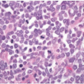
propria is often atrophic (Figure 19.1).2,4,8 Tumors are composed of a villous or flat proliferation of mucinous epithelial cells with abundant intracytoplasmic mucin (Figure 19.2). Deeper glands typically have straight luminal edges, although serrated architecture may also be present, mimicking the appearance of a serrated polyp, as discussed in Chapter 20.1,2 Mucin accumulation frequently produces cystic dilatation of the appendix, in which case the term cystadenoma may be applied. Cystadenomas mostly contain a flat, undulating epithelial cell lining with infrequent villous areas that distinguish these lesions from retention cysts (Figure 19.3).2,4,8,9

Appendiceal mucinous adenomas display a spectrum of cytologic atypia, which is classified as low- or high-grade dysplasia in the same manner as in other parts of the gastrointestinal tract. Most tumors are low grade with nuclear hyperchromasia, pseudostratification, rare mitoses, and apoptotic nuclear debris.4,9,10 High-grade tumors show a greater degree of nuclear pleomorphism with stratification and loss of cell polarity equivalent to intraepithelial carcinoma (Figure 19.4). However, appendiceal mucinous adenomas have no potential for aggressive clinical behavior and are cured by appendectomy. Mucinous neoplasms associated with mural changes, such as disruption of the muscularis mucosae or replacement of the muscularis mucosae by fibrous tissue, or with extraappendiceal mucin are better classified as LAMN to convey some degree of uncertainty regarding the biologic potential of the lesion.


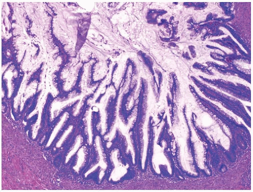
FIGURE 19.1: An appendiceal villous adenoma is composed of slender villi lined by mucinous epithelial cells with an intact muscularis mucosae. The appendiceal lymphoid tissue is atrophic.


FIGURE 19.2: The villi of an appendiceal mucinous adenoma are lined by columnar mucinous epithelial cells containing tall mucin vacuoles. The nuclei show mild hyperchromasia and nuclear pseudostratification, similar to low-grade dysplasia in other parts of the gastrointestinal tract.


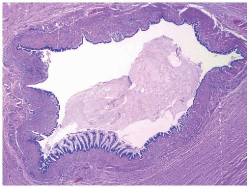
FIGURE 19.3: An appendiceal mucinous cystadenoma contains an undulating, flat lining of mucinous epithelial cells with only a small area of villous projections (lower left). The muscularis mucosae is intact.


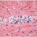
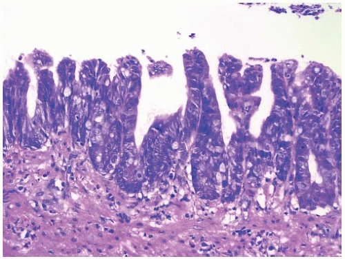
FIGURE 19.4: A mucinous adenoma with high-grade dysplasia is confined to the mucosa but shows nuclear hyperchromasia, nuclear disarray, and fullthickness nuclear stratification.


LOW-GRADE APPENDICEAL MUCINOUS NEOPLASM

LAMN resemble appendiceal adenomas but have the propensity to spread to the peritoneum, recur, and result in the death of the patient. They contain mucinous epithelium and display a villous architecture or undulating, flat luminal surface. Unlike adenomas, LAMN show pushing invasion through the muscularis mucosae, hyalinizing fibrosis of the muscularis mucosae, or frank rupture of the appendix with extruded mucin. Unfortunately, criteria for mural invasion in appendiceal mucinous neoplasms are not well defined. As a result, they have been variably classified as ruptured adenomas, tumors of uncertain malignant potential, and adenocarcinomas reflecting the difficulty of classifying them as benign or malignant (Table 19.1).1, 2 and 3,5,7, 8 and 9,11,12 The most recent World Health Organization classification scheme denotes low-grade mucinous tumors with pushing invasion as LAMN, whereas the term mucinous adenocarcinoma is reserved for high-grade tumors or those with conventional invasion.7 Both low- and high-grade mucinous neoplasms that extend beyond the muscularis mucosae are staged as adenocarcinomas in the seventh edition of the American Joint Committee on Cancer (AJCC) Staging Manual.13

May 22, 2016 | Posted by in ONCOLOGY | Comments Off on Appendiceal Mucinous Neoplasms and Pseudomyxoma Peritonei

Full access? Get Clinical Tree

Get Clinical Tree app for offline access