malignant potential. Others define the uncertain malignant potential category as tumors with involvement of the proximal margin, tumors with mucin or epithelium in the appendiceal wall that is not clearly invasive, and cases in which the possibility of extraappendiceal epithelium cannot be excluded.5,6 Some have suggested that, to avoid nomenclature confusion, the entire spectrum of mucinous tumors, from adenomas to mucinous tumors with presumed pushing invasion and pseudomyxoma peritonei, be classified using a single-term, low-grade appendiceal mucinous neoplasms (LAMN) and that the pathology report describes the stage of disease.4 In its most recent classification of gastrointestinal tumors, the World Health Organization adopted this terminology to describe low-grade carcinomas but maintained the term adenoma for tumors confined to the mucosa.7
Table 19.1 Classification Schemes for Mucinous Neoplasms of the Appendix | ||||||||||||||||||||||||||||||||||||||||
|---|---|---|---|---|---|---|---|---|---|---|---|---|---|---|---|---|---|---|---|---|---|---|---|---|---|---|---|---|---|---|---|---|---|---|---|---|---|---|---|---|
| ||||||||||||||||||||||||||||||||||||||||
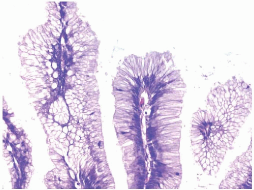
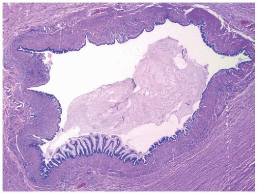
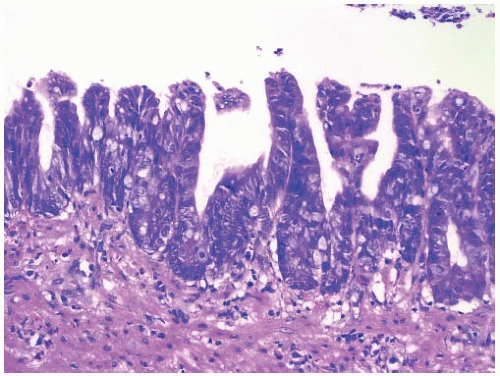
propria is often atrophic (Figure 19.1).2,4,8 Tumors are composed of a villous or flat proliferation of mucinous epithelial cells with abundant intracytoplasmic mucin (Figure 19.2). Deeper glands typically have straight luminal edges, although serrated architecture may also be present, mimicking the appearance of a serrated polyp, as discussed in Chapter 20.1,2 Mucin accumulation frequently produces cystic dilatation of the appendix, in which case the term cystadenoma may be applied. Cystadenomas mostly contain a flat, undulating epithelial cell lining with infrequent villous areas that distinguish these lesions from retention cysts (Figure 19.3).2,4,8,9
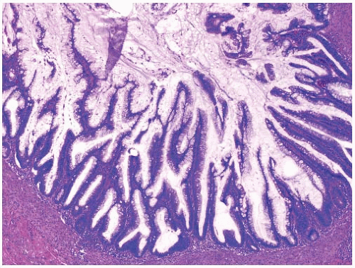 FIGURE 19.1: An appendiceal villous adenoma is composed of slender villi lined by mucinous epithelial cells with an intact muscularis mucosae. The appendiceal lymphoid tissue is atrophic. |
Stay updated, free articles. Join our Telegram channel

Full access? Get Clinical Tree


